
Abstract
This study investigated combined chemopreventive potential of curcumin and resveratrol during benzo(a)pyrene (BP)-induced lung carcinogenesis in mice. The mice were segregated into five groups that included normal control, BP-treated, BP + curcumin-treated, BP + resveratrol-treated, and BP + curcumin + resveratrol-treated groups. A statistically significant increase in the levels of lipid peroxidation (LPO) was observed in the lungs of mice after 22 weeks of single dose of benzo(a)pyrene. Further, BP treatment also resulted in a significant increase in the enzyme activities of aryl hydrocarbon hydroxylase as well as drug-metabolizing enzymes, namely cytocrome P450 and cytochrome b5. On the other hand, reduced glutathione (GSH) levels, the activities of superoxide dismutase (SOD), glutathione reductase (GR), and glutathione-S-transferase (GST) were found to be significantly decreased following BP treatment. Supplementation with curcumin and resveratrol to BP-treated mice significantly decreased the LPO levels, GSH levels, and enzyme activities of drug-metabolizing enzymes. Further, treatment of curcumin and resveratrol to BP-treated mice significantly elevated the activities of SOD, GR, and GST. Histoarchitectural studies showed well-differentiated signs of lung carcinogenesis following BP administration to mice. However, combined treatment with curcumin and resveratrol resulted in a noticeable improvement in the lung histoarchitecture. This study, therefore, concludes that curcumin and resveratrol when supplemented in combination regulate drug-metabolizing enzymes as well as antioxidant enzymes during lung carcinogenesis in mice.
Introduction
Polycyclic aromatic hydrocarbons (PAH) are ubiquitous environmental agents that significantly contribute to human cancers. 1 PAHs are metabolized enzymatically to various reactive metabolites by the cytochrome P450 and cytochrome b5 enzymes, which are known to be involved in carcinogenic metabolism. 2 Benzo(a)pyrene (BP), which is a major carcinogenic pollutant, 3 comes from the family of PAH and is metabolized to ultimate carcinogen by metabolic activation and, thereby, intercalates in DNA leading to formation of DNA adducts. DNA adducts finally end in mutations that ultimately lead to cancer.
Cancer chemoprevention, using two chemopreventives in combination, is an upcoming approach to fight this noxious disease. Dietary constituents, if taken in adequate amounts in combination, may offer efficient chemoprevention and could delay the process of carcinogenesis. 4 –7 Among various chemopreventive agents, phytochemicals have shown great potential in combating cancer and other chronic diseases that result from oxidative stress induced by free radicals. 8
In this study, curcumin and resveratrol are the phytochemicals of interest. Although both phytochemicals have desirable biological activities against cancer, there is a paucity of information with regard to their efficacy in modulating drug metabolizing enzymes during carcinogenesis. So this study is an attempt to explore the combined prophylactic action of these two well-known phytochemicals on modulation of cytochrome enzymes as well as antioxidant indices during benzo(a(pyrene against carcinogenesis.
Materials and methods
Chemicals
BP, curcumin, and resveratrol were procured from Sigma Aldrich company (St Louis, Missouri, USA). Nicotinamide adenine dinucleotide phosphate (NADPH), glutathione (GSH), nitroblue tetrazolium (NBT), and 5,5′-dithiobis-(2-nitrobenzoic acid) (DTNB) were procured from Merck chemicals (Shanghai, China).
Animals
Male Laka mice in the weight range of 18–20 g were procured from the central animal house, Southeast University, Xuzhou, Jiangsu, China. The animals were housed in polypropylene cages under hygienic conditions in the departmental animal house by strictly following the guidelines as outlined by the institutional ethical committee.
Experimental design
Animals were segregated equally and randomly into five treatment groups. Animals in group 1 served as normal controls and were also administered corn oil intraperitoneally (IP), which was used as a vehicle for the BP-treated animals. Animals in group 2 were given a single intraperitoneal injection of BP in corn oil at a dose level of 100 mg/kg body weight. 4 Group 3 animals were given curcumin orally in drinking water at a dose level of 60 mg/kg/body weight, 4 thrice a week. Animals in group 4 were given resveratrol orally at a dose level of 5.7 μg/ml drinking water, thrice a week. 4 Both the phytochemicals were given to animals using intubation gavage technique. Animals in group 5 were given a combined treatment of curcumin and resveratrol in a similar manner as was given to animals of groups 3 and 4, respectively. The animals were subjected to treatment with phytochemicals, 10 days prior to BP injection. All the animals had free access to the diet and water, and the treatments continued for a total duration of 22 weeks.
Preparation of lung homogenate
Lung homogenates were prepared by following the method of Malhotra et al. 5 After killing the animals, the lungs were removed immediately and washed with ice-chilled saline. Then, 10% lung homogenates were prepared in ice-cold Tris buffer (pH 7.4) using mechanically driven Teflon-fitted Potter– Elvejhem-type homogenizer for a few minutes till the cells disrupt completely. Homogenates were centrifuged at 1000g for 10 min at 4°C. Pellets were discarded, and supernatant were used for the estimation of lipid peroxidation (LPO) and reduced GSH levels. A portion of the above supernatant was again centrifuged at 10,000g for 20 min to obtain postmitochondrial fraction that was utilized for the rest of biochemical estimations.
Drug-metabolizing enzymes
Aryl Hydrocarbon Hydroxylase (AHH) by the method of Nebert and Gelbion. 9 BP was used as substrate, which on reaction with AHH get hydroxylated to 3-hydroxy benzo(a)pyrene, which was estimated using spectroflourimeter.
Cytochrome P450 by the method of Omura and Sato. 10 Cytochrome P450 was measured by the carbon monoxide difference spectrophotometry of dithionite-reduced samples.
Cytochrome b5 by the method of Omura and Sato. 11 Cytochrome b5 was measured by the carbon monoxide difference spectrophotometry of nicotine amide dinucliotide-reduced samples.
Antioxidant defense system enzymes and LPO
LPO assay was done by the method of Wills et al. 12 Briefly, 0.5 ml of tissue homogenate (10% w/v) was diluted to 1.0 ml with ice-cold 10% trichloroacetic acid (TCA) and after a thorough mixing, the reaction mixture was centrifuged at 800g for 10 min. To 0.5 ml of supernatant, 0.5 ml of 0.67% Thiobarbituric acid (TBA) was added, and the color was developed by boiling at 100°C for 10 min. Samples were cooled, and the absorbance was read at 532 nm.
Reduced GSH
Estimation of GSH was performed in the tissue homogenates of lung by following the method of Ellman. 13 Briefly, 0.1 ml of 25% TCA was added to 0.5 ml of lysate. After protein precipitation by TCA, the samples were centrifuged to obtain the supernatant. Then, 0.1 ml of supernatant was incubated with 2.0 ml of freshly prepared 0.6 mmol/l DTNB. Optical density of the yellow complex was measured at 412 nm.
Catalase
The method of Luck 14 was used for the estimation of catalase. Briefly, the reaction mixture contained 50 mmol/l potassium phosphate buffer (pH 7.0), 1.25 × 10⁻² mol/l hydrogen peroxide (H2O2), and the sample. Every sample was analyzed with appropriate blanks without H2O2. The decrease in the absorbance was measured at 240 nm, and enzyme activity was expressed as micromoles of H2O2 decomposed per minute per milligram protein.
Superoxide dismutase
The activity of superoxide dismutase (SOD) was estimated by using the method of Kono. 15 The method is based on the principle of the inhibitory effect of SOD on reduction of NBT dye by superoxide anions, which are generated by photooxidation of hydroxylamine hydrochloride.
Glutathione reductase
The enzyme was assayed by following the method of Carlberg and Mannervik. 16 Briefly, 2.6 ml of 0.2 M phosphate buffer (pH 7.0), 0.15 ml of 2 mM reduced NADPH, and 0.15 ml of 20 mM oxidized GSH were added. The reaction was initiated by the addition of 0.1 ml of lung homogenate to the cuvette and the decrease in absorbance was recorded at 340 nm.
Glutathione-S-transferase
This enzyme was assayed by using the method of Habig et al., 17 which involved the use of 1-chloro-2,4-dinitrobenzene, 1,2-dichloro-4-nitrobenzene, and ethacrynic acid as substrates for glutathione S-transferase (GST). The increase in absorbance due to formation of glutathione conjugates by GST with different substrates was measured in a double-beam spectrophotometer.
Protein
Protein assay was done according to the method of Lowry et al. 18 Briefly, the samples were diluted with 100 mmol/l phosphate buffer (pH 7.5) to a volume of 0.5 ml. The reactions were diluted with 0.5 ml of 1.0 N sodium hydroxide followed by the addition of 5.0 ml of reagent mixture (containing 48 ml of 2% sodium carbonate, 1 ml of 1% copper sulfate, and 1.0 ml of 2% sodium potassium tartarate). After 10 min of incubation at room temperature, the color was developed by the addition of 1.0 N Folin’s reagent, and absorbance was measured using a spectrophotometer at 750 nm.
Histopathological studies
For the histopathological observations at light microscopic level, fresh tissue pieces of mice were immersion fixed in formalin. Following an overnight fixation, the specimens were dehydrated in ascending grades of alcohol, cleared in benzene, and embedded in paraffin wax. Blocks were made and 5–7-μm thick sections were double stained with hematoxylin and eosin and were observed under the light microscope at 40×.
Statistical analysis
The statistical significance of the data has been determined using one-way analysis of variance, followed by a multiple post hoc least significant difference test. The results are represented as means ± SD.
Results
The results obtained from various experiments conducted in this study are depicted in Tables 1 to 3 and light micrographs (LMG 1–5). The data from various treatment groups have been compared with the normal control animals. Further, results obtained from BP + curcumin-, BP + resveratrol-, and BP + curcumin + resveratrol-treated groups were compared with BP-treated group, and the results of BP + curcumin- and BP + resveratrol-treated groups were also compared with BP + curcumin + resveratrol combined treatment group.
Effect of 22 weeks of curcumin and resveratrol treatments on the activity of aryl hydrocarbon(AHH) hydroxylase in lungs of mice subjected to benzo(a)pyrene treatment.

Data are expressed in Mean
a P ≤ 0.05, b P ≤ 0.01 and c P ≤ 0.001 by Least Significance Difference test when values are compared with normal control group.
x P ≤ 0.05, y P ≤ 0.01, z P ≤ 0.001 by Least Significance Difference test when values of Groups III, IV & V are compared with Group II.
Effect of 22 weeks of curcumin and resveratrol treatments on the activies of cytochrome P450 and b5 in lungs of mice subjected to benzo(a)pyrene treatment.

Data are expressed in Mean
a P ≤ 0.05, b P ≤ 0.01 and c P ≤ 0.001 by Least Significance Difference test when values are compared with normal control group.
x P ≤ 0.05, y P ≤ 0.01, z P ≤ 0.001 by Least Significance Difference test when values of Groups III, IV & V are compared with Group II.
Effects of curcumin and resveratrol treatment of 22 weeks on antioxidant enzymes in lungs of mice subjected to benzo(a)pyrene treatment.
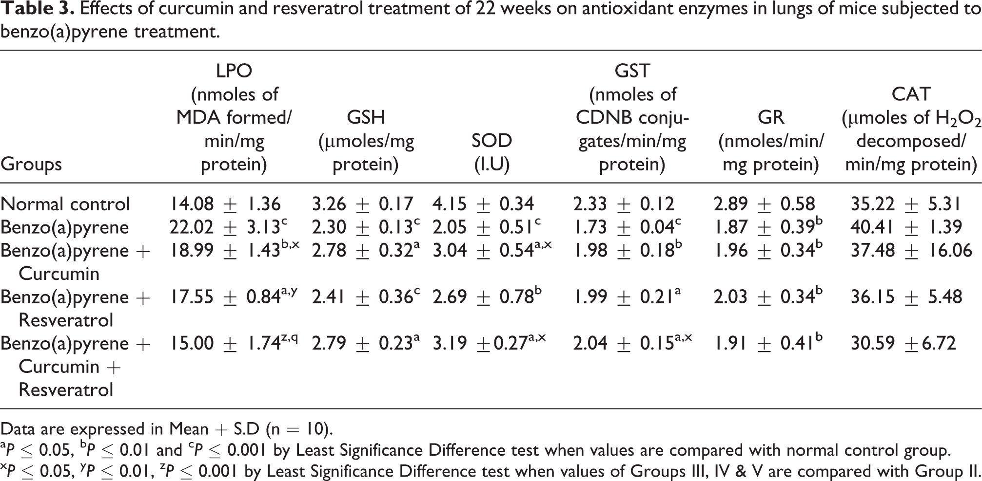
Data are expressed in Mean
a P ≤ 0.05, b P ≤ 0.01 and c P ≤ 0.001 by Least Significance Difference test when values are compared with normal control group.
x P ≤ 0.05, y P ≤ 0.01, z P ≤ 0.001 by Least Significance Difference test when values of Groups III, IV & V are compared with Group II.
A statistical significant increase in the enzyme activity of AHH (Table 1) was observed in the lungs of BP-treated mice. Further, combined treatment of curcumin and resveratrol significantly brought decrease in the enzyme activities of AHH in the BP-treated mice. The enzyme activity of cytochrome p450 (Table 2) was found to be significantly elevated in the lungs of the mice subjected to BP treatment. Supplementation with curcumin and resveratrol in combination significantly moderated the enzyme activities in the BP-treated mice. BP treatment also resulted in the significant increase in the enzyme activity of cytochrome b5 (Table 2). Supplementation with curcumin and resveratrol in combination to BP-treated mice significantly brought an appreciable decrease in the enzyme activity of cytochrome b5.
A statistically significant increase in the levels of LPO was observed in the lungs of mice in BP-treated mice (Table 3). Supplementation with phytochemicals resulted in a significant decrease in LPO levels in BP-treated mice. On the other hand, a statistically significant decrease was observed in the levels of reduced GSH as well as in the enzyme activities of SOD, GST, and GR (Table 3) in the BP-treated group. However, supplementation with curcumin and resveratrol to BP-treated mice resulted in improvement in the reduced GSH levels and in the enzyme activities of SOD as well as GST.
The histopathological observations (LMG 1–5) showed combined supplementation of curcumin and resveratrol under the conditions of the experiment and greatly prevented lung carcinogenesis by altering the efficacy of BP to cause histological changes. However, individual treatment of resveratrol and curcumin, respectively, also brought improvement in the histoarchitecture of BP-treated mice but improvement was much more in combined treatment. LMG 1 shows tissue section from normal control mice showing a normal histoarchitecture. Histological alterations evident of lung carcinogenesis were observed in BP-treated section (LMG 2). In the animals given curcumin and resveratrol treatments (LMG 3–5), histoarchitecture revealed significant improvements (Figure 1).

Histoarchitectural changes.
Discussion
Chemoprevention using combination of curcumin and resveratrol is a well-established comprehensive strategy to contain cancer. 4 –7 The study clearly observed that simultaneous administration of curcumin and resveratrol to the BP-treated animals appreciably improved the alterations in the drug-metabolizing enzymes as well as antioxidant enzymes. The study provided another evidence to prove the efficacy of this combination approach in chemoprevention during lung carcinogenesis.
AHH is an inducible enzyme system found in the endoplasmic reticulum of many tissues that transform carcinogenic polycyclic hydrocarbons, including BP, to metabolites with carcinogenic properties. In this study, a significant increase was observed in the enzyme activity of AHH in BP-treated mice indicating its extensive contribution toward conversion of pracarcinogen (BP) to ultimate carcinogen (bezo(a)pyrene 7,8 diol 9,10 epoxide). 19 Moreover, a statistically significant increase in the enzyme activities of cytochrome p450 and cytochrome b5 was noticed in the BP-treated mice, which further resulted in metabolic activation of BP. The observed increase in the enzyme activities of cytochromes have also been reported earlier by various researchers. 20 –22 On the other hand, upon supplementation with curcumin and resveratrol in combination, significant moderation was noticed in the enzyme activities of BP-treated mice that could be owed to the ability of phytochemicals in combination to induce apoptosis in the cancer cells as confirmed in various studies. 4,6,23–24
BP has the ability to induce formation of free radicals, which in turn react with lipids to cause increase in the levels of LPO. 25 In this study, an increased LPO in the lungs following BP treatment is based on the appraisal of malondialdehyde (MDA) levels formed during oxidative degradation of membranous polyunsaturated fatty acids. Further, an increase in LPO has also been reported during lung carcinogenesis. 19 Interestingly, simultaneous treatment with phytochemicals to BP-treated animals showed moderation in the MDA levels that could be owed to their antioxidant nature and their ability to scavenge reactive oxygen species. 26 In addition, curcumin and resveratrol have been reported to maintain normal zinc levels that have the ability to stabilize membranes. 27
The observed increased in LPO is associated with a decrease in GSH levels as reduced GSH is consumed by the GSH-related enzymes to detoxify peroxides formed by LPO. 28 Also, sequestration of antioxidants like GSH is reported as an essential demand for the growth of tumors. 29 The antioxidant enzyme SOD is considered to be the primary enzyme as it is involved in the direct elimination of reactive oxygen species. This enzyme works in coordination with other antioxidant enzymes to eliminate reactive oxygen species Also it is a chain-breaking antioxidant and plays an important role in protection against deleterious effects of LPO. 30 In this study, SOD activity was found to be significantly decreased following BP treatment, which in turn promoted the growth of cancerous tissue and its infiltration into the surrounding tissues, which is important for invasion and metastasis. 31 Supplementation with curcumin and resveratrol improved the activity of SOD. It is quite probable that phytochemicals supplementation made zinc available for the optimum functioning of the SOD against oxidative stress as phytochemicals have been reported to maintain normal zinc levels during carcinogenesis. 32
Histoarchitectural studies confirmed lung carcinogenesis in BP-treated mice (LMG 2) as evidenced by the presence of enlarged nuclei, thickening of epithelium, and structureless masses of cells. Nuclear pleomorphism with decreased cytoplasmic contents was also observed in BP-treated mice. Also cells were hyperchromatic and showed increased mitotic activity (LMG 2). Supplementation with phytochemicals resulted in significant decrease in the thickness of epithelial linings (LMG 3 and 4). The size and shape of the cells appeared near normal (LMG 4). Occasionally, hyperchromatic nucleus was evident. On the other hand, tissue from combined treatment of resveratrol and curcumin (LMG 5) showed marked improvement in the histoarchitecture of lungs. Structureless masses of cells were visible, occasionally. Nuclear pleomorphism and enlarged nuclear/cytoplasmic ratios were apparently not visible. Also signs of alveolar vocuolizations were not visible. The ability of phytochemicals treatment to restore the histological changes induced by BP indicated combined chemopreventive efficacy of phytochemicals during lung carcinogenesis. This study, therefore, concludes that curcumin and resveratrol in combination regulate drug-metabolizing enzymes as well as antioxidant indices to prevent lung carcinogenesis in mice.
Footnotes
Authors’ Note
Authors YL and Y-M Wu contributed equally to this work.
Funding
This research received no specific grant from any funding agency in the public, commercial, or not-for-profit sectors.