
Abstract
Background:
The activation of the PI3K/AKT/mTOR pathway has been proved to be associated with survival as well as proliferation of various tumour cells in multiple cancer types, including epithelial ovarian cancer (EOC).
Purpose:
Moreover, the activation of the PI3K/AKT/mTOR pathway is the key mechanism responsible for higher invasiveness and migratory capacities of ovarian cancer cells. Furthermore, PI3K is crucial for activation of the PI3K/AKT/mTOR pathway; therefore, its inhibition might be an effective strategy against cancer.
Research Design:
The combination approach is now an established strategy against cancer. So, the present study evaluated molecular mechanics behind the synergistic effects of curcumin and resveratrol along with cisplatin treatment on inhibition of the PI3K pathway in ovarian cancer cells.
Results:
The present study confirmed significant inhibition of the PI3K/AKT/mTOR pathway as observed by Matrigel invasion assay, Western blot expression of important molecular markers and apoptotic markers.
Conclusion:
The present study concludes that the combination of curcumin and resveratrol significantly sensitized the EOC cells to cisplatin treatment, thereby inhibiting chemoresistance in ovarian cancer cells by significant inhibition of the PI3K/AKT/mTOR pathway.
Introduction
Globally, ovarian cancer (OC) is the seventh most common cancer and the eighth most common cause of death due to cancer in women. 1 Moreover, OC is the leading cause of female reproductive tract cancer–related death in the world. 2 The invasiveness of the disease is the result of multiple factors including hereditary, hormonal, reproductive, lifestyle and geographical factors. 3 Cancerous ovarian tumours arise from three common cell types: surface epithelium, germ cells and stromal cells. Further, epithelial ovarian cancer (EOC) accounts for over 95% of malignant ovarian neoplasms. Besides this, the development of chemoresistance is quite common in advanced stages of the majority of EOC patients.3,4 There are multiple molecular mechanisms behind chemoresistance including presence of cancer stem cells (CSCs), epithelial-to-mesenchymal transition (EMT) and PI3K/AKT/mTOR pathway.5–7
The PI3K/AKT/mTOR pathway plays a crucial role in normal homeostasis of the cellular system that includes cell growth, proliferation, metabolism, motility, survival and apoptosis. 8 However, the deregulation of the PI3K/AKT/mTOR pathway has been known to be associated with survival and proliferation of tumour cells in various cancer types, including ovarian cancers.9–10 Therefore, the frequent PI3K/AKT/mTOR alterations in EOC patients are research hotspots for studying their molecular effects and elucidation of novel strategies against ovarian cancer.
The combination of curcumin and resveratrol has been proved earlier for their anticancer properties.11–14 The post-translational modifications (PTMs), including phosphorylation, ubiquitination, lysine acetylation and glycosylation, have been described to have pivotal roles in cellular physiology and diseases including cancer.15–16 Therefore, PTMs’ enrichment strategies have also seen tremendous progress in the past few years. Interestingly, both curcumin and resveratrol have been confirmed earlier to modulate these PTMs during various cancerous states. 17 So, the present study is the first of its kind to explore possible mechanisms behind combined action of curcumin and resveratrol on chemoresistance in cisplatin-resistant EOC cells.
Materials and methods
Chemicals
Curcumin and resveratrol were procured from Sigma Aldrich company. All antibodies used were obtained from different companies including Biovision (USA), Abcam, Santa Cruz. Cisplatin was procured from Shaanxi Iknow Biotechnology Co., Ltd. China. All other reagents were procured from Merck Chemicals and Loba chemicals Pvt. Ltd.
Cell lines
Epithelial ovarian cancer cell lines A2780 and A2780-cis were purchased from Sigma-Aldrich (Sigma, China). A2780 are cisplatin-sensitive cell lines (EOC-control), while A2780-cis are cisplatin-resistant cell lines (EOC-cis). All cell lines were cultured in RPMI-1640 medium supplemented with 10% (vol/vol) heat-inactivated FBS, 50 units/mL penicillin and 50 μg/mL streptomycin.
All cell lines were cultured in RPMI-1640 medium, supplemented with 10% (vol/vol) heat-inactivated FBS, 50 units/mL penicillin and 50 μg/mL streptomycin. Cisplatin 1 μmol/L was added to the media every 2–3 passages for A2780-cis cell line, and 3.3 μmol/L cisplatin was added to the media and exposed to IGROV1-cis for 1h before every passage. All cell lines were maintained in a humidified incubator at 370 C and 5% CO2. Sub-confluent cells cultured for 48 h without a change of medium were harvested by gently rinsing flasks twice with DPBS and then detached with 0.25% trypsin/0.05% EDTA at 370 C. cells were collected, re-suspended in the appropriate medium and used for various experimental analysis.
Matrigel invasion assay
Invasion ability of A2780 and A2780-cis cells was evaluated by commercial Matrigel or control transwell chambers (BD Bioscience, China). The invasion ability was calculated as follows: invasion ratio = [(mean cells invading through matrigel insert)/(mean cells migrating through control insert)] × 100%. This experiment was performed as previously described. 18 Briefly, 2 × 10⁴ cells in 500 μL serum-free RPMI-1640 medium were added to each transwell insert, respectively. 750 μL of complete medium was added to the outer wells to provide chemoattractant and prevent dehydration. Cells were incubated at 370C, and humidified 5% CO2 for 48 h, and then stained with a Diff-Quick staining kit (Allegiance Healthcare Corp, Illinois, USA). The number of stained cells that invaded through matrigel or control inserts was counted in five high-power fields by light microscope (Leica microscope, Nussloch, Germany)
Western blot assay
Protein expression levels were evaluated by WB as described. 19 Briefly, whole cell lysates were run on NuPAGE Novex 4–12% Bis-Tris gel and then transferred to the polyvinylidene fluoride membrane. Protein concentrations in these fractions were determined by the method of Lowry et al. 20 and were subjected to electrophoresis separation on SDS-PAGE followed by electro-transfer to PDVF membranes. The densitometric analyses of the bands were analysed using Image J software.
qRT-PCR
Primers used in the study.

Apoptosis detection
Cell apoptosis was detected using the AO/EB assay. 2 × 10⁵ A2780-cis cells were treated with vehicle control (saline), curcumin and resveratrol treatments at the dose rates of 30 µM and 70 µM,22–23 and dose of combination treatment for 48 h, respectively. Cells were then stained with the DNA-binding agents AO/EB (Sigma-Aldrich), according to manufacturer’s protocol and examined by FV300/FV500 laser scanning confocal microscope (Olympus, TKY, Japan). The method used was followed from an earlier study. 24
Statistical analysis
All experiments were repeated in triplicate. The statistical significance of the data has been determined using one-way analysis of variance, followed a multiple post-hoc least significant difference test. The results are represented as mean ± SD.
Results
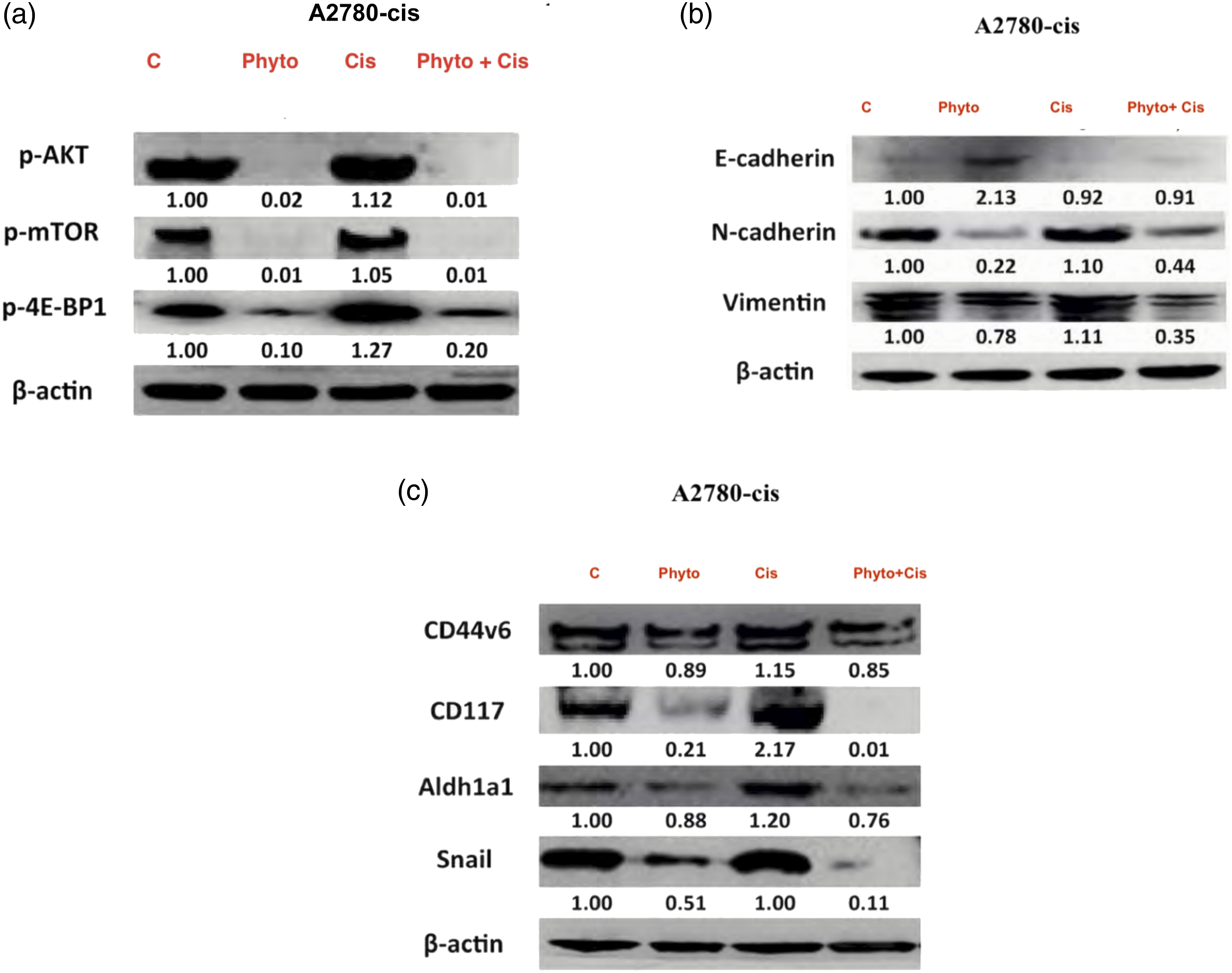
The results obtained from various experiments conducted in this study are depicted in Figures 1–3. The cells were incubated for 48 h for the invasion assay. The invasive potential in EOC-cis cell lines (A2780-cis) was significantly increased by 34.4% compared to the corresponding EOC-control cell lines (A2780), respectively (* p < 0.05). (a) Invasive Potential of EOC-cis cell lines in comprsion to controls (b) Representative images for cell invasion and control in EOC-control and EOC-cis cells by light microscope are shown. Magnification: 200x. The results were from three independent experiments (mean ± SD, n = 3). (c) qRT-PCR results for mRNA expressions of epithelial-to-mesenchymal transition markers (* p < 0.05, ** p < 0.01, *** p < 0.001). GAPDH was used as a loading control. The results were from three independent experiments (mean ± SD, n = 3). (d) The qRT-PCR results for expression of cancer stem cell markers in EOC-cis cells, and there is a significant difference in CD117, and ALDH1A1, and Snail between EOC-control and EOC-cis cell lines (* p < 0.05, ** p < 0.01, *** p < 0.001). GAPDH was used as a loading control. The results were from three independent experiments (mean ± SD, n = 3). EOC: epithelial ovarian cancer; GAPDH: glyceraldehyde 3-phosphate dehydrogenase. Effects of combination of curcumin and resveratrol with cisplatin treatment on the expression of PI3K/AKT/mTOR signalling proteins and epithelial-to-mesenchymal transition/cancer stem cell phenotype. The expression of those markers was significantly increased after cisplatin treatment compared to the EOC-cis cells without treatment. However, the combination treatment with cisplatin and phytochemicals reversed the activation induced by single cisplatin treatment. (a) Effects of combination of curcumin and resveratrol with cisplatin treatment on the expressions of the expression levels of p-Akt, p-mTOR and p-4EBP1. (b) Effects of combination of curcumin and resveratrol with cisplatin treatment on the expressions of N Cadherin and Vimentin proteins of EOC-cis cell lines. (c) Effects of combination of curcumin and resveratrol with cisplatin treatment on the protein expressions of CD44v6, CD117, Aldh1a1 and Snail. Effects of single or combination treatment on apoptosis in EOC-cis cell lines. (a) The expression of apoptosis biomarkers is shown as increased after cisplatin treatment compared to EOC-cis cells without treatment, whereby the combination treatment with cisplatin and phytochemicals further enhanced the apoptosis significantly. (b) The AO/EB experiment further indicates that the rate of cells undergoing apoptosis is obvious in cisplatin-treated cells and much higher in the combination therapy with cisplatin and phytochemicals. EOC: epithelial ovarian cancer.

Matrigel invasion of epithelial ovarian cancer cell lines
Epithelial ovarian cancer–cis cell lines showed substantially increased invasive potential compared to the corresponding parental EOC-control cell lines (* p < 0.05) (Figure 1(a) and (b)). The average percentage of invasion rate for A2780-cis was 58.3%, while the average percentage of invasion rate for A2780 was 10.9%, respectively. The invasive potential in EOC-cis cell lines (A2780-cis) was significantly increased by 34.4% compared to the corresponding EOC-control cell lines (A2780), respectively (* p < 0.05).
qRT-PCR results
The phenotypic changes of EMT as well as CSC phenotypes in EOC-cis cell lines were confirmed by qRT-PCR on mRNA levels (Figure 1(c) and (d)). Reduced expression of E-cadherin and increased expression of N-cadherin and vimentin in EOC-cis cells confirmed the EMT phenotype. On the other hand, there is a significant difference in CD117, and ALDH1A1, and Snail between EOC-control and EOC-cis cell lines confirming enhanced CSC phenotype. GAPDH was used as a loading control. The results were from three independent experiments (mean ± SD, n = 3).
Effects of combination of curcumin and resveratrol with cisplatin treatment on the expression of PI3K/AKT/mTOR signalling proteins and epithelial to mesenchymal transition/cancer stem cell phenotypes
The expression levels of p-Akt, p-mTOR and p-4EBP1 were significantly elevated after individual cisplatin treatment in EOC-cis cell lines compared to the control group. However, this change was abolished by the combination treatment of cisplatin with curcumin and resveratrol (Figure 2(a)).
To further investigate the association of the PI3K/AKT/mTOR signalling pathway with EMT and CSC phenotype, the expression levels of EMT and CSC markers were also examined in various treatments. The study results confirmed that with phytochemicals alone, treatment can reverse EMT phenotypes by elevating the expression of E-cadherin protein and decreasing the expressions of N-cadherin and vimentin proteins of EOC-cis cell lines (Figure 2(b)). Moreover, combined treatment of phytochemicals with cisplatin further reversed EMT phenotypes (Figure 2(b)). On the other hand, single cisplatin treatment enhanced the stemness of EOC-cis as noticed by the increased protein expressions of CD44v6, CD117, ALDH1A1 and Snail) compared to untreated control (Figure 2(c)). However, treatment with combination of phytochemicals and cisplatin significantly reversed protein expressions. In this way, the combination treatment of curcumin and resveratrol with cisplatin not only reversed the EMT phenotype of EOC-cis cells but also abolished the cisplatin-induced stemness enhancement (Figure 2(b) and (c)).These results suggested that combination of curcumin and resveratrol sensitised the EOC-cis cells to cisplatin through reversing the EMT and CSC phenotypes.
Effects of single or combination treatment on apoptosis in epithelial ovarian cancer–cis cell lines
The apoptosis is the main cell death pathway after chemotherapy. So, the present study investigated expression of two apoptosis-related biomarkers (p-PARP and a-caspase-3). The expression of apoptosis biomarkers is shown as increased after cisplatin treatment compared to EOC-cis cells without treatment, whereby the combination treatment with cisplatin and phytochemicals further enhanced the apoptosis significantly (Figure 3(a)). In addition, the AO/EB experiment further indicates that the rate of cells undergoing apoptosis is obvious in cisplatin-treated cells and much higher in the combination therapy with cisplatin (Figure 3(b)).
Discussion
The combination approach has been confirmed as an excellent and effective strategy against carcinogenesis in recent past. 25 It basically includes combined use two or more molecules (drugs/phytochemicals/chemo-preventive agents) to enhance efficacy of the treatment. Therefore, in the present study, we explored the molecular mechanics responsible for combined effects of curcumin and resveratrol with cisplatin chemotherapy against cisplatin-resistant EOC cells. The study clearly implied that the combined treatment of curcumin and resveratrol significantly sensitized EOC cells for cisplatin chemotherapy. The combination of phytochemicals effectively targeted key molecules of PI3K/AKT/mTOR pathway to reverse cisplatin resistance in cisplatin-resistant EOC cells.
Chemoresistance is the biggest obstacle in the pathway of any effective anticancer strategy. 26 Moreover, chemoresistance of a cell is directly related to its invasiveness as confirmed by Matrigel invasion in cisplatin-resistant EOC cells. 27 The chemoresistant cells showed a significantly higher invasion rate as compared to cisplatin-sensitive EOC cells. The prime reason for the above observation could be CSCs or EMT in tumour cells.28–29 The above results were corelated well with recorded elevated levels of expression levels of p-Akt, p-mTOR and p-4EBP in the cisplatin-resistant EOC cells. The stemness of EOC-cis was clearly evidenced by the increased protein expressions of CD44v6, CD117, ALDH1A1 and Snail). On the other hand, EMT was revealed by observed reduced expression of E-cadherin protein and increased expressions of N-cadherin and vimentin proteins in these cell lines. Interestingly, combined treatment with phytochemicals reversed the above changes, thereby making the cell more sensitive to cisplatin treatment and reduced invasive abilities. The above results could be owed to PTM modulation abilities of both resveratrol and curcumin in combination. 30 As a result of above post-translational modification abilities, the combination strategy significantly reduced stemness of cisplatin-resistant EOC cells. Also, they made them less invasive by inhibiting EMT in these cell lines, thereby making them further sensitive to cisplatin chemotherapy.
Apoptosis results also supported the efficacy of combined treatment of phytochemicals with cisplatin, as significantly higher apoptosis was observed in the cells treated with combined treatment. Earlier studies in the recent past have also confirmed apoptosis-stimulating abilities of both curcumin and resveratrol in the cancerous state.31–32 Both curcumin and resveratrol have been proved to stimulate apoptosis by both intrinsic as well as extrinsic pathways. 33 The observation is in corroboration with earlier reports which noticed the trigger of apoptosis by stimulation of the Bax gene and inhibition of the Bcl2 gene by curcumin and resveratrol during carcinogenesis. 34
Conclusion
The present study added another dimension to the existing knowledge of anticancer strategies against ovarian cancer. The study concludes that phytochemicals in combination significantly inhibit chemoresistance by targeting the PI3K/AKT/mTOR pathway in cisplatin-resistant ovarian cancer cells. The study may be explored further in-vivo for elucidation of further molecular details.
Footnotes
Declaration of conflicting interests
The author(s) declared no potential conflicts of interest with respect to the research, authorship, and/or publication of this article.
Funding
The author(s) disclosed receipt of the following financial support for the research, authorship, and/or publication of this article: This work was supported by Cancer Hospital of Xinjiang Uygur Autonomous Region, grant number XJZL210415 and the State Key Laborotory foundation of China No. SKL-HIDCA-2020-23.
Ethical approval
The present study and all protocols have been approved by Cancer Hospital of Xinjiang Uygur Autonomous Region animal ethical committee (Approved file No. XJZL210415).