
Abstract
To determine the no-observed-adverse-effect level (NOAEL) of exposure and target organs of neem oil for establishing safety criteria for human exposure, the subchronic toxicity study with neem oil in mice was evaluated. The mice (10 per sex for each dose) was orally administered with neem oil with the doses of 0 (to serve as a control), 177, 533 and 1600 mg/kg/day for 90 days. After the treatment period, observation of reversibility or persistence of any toxic effects, mice were continuously fed without treatment for the following 30 days. During the two test periods, the serum biochemistry, organ weight and histopathology were examined. The results showed that the serum biochemistry and organ coefficient in experimental groups had no statistical difference compared with those of the control group. At the 90th day, the histopathological examinations showed that the 1600 mg/kg/day dose of neem oil had varying degrees of damage on each organ except heart, uterus and ovarian. After 30-day recovery, the degree of lesions to the tissues was lessened or even restored. The NOAEL of neem oil was 177 mg/kg/day for mice and the target organs of neem oil were determined to be testicle, liver and kidneys.
Keywords
Introduction
Neem (Azadirachta indica) has been accepted universally as a “wonder tree” in India. 1 Neem oil, also called Margosa oil, is an extract from seeds or fruits of A. indica obtained through pressing or solvent extraction. Neem oil was widely used as a traditional medicine by Indians in India, Sri Lanka, Burma, Thailand, Malaysia and Indonesia and already has more than 2000 years of history. It is used mainly for external applications and was often administered orally for deworming, leprosy, constipation, rheumatism, ulcer, relieve itching and chronic skin diseases. 2,3 It contains mostly long- and medium-chain fatty acids (80–95%) and volatile sulfur compounds (4–20%) as well as a number of bioactive compounds, such as nimbin, nimbidin and nimbinin, 4 ,5 that have been demonstrated to have biocidal activity against nearly 200 medical and veterinary arthropods, without any adverse effects toward most nontarget organisms. 6,7 It was also found to have acaricidal, antibacterial, antifungal, antimalarial, antiparasitic, anti-inflammatory, promotion of wound healing and immunomodulatory properties in different animal species. 1,5,7 –13
Our previous research showed that the median lethal dose (LD50) of extract chloroform isolated from neem oil in Sprague Dawley rats was above 10,000 mg/kg, 14 the LD50 of neem oil in mice was 31,950 mg/kg, the accumulative coefficient (K) was above 5, it had low toxic effects. 15 The present study was thereby performed to evaluate the subchronic toxicity of neem oil according to the OECD test guideline 408 for “Repeated dose 90-day oral toxicity study in rodents” except for the lack of ophthalmological examination and functional observations during feeding experiment. 16 The study will provide information on the major toxic effects, indicate target organs, and can provide an estimate of a no-observed-adverse-effect level (NOAEL) of neem oil, and provide information on the possible health hazards likely to arise from people repeated exposure over a prolonged period of time.
Materials and methods
Plant material
Neem oil that was extracted from the seeds of the neem (A. indica) using carbon dioxide supercritical fluid extraction was supplied by a pesticide company (Green Gold Biological Science & Technology Co. Ltd, Chengdu, PR China). All chemicals we used in the test were of analytical grade (>99.7%).
Animals and treatments
Kunming strain male and female mice (a closed strain coming from Kunming, Yunnan Province, PR China) were obtained from the Chengdu Dossy Experimental Animals Co., Ltd (Sichuan provence, Chengdu, P.R. China, License No.SCXK (Sichuan) 2008-24), weighing 13–15 g, kept at room temperature of 22°C. Mice were fed with a standard diet from Nuvital Nutrientes (Colombo/PR, Brazil) and allowed free access to water and have been acclimated to laboratory conditions for 7 days.
Three groups of 20 mice, each containing 10 females and 10 males, received a daily dose of 177 mg/kg of body weight (b.w.; group II), 533 mg/kg b.w. (group III) and 1600 mg/kg b.w. (group IV) of neem oil mixed with 1% carboxymethyl cellulose sodium, a vehicle-control group (group I) formed by 20 mice received a daily dose of 533 mg/kg b.w. of 1% carboxymethyl cellulose sodium during a 90-day period. In each case, the product volume administered by gavage was 2 mL/100 g b.w. Each mouse was marked with a unique identification number using trinitrophenol. Body weight was measured once a week and the behavior was observed daily during the trial period. At the end of the treatment, mice were continuously fed without treatment for 30 days to detect persistence of or recovery from toxic effects.
Clinical biochemistry analyses
At the end of experimentation (the 90th and 120th day), half of the total amount of mice per sex, respectively, underwent overnight fasting prior to collection of the blood sample. Blood of each mouse was collected by retro-orbital bleeding and subjected to clinical biochemical tests. For the hepatic function, serum alkaline phosphatase, alanine aminotransferase and aspartate aminotransferase were determined, while for the renal function, serum urea nitrogen and serum creatinine were evaluated. Serum glucose was accessed for carbohydrate metabolism analysis. Total protein, albumin, globulin, albumin/globulin ratio (A/G), total bilirubin and cholesterol were also measured. All these biochemical parameters were determined as described previously by Lincopan et al. 17
Organic coefficient and histopathological analysis
On the 90th day and 120th day after blood collection for biochemical analysis, all the animals were euthanized, detailed gross necropsy was carefully examined. Extracted heart, liver, spleen, lungs and double kidneys were trimmed of any adherent tissue, their wet weight taken as soon as possible after dissection to avoid drying to cipher organic coefficient (ratio of organ weight to body weight was calculated). The principal vital organs (heart, liver, spleen, lung, kidney, testis, ovary and uterus) were preserved in fixation medium of 10% solution of buffered formalin (pH 7.4) and enclosed in paraffin-intended subsequent histopathological examination. A section of each organ tissue of 5 µm was stained with hematoxylin and eosin (H&E). Each section was examined under an optical microscope.
Statistic evaluation
All results were expressed as mean ± SD (
Results
General observation, effects on clinical signs and food consumption
There was no treatment-related mortality in animals treated with neem oil for 90 days at any dose tested. The group at the dose of 1600 mg/kg/day showed treatment-related clinical signs, such as rough fur and loss of appetite in the last 2 weeks. No treatment-related clinical signs were observed in other experimental groups.
The food consumption result is shown in Table 1. The food consumption of all the test groups had no statistical difference compared with that of the control group in month 1 and recovery month (p > 0.05), while that of the dose 1600 mg/kg/day group was very significantly decreased in months 2 and 3 (p < 0.01).
Food consumption (g/100 g) of mice treated with neem oil.a

ANOVA: analysis of variance.
aData shown as mean ± SD were analyzed by ANOVA followed by SPSS 17.0.
bSignificantly different from control p < 0.01.
cSignificantly different from control p < 0.05.
Biochemical examination
The serum biochemistry parameters were examined after 90 days of oral administration with neem oil. No significant differences were noted between the mice-treated groups with the 177, 533 and 1600 mg/kg/day dose of neem oil and the vehicle-control group. Similar results were obtained 30 days after recovery (Table 2).
Serum biochemistry parameters of mice treated with neem oil.a

ALT: alanine aminotransferase; AST: aspartate aminotransferase; BUN: urea nitrogen***; CRE: creatinine; GLU: glucose; TP: total protein; ALB: albumin; GLO: globulin; TBIL: total bilirubin; CHO: cholesterol; ANOVA: analysis of variance.
aData shown as mean ± SD were analyzed by ANOVA followed by SPSS 17.0. No significant difference from the control group at p > 0.05.
Effects on organ coefficient
The results of organ coefficient are summarized in Table 3, the organ coefficient of heart, liver, spleen, lung and kidneys in the experimental groups with 177, 533 and 1600 mg/kg/day dose of neem oil for 90 days had no statistical difference compared with that of the vehicle-control group, the same result was also shown 30 days after recovery (p > 0.05).
Organic coefficient (g/100 g) of mice treated with neem oil.a
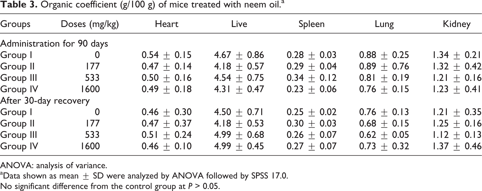
ANOVA: analysis of variance.
aData shown as mean ± SD were analyzed by ANOVA followed by SPSS 17.0. No significant difference from the control group at P > 0.05.
Histopathological findings
At the 90th day, the histopathological examination showed that only the 1600 mg/kg/day dose of neem oil had varying degrees of damage on each organ except heart, uterus and ovary. The consistent treatment-related histopathological changes were found in both sexes.
In the vehicle-control group (group I), the cross-section of liver showed normal appearance, hepatic artery, portal vein, bile duct, sinusoids (Sis) and hepatocytes (Hs), all clearly conserved (Figure 1(a)). Doses (177 and 533 mg/kg/day) of groups II and III, respectively, only showed central venous and Sis congestion in liver lobule, Hs occurred granular and slight vacuolar degeneration (Figure 1(b) and (c)), while at the dose (1600 mg/kg/day dose) of group IV, central venous congestion, granular and vacuolar degeneration, karyorrhexis were observed in Hs (Figure 1(d)).

Effect of neem oil on the microstructures of liver of mice after administration for 90 days. Panel A: group I (0 mg/kg, HE 200×); panel B: group II (177 mg/kg, HE 400×); panel C: group III (533 mg/kg, HE 400×); Panel D: group IV (1600 mg/kg, HE 400×). Photomicrographs of the liver from mice treated with 0, 177, 533 and 1600 mg/kg in a 90-day subchronic oral toxicity evaluation of neem oil. Cross-sections were stained with hematoxylin and eosin. Observation was made at different amplified levels. Group I, the cross-section showed the normal appearance of liver, hepatic artery, portal vein, bile duct, Sis, Hs, all clearly conserved. Groups II and III, showed central venous (CV) and Sis congestion in liver lobule (→); group IV, central venous congestion (→), granular and vacuolar degeneration (↑), karyorrhexis (K) were observed in Hs. Si: sinusoid; H: hepatocyte.
In group I, the cross-section of the spleen showed normal appearance, spleen trabecula, red pulp (RP), white pulp and germinal centers, all clearly conserved (Figure 2(a)). Groups II and III showed the normal characteristic of spleen associated with infiltration of multiple giant cells (Figure 2(b) and (c)), while in group IV severe hyperemia of RP and infiltration of multinucleate giant cells in spleen were observed (Figure 2(d)).

Effect of neem oil on the microstructures of spleen of mice after administration for 90 days. Panel A: group I (0 mg/kg, HE 200×); panel B: group II (177 mg/kg, HE 400×); panel C: group III (533 mg/kg, HE 400×); panel D: group IV (1600 mg/kg, HE 400×). Group I, the cross-section showed normal appearance of the spleen, spleen trabecula, RP, white pulp, germinal centers, all clearly conserved. Groups II and III, showed the normal characteristic of spleen, infiltration of multinucleate giant cells in spleen (→); group IV, severe hyperemia of RP (↑) and infiltration of multinucleate giant cells (→) in spleen were observed. RP: red pulp.
In group I, the cross-section of the lung showed normal appearance (Figure 3(a)). In group II, the alveolar walls were thickened, the capillaries in the alveolar walls and interstitial were congested with many red blood cells (Figure 3(b)), while in groups III and IV, the alveolar walls were thickened, the capillaries in the alveolar walls and interstitial were severely congested, a serious alveolar cavity hemorrhage was observed (Figure 3(c) and (d)).

Effect of neem oil on the microstructures of lung of mice after administration for 90 days. Panel A: group I (0 mg/kg, HE 400×); panel B: group II (177 mg/kg, HE 400×); panel C: group III (533 mg/kg, HE 400×); panel D: group IV (1600 mg/kg, HE 400×). Group I, the cross-section showed the normal appearance of lung. Alveolus (A), alveolar ducts (AD), alveolar sac (AS), all clearly conserved. Group II, showed the alveolar walls were thickened (↑), the capillaries in the alveolar walls and interstitial were congested with many red blood cells (→); groups III and IV, the alveolar walls were thickened (↑), the capillaries in the alveolar walls and interstitial were severe congested, alveolar cavity hemorrhage serious were observed (→).
In group I, the cross-section of the kidneys showed normal appearance, glomerulus and renal tubule structure was normal (Figure 4(a)). Groups II and III showed capillary of glomerulus and interstitial angiectasis hyperemia, renal tubular epithelial cells swelling, granular degeneration and some of them were separated from the basement membrane (Figure 4(b) and (c)). In group IV, massive inflammatory cells especially neutrophilic granulocyte infiltrated in the glomeruli and nephric tubules, RETC degeneration and necrosis, segregated with basilar membrane. The renal tubule revealed protein cast (Figure 4(d)).

Effect of neem oil on the microstructures of kidneys of mice after administration for 90 days. Panel A: group I (0 mg/kg, HE 200×); panel B: group II (177 mg/kg, HE 400×); panel C: group III (533 mg/kg, HE 400×); panel D: group IV (1600 mg/kg, HE 400×). Group I, the cross-section showed the normal appearance of kidney, glomerulus (G), proximal tubule (PT) and distal tubule (DT), all conserved. Groups II and III, capillary of renal interstitium was hemolysis (→), renal tubular epithelial cells swelling, granular degeneration, separated from basement membrane; group IV, massive inflammatory cells (↓) infiltrated in the glomeruli and nephric tubules, RETC degeneration and necrosis, segregated with basilar membrane. The renal tubule revealed protein cast.
In group I, the cross-section showed the normal characteristic of testicle, the seminiferous tubules structure was intact, all levels of spermatogenic cells (SCs) were arranged in order (Figure 5(a)). In Groups II and III showing the decrease of SCs and blister degeneration, in some more serious cases, SCs occurs ballooning degeneration (Figure 5(b) and (c)). Group IV, the basic structure of seminiferous tubule was destroyed, SCs were seriously dissolved and disappeared, sperm within the seminiferous lumen almost completely disappeared (Figure 5(d)).

Effect of neem oil on the microstructures of testicle of mice after administration for 90 days. Panel A: group I (0 mg/kg, HE 200×); panel B: group II (177 mg/kg, HE 400×); panel C: group III (533 mg/kg, HE 400×); panel D: group IV (1600 mg/kg, HE 400×). Group I, the cross-section showed the normal appearance of testicle, the seminiferous tubules, SCs at all levels, spermatozoon (Sz), testicular interstitial cells (IC) in stroma (St) surround the seminiferous tubules, all conserved. Groups II and III, the SCs abscission, quantity decrease, blister degeneration, serious turned into ballooning degeneration; group IV, the basic structure of seminiferous tubule was destroyed, SCs were seriously dissolved and disappeared, sperm within the seminiferous lumen was almost completely disappeared. SC: spermatogenic cell.
Thirty days after recovery, the degree of injury to the tissues was lessened or even restored.
Discussion
Food consumption is a key parameter for determining the dose of feeding and an important indicator of toxic effects of chemical substances. 18 The results obtained from the study demonstrated that the food consumption of mice at the dose of 1600 mg/kg/day decreased very significantly in months 2 and 3, while those from the other experimental groups had no significant changes compared with the control group. And this was consistent with the general clinical manifestations observed and recorded for group IV, so the clinical dosage of neem oil should be lower than 1600 mg/kg/day.
The results from the subchronic oral toxicity study with neem oil on male and female young adult mice showed that all the serum biochemistry parameters of experimental groups had no statistically significant difference compared with those of the control group (p > 0.05) after oral administration with neem oil for 90 days. Similar results were obtained 30 days after recovery. It indicated that oral administration of neem oil in mice for 90 days at and below 1600 mg/kg/day dose had no damage on the serum biochemistry parameters. And this was consistent with the result of the studies of Rukmini et al. and Lakshminarayana. 19 ,20
Organ coefficient can reflect the degree of organs’ damages. Excluding the loss of water, age, gender, the effect of malnutrition and other factors before being weighed, the increase in the organ coefficient indicates that there are changes in congestion, edema, hyperplasia, hypertrophy in organs, while there are changes in shrink and degeneration when it is decreased. 21 In the present study, all the examined organ coefficients had no statistical difference compared with those of the vehicle group, which indicated that neem oil had mild or no damages on organs of mice. And this was consistent with the result that the oral administration with neem oil for 90 days had varying degrees of damages on organs, but this was lessened or even to be restored 30 days after recovery.
To determine the NOAEL and target organ toxicity of neem oil, the pathological examination of principal vital organs in mice at different doses of neem oil were observed under a microscope. The results of oral administration with neem oil for 90 days from the study showed that neem oil had no damage on heart. The 177 and 533 mg/kg/day doses of neem oil had mild damages on liver, spleen, lung, kidneys and testicle, such as slight vascular congestion, while 1600 mg/kg/day dose of neem oil had varying degrees of damages on each organ, mainly granular and vacuolar degeneration in cells and vascular congestion, in addition, in the lung, alveolar walls were thickened and hemorrhage damage was shown. All the damages on organs were lessened or even to be restored 30 days after recovery. So the pathologic damage on mice with neem oil was reversible, the damage may be gradually restored after the discontinuation of treatment in the long run.
In the present study, neem oil had mild damages on organs at a dose of 177 mg/kg/day, and the damages were restored after the discontinuation of treatment for 30 days, so the NOAEL for males and females was considered to be 177 mg/kg/day. The effect of neem oil on the liver, kidneys and testicle was showing a good dose–response relationship, indicating that the target organs of neem oil toxicity were the liver, kidneys and testicle.
The testicle has been proved to be the target organ, which was consistent with the findings of Yin et al. 22 ,23 The mechanism of the antifertility effect of extract chloroform from neem oil on mice may be that the replacement process of testicular sperm nuclear protein was blocked, which led to abnormal epididymal sperm nucleoprotein and sperm not sufficiently differentiated to mature. Further study needed to do to explore the toxic mechanism of neem oil on liver and kidney.
Footnotes
Funding
This work was supported by the Special Fund for Agro-scientific Research in the Public Interest (201203041); National Natural Science Foundation of China (Grant no. 31272612); National Science & Technology Program in Rural Areas During the 12th Five Year Plan Period (2011BAD34B03-4); the Doctoral Program of Higher Education Research Fund (Instructor Dr Class 20105103110001).
Authors’ Note
The first three authors contribute equally to this work and should be considered as first author. CW and MC contributed equally to this work.
Acknowledgments
The authors thank Green Gold Biological Science & Technology (Chengdu, PR China) for supplying neem oil.