
Abstract
Hypoglycemic effect of ethanol extracts of Peganum harmala (commonly known as ‘Harmal’) seeds has been reported on normal and streptozotocin-induced diabetic rats. In the present study, the authors determine anti-diabetic and anti-oxidative properties of 4-hydroxypipecolic acid (4-HPA) isolated from seeds of P. harmala in C57BL/KsJ-db/db mice. Twelve week old male mice were administered 50 mg/kg body weight (4-HPA suspension were made in 1% gum acacia) for the period of 10 days, and a significant reduction in the fasting blood glucose, plasma triglycerides (TG), cholesterol, free fatty acid, low-density lipoprotein-cholesterol and a significant increase in high-density lipoprotein-cholesterol level was observed with respect to vehicle-treated db/db mice. The anti-oxidant activity of 4-hydroxypipecolic acid was studied in liver and kidney tissues by assessing malondialdehyde levels for lipid peroxidation and enzyme activity of catalase (CAT), glutathione peroxidase (GSH-Px) and superoxide dismutase (SOD). Treatment of 4-HPA significantly lowered the lipid peroxidation in hepatic and renal tissue and increased the activity of CAT, GSH-Px and SOD in treated mice.
Introduction
The increasing worldwide incidence of diabetes in adults constitutes a global health burden. According to World Health Organization and American Diabetes Association, the worldwide prevalence of diabetes among adults (aged 20–79 years) will be 6.4%, affecting 285 million adults in 2010, and will increase to 7.7% and 439 million adults by 2030. Between 2010 and 2030, there will be a 69% increase in numbers of adults with diabetes in developing countries and a 20% increase in developed countries. 1 Approximately 90–95% of diabetic patients have type 2 diabetes mellitus, which is a complex, heterogeneous, polygenic disease characterized mainly by insulin resistance and pancreatic β-cell dysfunction. Persistent hyperglycemia in diabetes leads to increased production of free radicals through auto-oxidation of glucose and non-enzymatic protein glycation.2,3 These highly reactive free radicals exert their cytotoxic effects on membrane phospholipids and cause a wide spectrum of cell damage, including lipid peroxidation, inactivation of enzymes, alteration of intracellular oxidation–reduction state and DNA damage.4,5 This oxidative stress is believed to be a pathogenetic factor in the development of diabetic complications. The use of medicinal plants has flourished as an alternative for the treatment of diabetes because modern medicines are tagged with several side effects and are also expensive. A multitude of herbs and medicinal plants and some compounds purified from them have been studied for the treatment of diabetes throughout the world as they might provide a basis of new synthetic anti-diabetic analogues with potent activity.6–8 Therefore, investigation on such agents from traditional medicinal plants has become more important. 9
Peganum harmala (Family: Zygophyllaceae), commonly known as ‘harmal’, grows wildly in semi-arid areas of Indo–Pak subcontinent, Iran and Africa. It is one of the most important medicinal plants of India and used for the treatment of variety of human ailments. The β-carboline alkaloids isolated from this plant are reported to possess anti-depressant and anti-hallucinogenic activity.10,11 Recently, our group reported the hypoglycemic effect of ethanol extracts of P. harmala seeds on normal and streptozotocin (STZ)-induced diabetic rats. 12 In the present study, we evaluated the anti-diabetic and anti-oxidative effects of 4-hydroxypipecolic acid in C57BL/KsJ-db/db mice, which displays many of the characteristics of type 2 diabetic human patients including hyperphagia, hyperglycemia, dyslipidemia, hyperinsulinemia and obesity.
Materials and methods
Plant material
The seeds of P. harmala were collected from Kashmir in the month of April. The reference material is deposited in the medicinal and process chemistry division of the central drug research institute, Lucknow, India.
Extraction/fractionation/isolation procedure
Powdered seeds (6 kg) of P. harmala were extracted with 25 L of 95% ethanol in pilot plant. Ethanolic extract was concentrated under vacuum using rotavapour at 50°C. The ethanolic extract weighed 305 g and was partitioned between chloroform and water; the resultant aqueous fraction (170 g) was concentrated under reduced pressure and subjected to gross silica-gel column chromatography. The fractions eluted with chloroform-aqueous methanol (1:1) yielded a crystalline compound which was characterized as 4-hydroxypipecolic acid (Figure 1 ). Stereochemistry was determined by chemical studies and was shown to be trans-4-hydroxy-L-pipecolic acid; m.p. 285–286°C (decomp.); MS (ESI) m/z 146 (M+1); IR (KBr) 3536 cm−1(OH), 1608 cm−1(CO); 1 H-NMR (200 MHz, D2O) δ 1.89–1.98 (m, 2H, H-5), 2.00 (ddd, 1H, H-3a), 2.22 (ddd, 1H, H-3b), 3.31 (dd, J = 4.6, 4.0, 2H, H-6), 3.92 (dd, J = 3.7, 3.7, 1H, H-4), 4.23 (dd, J = 4.0, 2.6, 1H, H-2). 13 C-NMR (50 MHz, D2O) δ 30.7 (C-5), 35.6 (C-3), 41.3 (C-6), 56.7 (C-2), 64.6 (C-4), 177.1 (C-1). Anal. calcd. for C6H11 NO3: C, 49.65; H, 7.64; N, 9.65; O, 33.07.
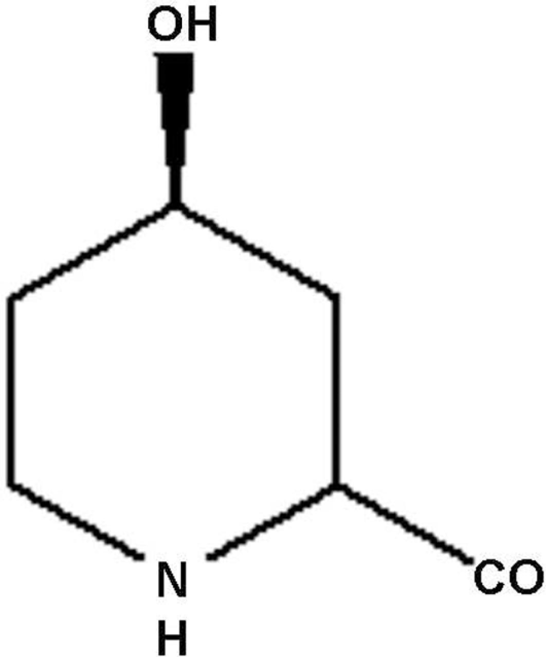
4-Hydroxypipecolic acid.
In vitro experiment
Cell culture
L6 skeletal muscle cell lines were procured from National Center for Cell Science (NCCS), Pune. The cells were maintained in Dulbecco’s modified eagles medium (DMEM) with 10% fetal bovine serum (FBS) supplemented with penicillin (100 units/ml), streptomycin (200 μg/ml) and gentamycin (50 μg/ml) in a humidified atmosphere of 5% CO2 at 37°C. For differentiation, myoblasts were transferred to DMEM with 2% FBS and allowed to reach confluency and aligned to form myotubes before being used for experiment.
Glucose uptake in differentiated myotubes
Glucose uptake was estimated by the method of Klip et al. 13 L6 myoblasts were differentiated post-confluency for 4–6 days till the cells aligned to form myotubes. The myotubes were then treated with drugs in low-glucose DMEM with 10% FBS for 18 h. The myotubes were washed with phosphate buffered saline (PBS) and incubated for 3 h in serum-free DMEM. The myotubes were washed with PBS and were incubated with Krebs Ringer phosphate hepes buffer (KRPH) containing 0.5% BSA and 100 nM insulin for 30 min. The myotubes were then briefly washed with KRPH containing 0.5% BSA. 2-Deoxy-D-[U- 14 C] glucose uptakes were carried out in KRPH buffer having 0.5 µCi of 2-DG for 10 min and the myotubes were washed thrice with cold PBS. The myotubes were then lysed in 0.5ml of 0.1 N NaOH followed by radioactivity measured (Beckman Coulter, CA, USA). All assays were performed in triplicates and results are expressed as picomoles/min/well.
In vivo experiment
Animals (db/db mice)
Male C57BL/KsJ-db/db mice (12-week-old, 35–45 g body weight) were procured from National Animal Laboratory Centre of Central Drug Research Institute (CDRI), Lucknow and all procedures were conducted in accordance with care and use of laboratory animals after necessary approval by institutional animal ethical committee. The animals were housed in polypropylene cages, in controlled environment (temperature 25 ± 2°C; humidity 50–60%; light 300 lux at floor level with regular 12 h light–12 h dark cycle; noise level 50 dB; ventilation 10–15 air changes per hour). They were provided with a standard laboratory diet unless stated otherwise and have free access to water.
Experimental design
Diabetic db/db mice showing blood glucose values from 10 to 20 mM (180–360 mg/dl) were selected for this study and divided into various groups of five mice in each. Group one served as diabetic control and was given vehicle (1% gum acacia), while the other experimental groups were orally administered with 4-HPA or standard drug metformin at a dose 50 mg/kg body weight once daily for a period of 10 days. The fasting blood glucose (FBG) levels were measured after animals fasted for 4 h (starting from 9:00 a.m.) on day 0 (before treatment), day 5 (during treatment) and day 10 (last day of treatment). Blood glucose levels were measured by Glucometer (ACCU-CHEK II; Roche Diagnostics, IN, USA) as per manufacturer’s instructions.
An oral glucose tolerance test (OGTT) was performed at last day of treatment after overnight fasting. Blood was sampled from the tail vein of mice at time 0 min (baseline) and at 30, 60, 90 and 120 min after an oral glucose load of 3.0 g/kg of body weight. Food (but not water) was withheld from the cages during the course of fasting. The area under a curve (AUC) of experimental groups was compared with that of vehicle-treated control group and the percent anti-hyperglycemic activity was determined.
Biochemical analysis
Estimation of plasma lipids and biomarkers
At the end of the experiment (on day 11) blood samples of overnight fasted db/db mice of various groups were collected from retro-orbital venous plexus by using 5 µl heparinized glass capillary. Plasma was separated from the collected blood and used for the estimation of plasma cholesterol (CHOL), TG, low-density lipoprotein cholesterol (LDL-C), free fatty acid (FFA) and high-density lipoprotein cholesterol (HDL-C) levels by respective assay kits (Roche Diagnostics, Germany). Plasma insulin and glucagon were also measured by radioimmunoassay (BRIT, Mumbai, India). The animals were killed at the end of experiment by cervical dislocation and liver and kidney were excised and rinsed with normal saline, immediately frozen in liquid nitrogen, and stored at −70°C for various assays.
Estimation of lipid peroxidation
Lipid peroxidation in liver and kidney tissue was determined by measuring malondialdehyde (MDA) using the method described by Colado et al. 14 Briefly, the liver and kidney tissue were washed with 1% (w/v) potassium chloride and subsequently homogenized in the same. The homogenized tissue was centrifuged at 1000 g for 15 min and supernatant was used for the estimation of MDA. The supernatant (0.5 ml) was added to a reaction mixture consisting of 30% TCA, 5 M HCl and 2% (w/v) thiobarbituric acid in 0.5 M NaOH. The reaction mixture was heated in a water bath at 95°C for 15 min and centrifuged at 12,000 rpm for 15 min. Absorbance of the supernatant was read at 532 nm with spectrophotometer (Perkin Elmer’s, MA, USA). The amount of MDA produced was calculated using 1, 3, 3-tetra methoxy propane as a standard and the result of peroxidation was expressed as mM of MDA formed per mg of protein.
Assay of catalase activity
Catalase (CAT) was estimated by the method of Aebi. 15 The reaction mixture contained 0.1M sodium phosphate buffer (pH 7.2), 4 mM H2O2 and suitably diluted enzyme. Reaction was started by addition of H2O2 and the rate of change in H2O2 concentration was followed by observing the decrease in optical density (OD) at 240 nm for 3 min. One unit of CAT is defined as the amount of enzyme that reduces 1 μmol of H2O2 per minute. Values of this and other enzymes are expressed as specific activity (units/mg protein). Protein concentration was determined by Lowry et al. 16
Assay of glutathione peroxidase activity
GSH-Px activity was assayed by the method of Forstrom et al. 17 The reaction mixture contained 25 mM Tris-HCl buffer (pH 7.6), 0.12 mM NADPH, one unit/ml Glutathione reductase, 1 mM reduced glutathione, 0.1 mM hydrogen peroxide, and 160 μl of suitably diluted enzyme. The reaction was started by the addition of hydrogen peroxide and depletion of NADPH was measured spectrophotometrically at 340 nm for 3 min. One unit of GSH-Px is defined as the amount of enzyme that oxidizes one μmol of NADPH per minute.
Assay of superoxide dismutase activity
SOD activity was determined according to the method of Misra and Fridovich. 18 The reaction mixture contained 100 µl tissue homogenate, 880 μl of (0.05 M), carbonate buffer (pH 10.2), 20 μl of 30 mM epinephrine in 0.05% acetic acid was added to the mixture and absorbance was observed for 4 min at 480 nm with a spectrophotometer. One unit of SOD is defined as the amount of enzyme that results in 50% inhibition in rate of epinephrine auto-oxidation.
MTT assay
Cytotoxic effect of in vivo active pure compounds on L6 skeletal muscle cell was evaluated by MTT (3-(4, 5-Dimethylthiazol-2-yl)-2, 5-diphenyltetrazolium bromide) assay. 19 Cells were seeded at 1 × 10 4 cells/well in 96-well culture plate in the same medium for 24 h. Compound solution was added into each well at different concentration (1.0 ng/ml to 100 μg/ml) in triplicate and incubated at 37°C for 24 h. About 20 μl of MTT solution (5 mg/ml in PBS) was added into each well and incubated at 37°C for 4 h. The absorbance was measured at 540 nm using ELISA plate reader (Molecular Devices, CA, USA).
Statistical analysis
Student t test or one-way ANOVA were used where ever appropriate. All the statistical analyses were performed with Graph Pad Prism software (ver 3.03). A p value of <0.05 was considered statistically significant.
Results
4-HPA stimulates glucose uptake in vitro
Differentiated L6 myotubes treated with 4-HPA at 10 µM resulted a significant increase in 2-deoxyglucose uptake (2-DG) by 98.6% (p < 0.01), whereas metformin induces 50.5%, (p < 0.05) 2-DG uptake at 400 µM concentration compared to vehicle-treated control. While insulin alone increased the glucose uptake by 38.3% at 1 μM concentration compared to vehicle-(DMSO) treated control. However in presence of insulin, 4-HPA showed 142.8% (p < 0.001) 2-DG uptake in differentiated myotubes to that of vehicle-treated control. This indicates that 4-HPA illustrates synergistic effect on 2-DG uptake in L6 myotubes in the presence of insulin (Figure 2 ).

Effect of 4-HPA on 2-DG uptake in differentiated L6 myotubes. L6 myotubes were incubated for 24 h with 4-HPA 10 µg/ml concentration or 400 µM of metformin at 37°C. About 1 µCi/well of 3 H-2-deoxyglucose (10 µM) was added for 10 min and uptake was measured. Values are mean ± SE of three independent experiments. *p < 0.05, **p < 0.01 and ***p < 0.001 as compared to vehicle-treated control.
In vivo anti-hyperglycemic effect of 4-HPA
The repeated oral administration of 4-HPA for the period of 10 days, significantly reduced the FBG concentration by 29.1% (p < 0.01) on day 5 and 62.0% (p < 0.001) on day 10 in comparison to vehicle-treated control group (Figure 3B ). The results are very much comparable to those of metformin-treated group, which was decreased the FBG level by 23.0% (p < 0.05) on day 5 and 65.0% (p < 0.001) on day 10, respectively. The 4-HPA treatment also improved the glucose tolerance by 46.8% (p < 0.01) in treated db/db mice, whereas standard drug metformin induced 41.8% (p < 0.01) improvement in glucose tolerance (Figure 4 ). Oral administration of 4-HPA, significantly reduced the body weight (from 39.8 ± 2.1 g to 28.3 ± 1.5 g, p < 0.05) of treated db/db mice (Figure 3A).
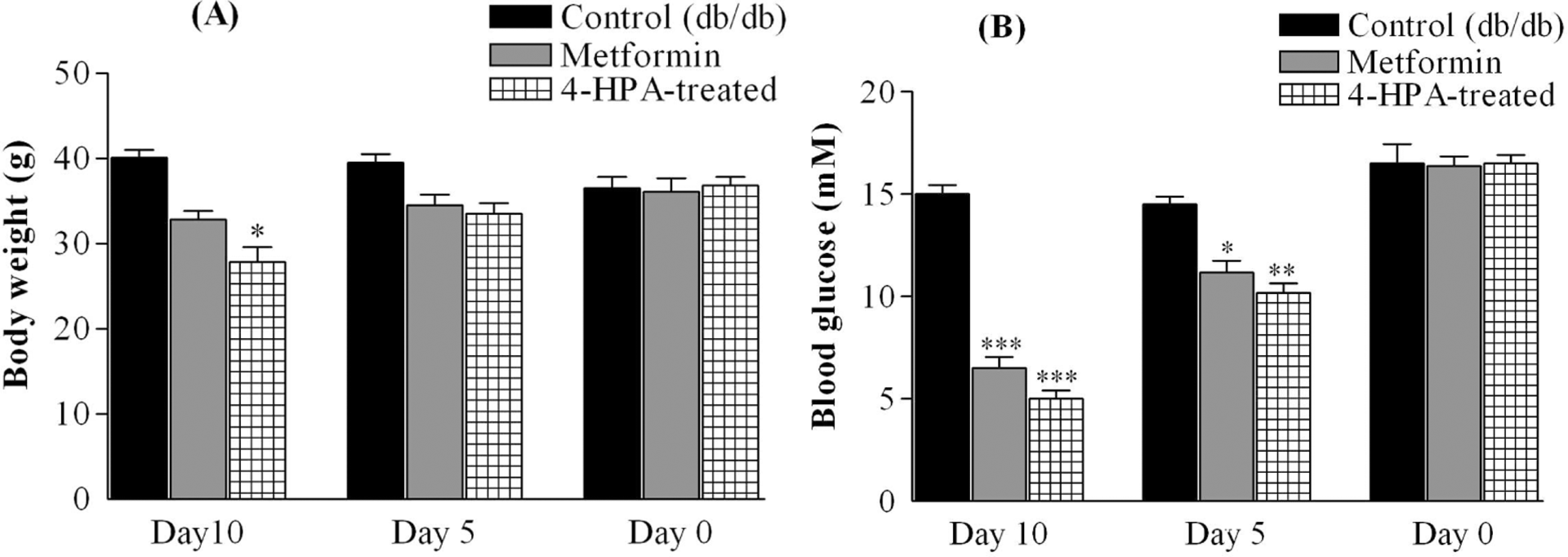
Effect of 4-HPA on (A) Body weight and (B) Fasting blood glucose level in db/db mice post 10-day treatment. Values are expressed as mean ± SE; (*p < 0.05, **p < 0.01 and ***p < 0.001 vs. vehicle-treated control group).

Effect of 4-HPA on glucose tolerance in db/db mice post 10-day treatment. (**p < 0.01 as compared to vehicle-treated control group).
Impact of 4-HPA on plasma lipids and biomarkers
Repeated oral administration of 4-HPA significantly lowered the level of plasma TG by 27.0% (p < 0.01), cholesterol by 26.5% (p < 0.01), FFA by 24.8% (p < 0.01) and low-density lipoprotein cholesterol by 28.3% (p < 0.01), respectively (Figure 5A and B ), beside it also enhanced cardio protective high-density lipoprotein cholesterol by 29.1% (p < 0.01) compared to vehicle-treated group (Figure 5C). Administration of 4-HPA also significantly lowered the plasma insulin and glucagons levels by 46.1% (p < 0.01) and 35.5% (p < 0.01) respectively in comparison to control group (Figure 5D).
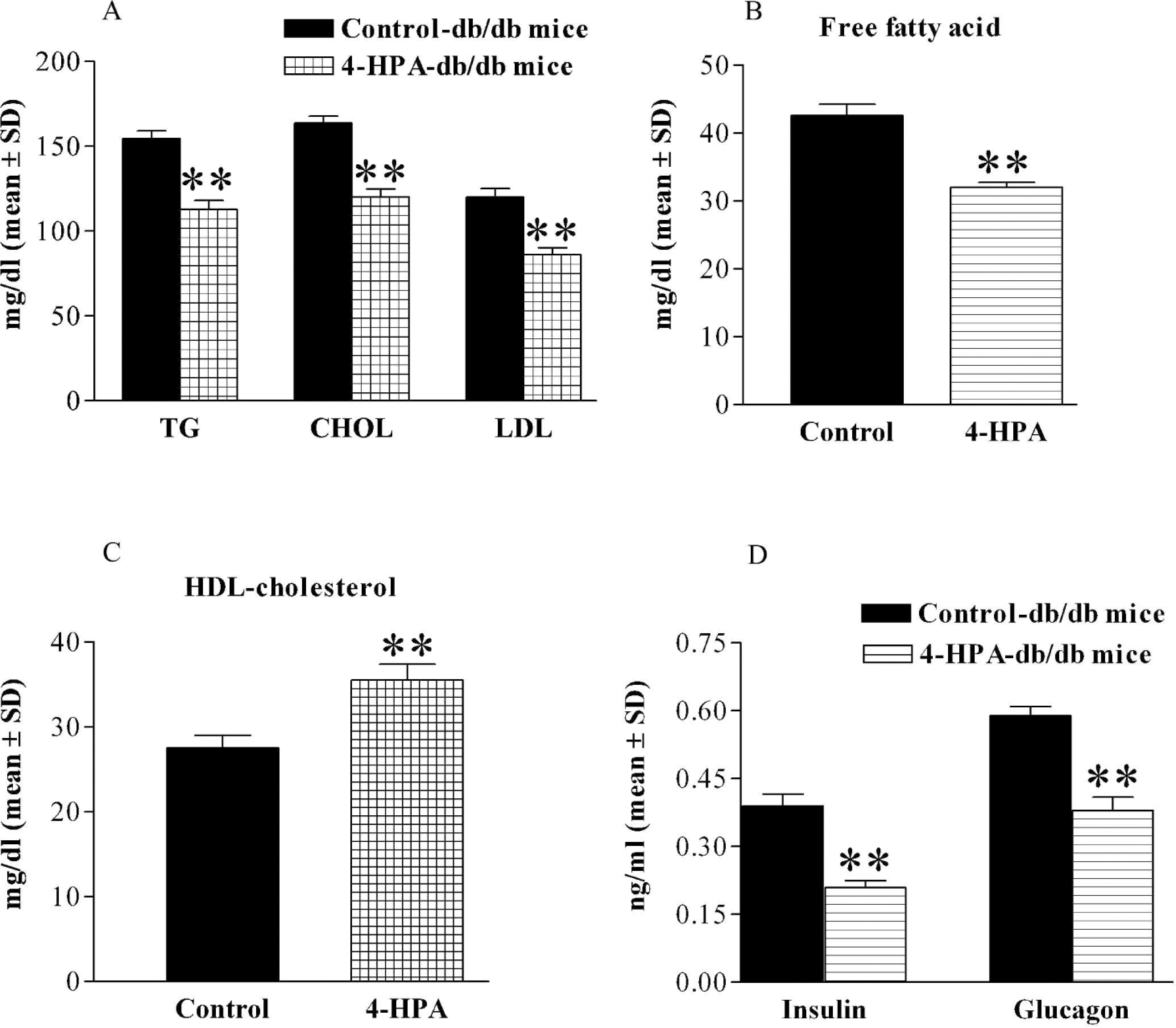
Effect of 4-HPA on plasma lipid profiles and biomarkers in db/db mice. Results are expressed as mean ± S.E (**p < 0.01 as compared to vehicle-treated group control).
4-HPA lowered the MDA concentration and increases CAT, GSH-Px and SOD in liver and kidney
Mice fed with 4-HPA had significantly lowered the MDA concentration by 25.6% (p < 0.01) in liver and 26.5% (p < 0.01) in kidney tissue respectively, compared to vehicle-treated group (Figure 6 ). It also increased CAT activity by 60.4% (p < 0.001); GSH-Px activity by 22.9% (p < 0.01) and SOD activity by 41.0% (p < 0.01) in liver tissue (Figure 7A ). Similar increment was also observed in kidney tissue. There is 53.5% (p < 0.01), 47.9% (p < 0.01) and 36.7% (p < 0.01) increase in the activity of CAT, GSH-Px and SOD, respectively (Figure 7B).

Effect of 4-HPA on lipid peroxidation in db/db mice. (**p < 0.01 as compared to vehicle-treated control group).

Effect of 4-HPA on activity of CAT, GSH-Px and SOD enzymes in liver and kidney tissue of db/db mice post 10-day treatment. Enzyme activities are expressed in specific activity (units/mg protein) ± SD. (**p < 0.01 and ***p < 0.001 as compared to vehicle-treated group).
Cytotoxic effect of 4-HPA
In order to evaluate any cytotoxic effect of the 4-HPA, L6 skeletal muscle cells were incubated with 4-HPA for 24 h. The effect was monitored by MTT assay. The compounds at various concentrations (1 ng/ml to 100 µg/ml) did not show any effect on cell viability of L6 skeletal muscle cells and found safe at this concentration range (Figure 8 ).
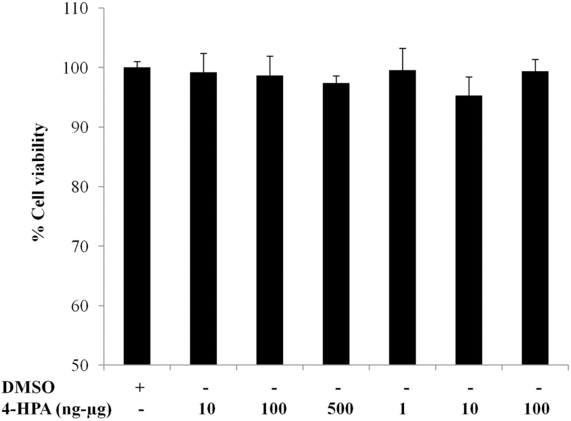
Cytotoxic effect of 4-HPA was tested on L6 skeletal muscle cells by using MTT assay (mean ± SE, n = 3).
Discussion
Diabetes is a chronic metabolic disorder affecting a major population worldwide. A sustained reduction in hyperglycemia can decrease the risk of developing micro vascular diseases. 20 The conventional pharmacological therapies have various side effects as well as high risk of secondary failure. However, medicinal herbs are expected to have a similar degree of efficacy without the troublesome side effects associated with conventional drug treatment. After finding encouraging results in crude extracts of P. harmala, we determine the effect of active component 4-HPA in C57BL/KsJ-db/db mice.
The glucose uptake study performed in L6 myoblasts cells have clearly demonstrated that the 4-HPA stimulates glucose uptake in differentiated L6 myotubes and showed the synergistic effect in the presence of insulin. Since skeletal muscle is the major tissue responsible for the maintenance of glucose homeostasis in vivo. 21 In type 2 diabetes, the capacity of skeletal muscle to take up glucose diminished. This phenomenon is observed at the level of both basal and insulin stimulated uptake. 22 Since skeletal muscle is a primary disposal site for glucose and thus is a major determinant of glycemia, it would be expected that interventions enhancing muscle glucose uptake would reduce glycemia in diabetic human and animals. 23
In vivo studies were carried out in db/db mice, which exhibited most of the human characteristics of type 2 diabetes including hyperglycemia during the fasting and fed states, hyperinsulinemia and insulin resistance. 24 The treatment of 4-HPA helped to reduce the FBG level in diabetic mice. 4-HPA along with FBG levels, improved the glucose tolerance and it also lowered the plasma insulin and glucagon concentration, while increased the ability of insulin to lower glucose in OGTT, demonstrating improved insulin sensitivity.
It has been observed mostly that hyperlipidemia has been reported to accompany hyperglycemia states and the most common lipid abnormality observed is hypertriglyceridemia. The dose of 50 mg/kg of the 4-HPA not only lowered the plasma levels of TG, CHOL, LDL and FFA levels but also enhanced the level of cardio protective HDL cholesterol. Several studies showed that an increase in HDL cholesterol is associated with a decrease in coronary risk and most of the drugs that decrease CHOL also decrease HDL cholesterol. It is important to note that in the present study, the 4-HPA not only decreased the CHOL but also increased the HDL cholesterol significantly after 10 days treatment as an additional advantage over the existing drugs. Since diabetes is associated with coronary complications, which is the major cause of morbidity and deaths in diabetic subjects,25,26 due to high levels of CHOL and more importantly LDL cholesterol, 4-HPA will therefore help in reducing the incidence of coronary event. Recently, FFAs have been suggested as a mediator of insulin resistance. This study demonstrated that 4-HPA treatment decreases in plasma FFA levels. Thus, 4-HPA may produce insulin sensitization by reducing plasma FFA levels.
It has been reported that reactive oxygen species (ROS) play an important role in death of β-cells in pancreas and development of diabetes in experimental animals. Furthermore ROS, generated as a result of hyperglycemia, possibly causes many secondary complications of diabetes such as nephropathy, retinopathy and neuropathy.27,28 Anti-oxidant enzymes like CAT, GSH-Px and SOD participate in the protective mechanism of cells. During chronic conditions, these enzymes seem to get worn out/ used up, as it has been reported that the levels of these enzymes are significantly reduced in diabetic condition. 29 In case of the anti-oxidant parameters, 4-HPA treated mice showed significant increase in the activity of SOD, CAT and GSH-Px with a decrease in MDA concentration in hepatic and renal tissue. These enzymes are known to quench the superoxide radical and thus prevent the damage of cells caused by free radicals. In cytotoxic evaluation on L6 skeletal muscle cell line, 4-HPA showed no adverse effect on cell viability in a concentration range of 1 ng/ml to 100 μg/ml and was found to be safe at this concentration gradient.
In conclusion, the study demonstrates that 4-HPA isolated from the traditional medicinal plant P. harmala has potential for anti-diabetic and anti-oxidative effects in db/db mice. Although the exact mechanism involved in the therapeutic effects of 4-HPA remained unexplored, it can be inferred from the results that it may control the hyperglycemia, hyperlipidemia and oxidative stress-mediated damage. Thus, 4-HPA could be considered as a natural lead for treatment of diabetes and associated complications, such as dyslipidemia and cardiovascular disease. Further study needs to be carried out in this direction to elucidate the mechanism of action(s) of 4-HPA.
Footnotes
Acknowledgements
ABS is thankful to CSIR, New Delhi, India for the financial support in the form of Senior Research Fellowship.
This study has been supported by the Council of Scientific and Industrial Research (CSIR) INDIA (Grant No. NWP-0032). CDRI Communication no: 7229.