
Abstract
The ameliorative effects of Opuntia vulgaris fruit extract (OE) were evaluated against methanol-induced haematological and biochemical toxicity in rats. The methanol-induced haematological and biochemical perturbation significantly decreased the levels of red blood cell (RBC), haemoglobin (Hb), haematocrit (Ht), serum total protein and increased glucose, cholesterol, and triglyceride levels in serum. Treatment of rats with methanol significantly increased lipid peroxidation (LPO) level and decreased the activities of superoxide dismutase (SOD), catalase (CAT) and glutathione peroxidase (GPx) in erythrocytes. OE treatment could increase significantly the levels of RBC, Hb, Ht and total protein, and decrease glucose, cholesterol and triglyceride levels in serum, and increase the activities of SOD, CAT and GPx in erythrocytes, when compared with methanol-treated group. Spleen histopathology showed that OE could significantly reduce the incidence of spleen lesion induced by methanol. These results suggested that OE could exhibit a potential source of natural antioxidants against methanol-induced haematological and biochemical disruption in rats. The protective effects of OE may be due to the modulation of antioxidant enzymes activities and inhibition of LPO.
Introduction
Methanol (M) is used as a solvent in many products such as antifreeze, pesticides, varnish and gasoline. Being first in the alcohol series, it is normally used as an industrial solvent and cleanser. People handling products that contain methanol may inhale the toxic vapor during its evaporation from the product surface. Accidental or intentional exposure to this alcohol can yield mild to severe health problems and, in extreme cases, coma and death. 1 The neurotoxicity of methanol (through acute, subacute or chronic poisoning) is attributed to its metabolite, the formic acid or formate, which inhibits the cytochrome oxidase system, necessary for ATP production. 2 Formic acid is the toxic metabolite responsible for the metabolic acidosis observed in methanol-intoxicated humans 3 and nonhuman primates. 4
Methanol poisoning is a public health problem in Tunisia. Sixteen patients with acute methanol poisoning admitted to intensive care unit denoted that patients developed headache, gastrointestinal symptoms, visual disturbances and metabolic acidosis.5,6 Two cases of acute methanol intoxication, 41 and 16 h after the voluntary ingestion of an unknown amount of methanol were treated by oral doses of 4-methylpyrazole. 7 In other cases of intoxication, fomepizole, a competitive alcohol dehydrogenase inhibitor, was utilized to treat methanol-poisoned patients from 1987 to 1999 (n = 14), as demonstrated by an alteration in toxicokinetics of methanol. 8
Now methanol is an ever-more recognized substance that damages the liver cells where it is oxidized to formaldehyde and later to formate. 9 These processes are accompanied by the elevation of NADH level and the formation of superoxide anion that may be involved in lipid peroxidation (LPO). 10 LPO products are harmful to cells, finally causing their death and can act as second toxic messengers of the complex chain reaction. 11 In addition, immune cells are sensitive to changes in the antioxidant status, as they carry out important functions through the generation of a high number of oxygen free radicals. 12 Erythrocytes are constantly being subjected to various types of oxidative stress, ingested chemicals and accidentally methanol. Although rat erythrocytes contain an abundance of catalase (CAT), they are incapable of oxidizing chemicals. 13 Methanol toxicity, either acute or chronic, is characterized by a severe dearrangement of subcellular metabolism and structural alteration of different cells. Systematically, methanol is moderately toxic to the liver and produces haematological effects. The mechanism by which methanol produces these effects is unknown. Previous data of Kadiiska and Mason. 14 mentioned that methanol induced oxidative stress in rats liver and kidney tissues. Red blood cell (RBC) membrane is rich in polyunsaturated fatty acids which are very susceptible to free radicals-mediated peroxidation. Eventually, haemolysis is induced by membrane LPO. 15 LPO is associated with a wide variety of toxicological effects, including decreased membrane fluidity and function.
O. vulgaris, a native species to Tunisia, is widely distributed in the center and the south of Tunisia, and its fruits known as prickly pears or cactus pears are an excellent source of betalain natural dye-stuff and functional compounds. Opuntia spp. fruits involve a lower risk of microbiological contamination, have no nitrate content, are highly flavoured, show adequate nutritional properties (e.g. high levels of calcium, magnesium and vitamin C), and contain interesting functional compounds like quercetin.16–19 The plant juice is used in the treatment of syphilis in Ayurveda. The aqueous extract of O. vulgaris on preliminary chemical analysis is found to contain saponin and alkaloid. 20 On the other hand, Opuntia spp. extracts have shown analgesic, anti-inflammatory, hypoglycaemic, physiological antioxidant, cancer chemoprevention and neuroprotective effects.21–23 Recent studies have shown that some phenolic compounds can prevent some chemical solvent-induced oxidative damage and the ability of phenolic compounds might be related to their antioxidant properties. Thus, the objective of this study was to evaluate the potential protective effects of aqueous extract of O. vulgaris fruit on methanol-induced haematological and biochemical toxicity in male Wistar rats.
Materials and methods
Plant
Preparation of O. vulgaris extracts (OE)
The O. vulgaris fruits were collected from a culture area located in Kasserine region, Tunisia. Fruit samples were ground, put in water and shaken (10 g/l, v/w) for 15–20 min, and then filtered using Whatman filter paper. The aqueous extract was given as beverage instead of tap water.
Animals
Adult male albino Wistar rats weighing 180–200 g were obtained from the Central Pharmacy of Tunisia (SIPHAT, Tunisia). The animals were quarantined and allowed to acclimatize for a week prior to experimentation. The animals were handled under standard laboratory conditions of a 12-h light/dark cycle in a temperature- and humidity-controlled room. Food and water were available ad libitum. Our Institutional Animal Care and Use Committee approved the protocols for the animal study, and the animals were cared for in accordance with the institutional ethical guidelines.
Treatment
After acclimatization, the rats were divided into two batches: 16 control rats (C) drinking tap water and 16 treated rats drinking O. vulgaris fruit extract (OE) for six weeks. Then, each group was divided into two subgroups and one of them was intraperitoneally injected (IP) daily, for four weeks, with methanol (2.37 g/kg b.wt.) according to Parthasarathy et al. 11 After treatment, eight rats of each group were sacrificed under anaesthesia by IP injection of chloral hydrate.
Determination of haematological parameters
After animal anaesthesia with chloral hydrate by intra-abdominal way, blood samples were collected with heparin by heart puncture to determine blood cell parameters (RBC, haemoglobin [Hb] concentration, haematocrit [Ht] value, mean corpuscular volume [MCV], mean corpuscular haemoglobin [MCH] and mean corpuscular haemoglobin concentration [MCHC]). A haematology analyzer Coulter MAXM (Beckman Coulter, Inc. Fullerton, USA) was used to determine these parameters.
Biochemical evaluation
Serum samples were obtained by the centrifugation of blood at 4000 rpm for 15 min at 4°C, and were then divided into eppendorf tubes. Isolated sera were stored at −20°C until used for analysis. The levels of serum glucose, total protein, cholesterol and triglycerides were measured using commercial kits according to the manufacturer’s directions.
Determination of antioxidant enzyme activities and lipid peroxidation
Other blood samples were collected without anticoagulant. The blood was centrifuged at 4000 rpm for 15 min (4°C) and erythrocyte was carefully sampled. They were then rinsed with ice-cold saline and homogenized (Ultra Turrax T25, Germany; 1:2, w/v) in 50 mmol l−1 phosphate buffer (pH 7.4). The homogenate and supernatant were frozen at −30°C in aliquots until used for antioxidant enzymes and LPO. The protein content of the supernatant was determined using the method of Lowry et al. 24
Measurement of lipid peroxidation levels
The thiobarbituric acid (TBA) method of Buege and Aust 25 was used to determine LPO by determining the amount of TBA reactive substances present in the erythrocyte homogenates obtained from rats. The principle of the method is spectrophotometric measurement of the color produced during the reaction to TBA with MDA. The absorbance of the solution was measured at 530 nm. The concentration of MDA was calculated by the extinction coefficient of MDA-TBA complex (1.56 × 105 cm−1 mol−1 l) and expressed in nanomoles per milligram of protein.
Determination of superoxide dismutase activity
Superoxide dismutase (SOD) activity was assayed by measuring its ability to inhibit the photoreduction of nitroblue tetrazolium ‘NBT,’ 26 50 μl of supernatant combined with 50 mM of phosphate buffer (pH 7.8), 39 mM of methionine, 2.6 mM of NBT and 2.7 mM of EDTA-riboflavin, was added last to obtain a concentration of 0.26 mM, and switching on the light started the reaction; changes in absorbance at 560 nm were recorded after 20 min. In this assay, the activity was expressed in relative units per milligram of protein (U mg−1). One unit of SOD activity is defined as the amount of protein that inhibits the rate of NBT reduction by 50%.
Determination of catalase activity
CAT activity was measured by the UV colorimetric method of Aebi 27 using H2O2 as substrate.
Determination of glutathione peroxidase activity
Glutathione peroxidase (GPx) activity was performed by estimating the content of oxidized glutathione formed by the action of GPx as described by Flohe and Gunzler, 28 but modified using the H2O2 as substrate with the presence of 5,5'-dithiobis (2-nitrobenzoate) (DTNB). One unit of activity catalyzes the oxidation by H2O2 of 1.0 μmol of reduced glutathione to oxidized glutathione per minute at pH 7.0 at 25°C. The specific enzyme activity was expressed in units per gram of protein (U g−1).
Histopathological observations
For histological studies, the spleen tissues were fixed in bouin solution, dehydrated in graded (50%–100%) alcohol and embedded in paraffin. Thin sections (4–5 μm) were cut and stained with routine hematoxylin-eosin (H&E). 29
Statistical analysis
All values are expressed as mean ± SEM. The results were analyzed by one-way analysis of variance (ANOVA) followed by Tukey test for multiple comparisons using SPSS for Windows (version 11). Differences were considered significant at p < 0.05.
Results
Haematological findings
The effects of methanol, OE and their combination on some haematological parameters in the rats are given in Table 1. Results showed that methanol alone caused a significant decrease (p < 0.01 vs controls) in RBC count, Hb level and Ht percentage. After treatment of rats with methanol plus OE, RBC count, Hb level and Ht percentage were normalized to their control values. However, no statistically significant change was observed in the other haematological parameters (MCV, MCH and MCHC levels) in methanol-treated group compared to control group. The present study showed that the administration of OE alone did not cause any significant alteration on the haematological indices.
Effects of methanol (M), Opuntia vulgaris fruit extract (OE) and their combinations (OE + M) on erythrocyte parameters in treated and control rats a

RBC: red blood cell, MCV: mean corpuscular volume, MCH: mean corpuscular haemoglobin and MCHC: mean corpuscular haemoglobin concentration.
Values are mean ± SEM for eight rats in each group. OE, M and OE + M treated groups vs control group.
a p < 0.01, M group vs (OE+M) group;
b p < 0.05,
c p < 0.01.
Biochemical findings
The effects of methanol, OE, and their combination on some biochemical parameters in the rats are given in Table 2. Results indicated that methanol caused a significant increase in glucose, cholesterol and triglycerids levels as compared to the control group. Results revealed that treatment with methanol caused a significant decrease (p < 0.01 vs controls) in the total protein level. At the same time, the decrease in total protein level induced by methanol alone was increased in the presence of OE with methanol.
Effects of methanol (M), Opuntia vulgaris fruit extract (OE) and their combinations (OE + M) on biochemical assays in treated and control rats

Values are mean ± SEM for eight rats in each group. OE, M and OE + M treated groups vs control group.
a p < 0.01,
b p < 0.001. M group vs (OE + M) group;
c p < 0.05,
d p < 0.01,
e p < 0.001.
Effect on lipid peroxidation
To explore the oxidative consequences of methanol treatment in erythrocytes of rats and to determine the possible protective effects of OE, we analyzed the LPO as a marker for membrane damage. Exposure to methanol significantly increased the LPO levels in the erythrocyte tissues samples as compared to control group (Figure 1). OE treatment prevented the LPO production induced by methanol. OE alone did not change the degree of LPO compared to controls.

Effects of methanol (M), Opuntia vulgaris fruit extract (OE) and their combinations (OE + M) on lipoperoxidation levels in erythrocyte of treated and control rats. Each value represents the mean ± SEM for eight rats in each group. OE, M and OE + M treated groups vs control group; ***p < 0.001, M group vs (OE + M) group; ##p < 0.01.
Effect on antioxidant enzymes
SOD activity in the erythrocyte tissues of experimental animals are shown in Figure 2. SOD levels were significantly reduced (p < 0.01) in the erythrocyte tissues of methanol-treated rats compared to control. Whereas, no significant change was observed in OE-treated rats, as well as in OE + M rats, showing the protective effects of OE against changes induced by methanol treatment. The CAT activities in the erythrocyte tissues of all experimental animals are shown in Figure 3. Methanol intoxication significantly decreased the CAT activities and treatment with OE revert the enzyme activity near to normal status. A significant decrease (p < 0.01) in GPx activities was observed in erythrocyte tissues of methanol-treated animals (Figure 4).
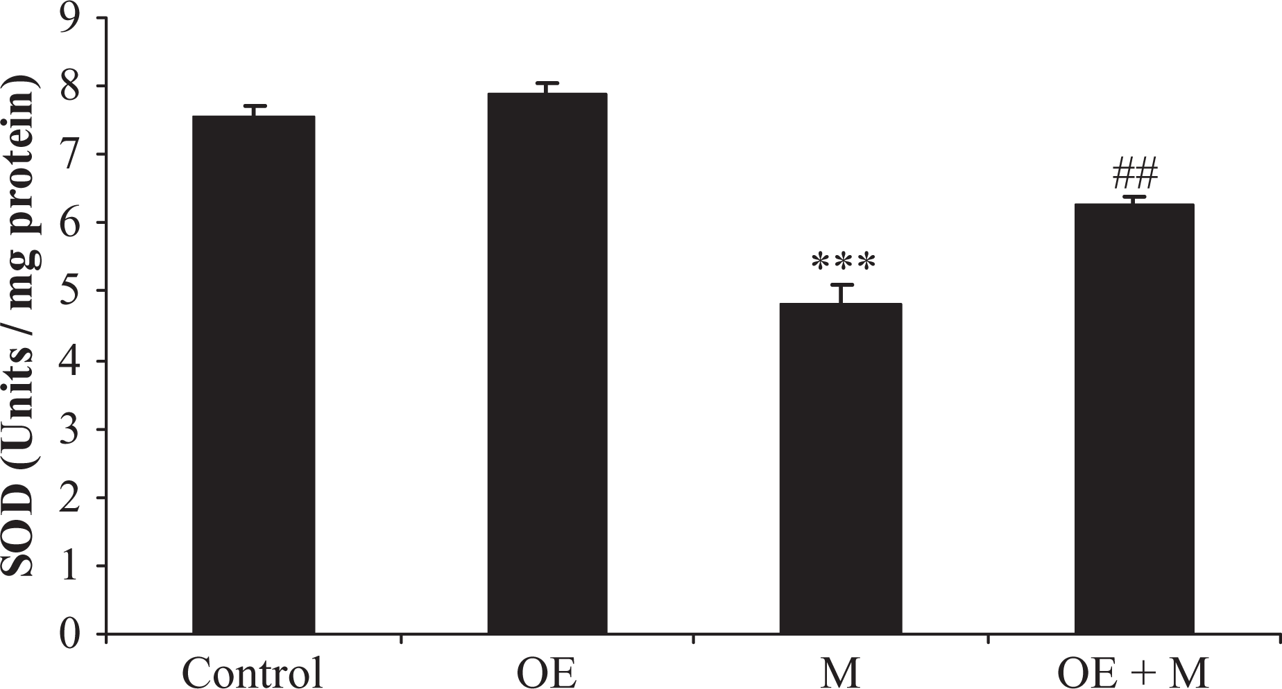
Effects of methanol (M), Opuntia vulgaris fruit extract (OE) and their combinations (OE + M) on superoxide dismutase activity in erythrocyte of treated and control rats. Each value represents the mean ± SEM for eight rats in each group. OE, M and OE + M treated groups vs control group; ***p < 0.001, M group vs (OE + M) group; ##p < 0.01.

Effects of methanol (M), Opuntia vulgaris fruit extract (OE) and their combinations (OE + M) on catalase activity in erythrocyte of treated and control rats. Each value represents the mean ± SEM for eight rats in each group. OE, M and OE + M treated groups vs control group; ***p < 0.001, M group vs (OE + M) group; ###p < 0.01.
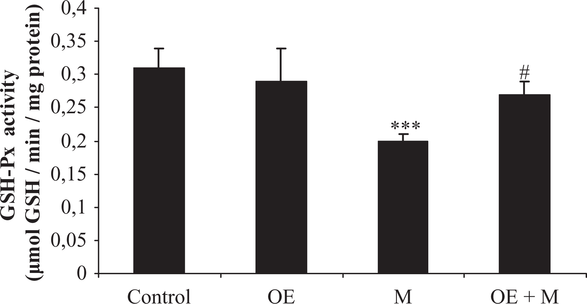
Effects of methanol (M), Opuntia vulgaris fruit extract (OE) and their combinations (OE + M) on glutathione peroxidase activity in erythrocyte of treated and control rats. Each value represents the mean ± SEM for eight rats in each group. OE, M and OE + M treated groups vs control group; ***p < 0.001, M group vs (OE + M) group; #p < 0.01.

Splenic tissue of rats: (A) control spleen showing normal architecture; (B, C and D) spleen from treated groups that received Opuntia vulgaris fruit extract (OE), methanol (M) and their combination (OE + M) showing proliferation and hypertrophy of reticuloendothelial cells (arrow), oedema and congestion of the pulp, and severe depopulation of the spleen follicle (arrowhead); original magnification ×400, H&E stain.
Histopathological findings
Histopathological studies of the spleen showed that methanol induced oedema and congestion of the pulp and severe depopulation of the spleen follicle in treated rats compared to controls. In addition, a massive proliferation and hypertrophy of reticuloendothelial cells were observed in methanol-treated rats. The histopathological disorders were in agreement with biochemical parameters. There were no histological alterations in the spleen of OE group when compared to control.
Discussion
The results of this study showed that methanol caused a significant decrease in some haematological parameters in rats, such as RBC counts, Hb level and Ht percentage. Haematological characteristics have been widely used in the diagnosis of variety of diseases and pathologies induced by industrial compounds, drugs, dyes, heavy metals, pesticides and several others. 30
The reduction in RBC count is considered to be due to the direct injurious action of the methanol on the animals. The erythropenia along with decreased Hb concentration is an indication of a decrease in oxygen carrying capacity in the animals, resulting in insufficient supply of oxygen to the tissues and causing adverse effects on animal health. The reduction in Hb level may be due to increased rate of red cells breakdown and/or reduction in the rate of RBC formation. In fact, Armutcu et al. 31 suggested that the decrease in RBC count is either indicative of excessive damage to erythrocytes or inhibition of erythrocyte formation. The results of this study indicated that OE given orally for 6 weeks attenuated the extensive changes of haematological parameters in methanol-treated rats. However, the exact mechanisms by which OE exert their protective effects against methanol-induced toxicity are not yet known. Nevertheless, the potential antioxidant source of OE (with direct or indirect actions) able to counteract or to minimize the undesirable effects induced by methanol might be evidenced. Previous studies of Tesoriere et al. 32 demonstrated that cactus pear (Opuntia) yield high values of important nutrients and exhibit antioxidant functions. Furthermore, cactus extracts exhibit antitumoral 33 and anti-inflammatory effects. 34 In the Mediterranean countries, cladodes are not a usual nutritional source for humans, but the fruits are largely consumed.16,35
Liver, muscle and brain are organs involved in glycogenesis, glycogenolysis, gluconeogenesis and glycolysis. In addition, the pancreas keeps hormonal control of glucose by the secretion of glucagon and insulin. The present study showed that the increase observed in glucose level in rats treated with methanol might be due to the effect of methanol on pathways involved in glucose homeostasis in the pancreas. Our results are in agreement with a study of Atrens et al. 36 which demonstrated that ethanol and tertiary butanol produced hyperglycaemic and hypothermic effects in treated rats. The present study showed that the significant increase in serum cholesterol and triglycerides which recorded in the methanol-administered group could be an indicator of biochemical and metabolic disruption. It seemed that high triglycerides and total cholesterol concentrations were linked with a greater risk for the development of coronary artery disease and other organ complications. 36 In this work, the decrease in total protein level might be explained by protein synthesis deficiency as a result of liver dysfunction induced by methanol.
The treatment of rats with methanol plus OE decreased serum glucose, triglycerides and cholesterol levels and increased total protein levels compared to the rats treated with methanol. In our experimental conditions, OE orally given to rats was found to inhibit the effects of methanol poisoning on the metabolism of lipids. Previous data of Wolfram et al. 37 ascribed the cactus antihyperlipidemic effects to the pulp pectin, which both reduced lipid absorption and increased faecal sterol excretion. Nevertheless, Fernandez et al. 38 claimed that the hypocholesterolemic effect of prickly pear pectin did not result from the reduction of cholesterol absorption but rather from an increase in apolipoprotein B/E receptor expression and changes in hepatic cholesterol homeostasis.
However, aerobic organisms generate superoxide anion radicals, hydrogen peroxide (H2O2) and hydroxyl radicals as a result of oxidative metabolism. Damage at the cell level by oxidants is attenuated by antioxidant enzymes such as SOD, CAT and GPx. Oxidative stress, generated by xenobiotics, induces disturbances in antioxidant enzyme systems. 39 The present study has demonstrated an increase of MDA in erythrocytes of rats treated with methanol alone showing excessive LPO. The significant increase of LPO levels in erythrocytes could indicate the damage caused in the examined tissues, as a result of free radicals generated by methanol. Methanol is oxidized via three main oxidative pathways among which alcohol dehydrogenase (folate dependent) and catalase peroxidative system have been extensively studied. 40 In rats, the oxidation of methanol is performed primarily by CAT. This enzyme forms the catalase-hydrogen peroxide system in the presence of H2O2, which intermediates the oxidation of various alcohol in to corresponding aldehydes.
The increase in the levels of MDA indicate enhanced LPO leading to tissue injury and failure of the antioxidant defense mechanisms to prevent the formation of excess free radicals. The present study also shows that the changes in LPO are accompanied by the concomitant decrease in the activities of antioxidant enzymes such as SOD, CAT and GPx in methanol exposed rats. Similar results have been previously reported by Dhabhar and McEwen 41 reported that methanol increased LPO by a direct effect or by decreasing the glutathione content. The reduced activity of CAT and SOD in the presence of methanol may cause the accumulation of O2−, H2O2 or the products of its decomposition. Loss of CAT and SOD activity results in oxygen intolerance and triggers a number of deleterious reactions. It has been proposed that the contribution of CAT might be enhanced if significant amounts of H2O2 become available through β-oxidation of fatty acids in peroxisomes. 42 Our results indicated that treatment of rats with methanol plus OE increased antioxidant enzymes (SOD, CAT and GPx), and decreased LPO as compared to the rats treated with methanol alone. This suggests that OE can modulate the balance of antioxidants and prooxidants.
The use of Opuntia has been recommended for its beneficial and therapeutic properties. 43 Previous studies reported that this plant exhibited diverse pharmacological actions, including emollient, hypocholesterolemic and hypoglycaemic effects. Inhibition of stomach ulceration and neuroprotective effects through antioxidant actions and anti-inflammatory effects showed to be also considered. 44
This study has clearly shown that, in addition to causing haematological and biochemical perturbation, and erythrocyte damage, methanol causes damage to spleen tissues in treated rats compared to those of control. The histopathologic evaluation of spleen showed that methanol-treated group had morphological defects. As a consequence of the haemolysis caused by methanol, an extensive filtration of damaged RBC by the reticuloendothelial system (RES) of the spleen as well as an effective haematopoietic process was observed. The major role and functions of the spleen is to remove damaged erythrocytes, and since such xenobiotics proved to damage erythrocytes by altering its antioxidant status, 45 it is expected that injured erythrocytes will be ultimately scavenged by the spleen generating ROS and subsequent tissue injury.
In summary, this study demonstrates that the administration of methanol at a dose of 2.37 g/kg b.wt./day for a period of 4 weeks caused significant changes in some haematological parameters (RBC, Hb and Ht), biochemical (glucose, cholesterol, triglyceride and total protein levels) and erythrocyte oxidative damages (LPO level, SOD, CAT and GPx activities) accompanied histopathological changes in spleen of treated group compared to controls. The use of OE was ascertained to alleviate the harmful effects of methanol in the mentioned parameters. In perspective, the antioxidant properties and the composition of O. vulgaris fruit juice are important to test the compounds separately and in combination to show whether O. vulgaris extract has additional protective properties compared to already established natural compounds. Improved knowledge of the composition, analysis and properties of O. vulgaris juice would assist in efforts for the industrial application of this fruit.
This study shows the potential value of O. vulgaris fruit as a good source of natural antioxidants; its consumption may contribute substantial amounts of antioxidants to the diet.
Footnotes
Acknowledgements
We thank the volunteers for their cooperation in the present study. This study was supported by the Ministry of Higher Education and Scientific Research in Tunisia.
This research received no specific grant from any funding agency in the public, commercial, or not-for-profit sectors.