
Abstract
The current study examined the impact of sub-chronic lead (Pb)-exposure upon global protein profile in rodent kidney (blood Pb levels ~50 μg/dL; 5 weeks oral Pb-acetate exposure). Utilizing 2D SDS-PAGE for kidney protein separation, greater than 500 protein spots were analyzed by densitometry following background noise removal, spot alignment, and intensity filtering. Approximately 100 protein spots were identified by ESI-MS/MS with mitochondrial, chaperone, antioxidant, and Pb-binding proteins included. Forty-eight protein spots exhibited significant alterations in abundance (18 identified by ESI-MS/MS) including the increased protein abundance of ketohexokinase, enolase, protein disulfide-isomerase, lamda crystallin, lactamase, and glycerol-3-phosphate dehydrogenase. Decreased protein abundances were observed for α-2 microglobulin, glutamate cysteine ligase, prohibitin, homogentisate 1,2-dioxygenase, alpha-ETF, argininosuccinate synthetase and ATP synthase (H+ transporting). These data support the hypothesis that protein profiles in the kidney are altered following sub-chronic physiologically relevant Pb-exposure.
Introduction
Environmental Pb toxicity remains a major health concern for adults and especially children. Despite the banning of Pb-containing gasoline, pesticides, and paints, Pb-exposure (in both adults and children; blood Pb levels >10 µg/dL) still occurs in the United States. 1 As documented by the National Health and Nutrition Examination Survey, 2 approximately 434,000 children aged 1−5 years within the United States were identified with blood Pb levels above the CDC recommended level of less than 10 µg/dL. Bone and/or blood Pb levels are significantly associated with hypertension-related disorders including cardiovascular disease, kidney failure and stroke decades after the initial exposure.3–12
The kidney is a primary target organ for absorbed Pb. 13 Chronic Pb exposure results in decreased glomerular filtration rate, nephropathy of proximal tubules, and accelerated kidney tumor growth.14–16 Chronic Pb exposure induces oxidative stress and inflammation and interrupts nitric oxide signaling pathways which ultimately leads to the observed nephrotoxicity.17,18
Alterations in rodent kidney proteome profiles due to acute Pb exposure (3 days ip) were previously investigated with a primary impact on Pb-binding protein and antioxidant balance noted. 19 The current study seeks to extend the existing knowledge of the effect of Pb-exposure on kidney biochemical homeostasis by examining the alterations in protein profiles of sub-chronically Pb-exposed rodents. The soluble proteome profiles from adult Fisher 344 rats, divided into two groups—unexposed (acidified water) and exposed (1500 ppm in acidified water) for 5 weeks—were examined for indications of persistent alterations in abundance of cellular metabolic enzymes, antioxidant enzymes, redox proteins, and chaperone proteins.
Methods
Animal exposures
Fisher 344 rats (75−100 g) were obtained from Charles River, fed and watered ad libitum, and maintained with a 12-hour light: dark cycle in the University of Missouri–Rolla animal facility. 18 The rats were cared for in accordance with the guidelines set forth by the Animal Research Advisory Committee of the National Institutes of Health. The University of Missouri-Rolla is an American Association for the Accreditation of Laboratory Animal Care-accredited institution. Following 1 week of acclimation, the 12 animals were randomized into two groups: Control, receiving distilled water acidified with glacial acetic acid for 5 weeks; and Pb, receiving 1500 ppm Pb acetate in acidified water for 5 weeks. At the end of 5 weeks, the animals were euthanized according to the University of Missouri–Rolla animal-handling protocols, blood was collected via cardiac puncture, and the organs were collected. The kidney tissues were stored at –80°C until analysis.
Tissue samples
The kidneys were removed, weighed, and shipped on dry ice to the Proteomics and Mass Spectrometry Facility at Montana State University. Three hundred micrograms of kidney tissue was homogenized with lysis buffer (7 M urea, 2 M thiourea, 4% CHAPS, protease inhibitor cocktail (Roche, Basel, Switzerland]) in a Dounce homogenizer. The homogenates were centrifuged at 3000 rpm for 10 minutes and the supernatant containing the soluble proteins was collected. 20 The insoluble protein fraction was saved for analysis but is not the subject of this paper. The soluble protein concentrations were determined by Bradford Assay (BioRad, Hercule, California, USA) using bovine serum albumin (BSA; 2 mg/mL, Sigma-Aldrich, St. Louis, Missouri, USA). 21 The samples were stored at −20°C prior to 2D-PAGE.
Pb Analysis
Blood Pb levels were determined by graphite furnace atomic absorption spectrophotometry (Varian SpectrAA) at the Springfield-Greene County Department of Public Health, Springfield, Missouri, USA. 18
2D-SDS-PAGE
Isoelectric focusing of the first dimension was accomplished on an IPGphor with non-linear, pH 3-10, 180 mm × 3 mm × 0.5 mm Immobiline DryStrips (GE Healthcare, Piscataway, New Jersey, USA) according to the manufacturer’s instructions. The samples were brought up in 350 µL total volume, containing 500 of µg protein, and loaded onto the strips by the standard protocol. Passive rehydration of the strips was accomplished in a 7 M urea, 2 M thiourea, and 4% CHAPS solution containing 2.8 mg/mL DTT and 2% pH 3-10 IPG buffer. The proteins were focused for 28,000 Vhrs. The strips were equilibrated first with equilibration buffer (1.5 M Tris pH 8.8 buffer containing 6 M urea, 34.5% glycerol, and 2% SDS) and 3.5 mg/mL DTT for 15 minutes and second with equilibration buffer and 45mg/mL iodoacetamide for another 15 minutes. The strips were then loaded onto 12% acrylamide gels for the second dimension and run at 70 volts for 24 hours at room temperature. The gels were fixed in 30% ethanol and 7% acetic acid for a minimum of 1 hour followed by staining with colloidal Coomassie Blue G-250 and washed in deionized water for at least 24 hours. 22 The images were collected using an Epson Expression 1680 desktop scanner with transparency adapter at 266 dpi and 16 bit grayscale pixel depth. Each kidney sample was prepared and separated by 2D gel electrophoresis in triplicate.
Image analysis
Gel images were analyzed using SameSpots image analysis software (Nonlinear Dynamics). The gels were aligned with the TT900 alignment algorithm and spots were detected and matched automatically with Progenesis background subtraction and INCA noise removal. Normalized spot volume data for Averaged Gels consisting of experimental replicates for each group were analyzed for significant up- or down-regulation in protein abundances. Technical replicates of each sample were analyzed for spot density variations. Each averaged gel data set contained 4−6 biological replicates (2 gels discarded due to poor IEF separation). Quantitation of differential protein abundances among the 2 groups was assessed via ANOVA with p < 0.05 identified as significant. In the case of comparison of 2 means, ANOVA will produce the same result as the t-test of independent means. We have kept the ANOVA terminology used by the SameSpots software for clarity. Only spots that were designated as meeting the ANOVA criteria in each of the triplicate technical replicate experiments are reported as significantly altered. 22
Trypsin digest and mass spectrometry
Protein spots were cut from the 2D gels and destained with 50% ethanol (EtOH) and 50 mM NH4HCO3 at room temperature. The gel pieces were dehydrated in ethanol and subjected to in-gel protease digestion with modified trypsin (10 ng/µL; Fisher Scientific) in 50 mM NH4HCO3 for 18 hours. Tryptic peptides were extracted via sequential steps of 50% EtOH in 0.1% formic acid followed by 95% EtOH in 0.1% formic acid. The extracted peptides were lyophilized and stored in −80°C until analysis. 23 The lyophilized peptides were resuspended in 20 µL of 5% acetonitrile (ACN), 95% water, 0.1% formic acid and transferred to auto-sampler vials. The peptides were separated by gradient elution ranging from 5% ACN in 0.1% formic acid to 90% ACN in 0.1% formic acid on a C18 microfluidics device (ChipCube interface) connected to an Agilent ESI-Iontrap-MS (XCTUltra). The instrument isolation, fragmentation, and accuracy were calibrated with ESI Tune Mix (Agilent) and sensitivity measured with 50 fmol trypsin digested bovine serum albumin immediately prior to analysis of samples. Proteins were identified by NCBInr database searching with an in-house Mascot Server (Matrix Scientific). Parameters for the search were Mammalian proteins, ±0.8 daltons for the parent peptide, ±0.4 daltons for fragmentation masses, 2 missed trypsin cleavage sites allowed, and carbamidomethylation of cysteines as a variable modification. The total MOWSE score is the absolute probability that the observed match is a random event with scores reported as the negative 10*LOG10(P), where p is the absolute probability of a correct score. A probability of 1020 thus becomes a score of 200. 24 A MOWSE score above 65 is generally considered significant although for our studies we require a minimum of two peptides with individual ion scores above 45 (measure of the agreement of MS/MS spectra with predicted b- and y-ion fragmentation series), which generally creates a MOWSE score cutoff of 85.
Results
After 5 weeks of oral treatment with 1500 ppm Pb acetate in acidified water, the blood Pb levels of Pb-exposed rats (53.4 ± 11.3 μg/dL) were significantly higher than controls (<1 μg/dL). Figure 1 is a representative 2D gel from a Control sample run with 500 μg of protein. The selected gel is representative of multiple control samples which contain over 500 aligned protein spots (filtered to remove noise, non-reproducible spots, and spots with density less than 200). The central region of the gel (pI range 4−8) is well focused, with a loss of focus outside this range (the acidic and basic edges) that can be alleviated by running narrow range isoelectric focusing strips (pI strips), which may be of interest for future studies.

Representative 2D-SDS PAGE gel image of urea/thiourea soluble kidney proteins from control Fisher 344 rat.
Proteins were identified from 105 spots numbered on the image. Table 1 identifies of the protein spots numbered in Figure 1. Only the dominant protein identification is given though low levels of secondary proteins were found in several spots. A ~90% success rate for identification of proteins from gel spots was found. Note that a series of spot trains (spots 18, 19, 92, 93) are the same protein with different isoelectric focusing (IEF) points, which is indicative of post-translational modification variants. Spots 2, 61, 62, 97, 98, and 101 are also the same protein though different molecular weights and IEF points were found thus indicating cleavage products and post-translational modifications. Dominant and faint protein spots (example: 18 and 66 respectively) were identified. Classes of proteins observed included mitochondrial proteins, chaperone proteins, antioxidant enzymes, and Pb-binding proteins. Significant amounts of the protein albumin (Spots 18, 19, 92, 93) are present on the gel as expected from vascularized tissues. The relative abundance of these albumin spots do not change with Pb exposure as compared to the control tissue (control and Pb; less than 7% variance in normalized spot density). The total abundance of albumin can act as a measure of gel-to-gel variation which is due to the degree of blood and serum contamination. A large degree of variability in albumin is indicative of non-reproducibility in biological replicates.
ESI-MS/MS protein spot identifications a


a Related to Figure 1.
As shown in Figure 2 , approximately 18 protein spots (outlined in black) were either increased or decreased in abundance in response to 5 weeks of 1500 ppm Pb exposure based on normalized spot densities (p < 0.05). In Table 2 , the protein identification for 18 of these spots, degree of variance in abundance within group (%CV), and relative change in percentage abundance between experimental groups are noted. The general trend of relative changes between 20% and 75% represents a moderate alteration of levels for affected proteins. In general, the normalized spot density variation within group was in the acceptable range under 20% CV except for α-2 microglobulin. All isoforms of this metal-binding protein (except Spot 101; a faint spot with high %CV) were found to be significantly decreased in kidney of the Pb-exposed animals. Consistent with impairment of kidney function noted by other studies, 17 a high degree of variability in abundance for the protein spots representing the α-2 microglobulin was observed in the Pb-exposed group with considerably lower within group %CV for the control group. The abundance of a rate-limiting enzyme in the synthesis of GSH and enzymes involved in arginine metabolism were also decreased. The abundances of a chaperone protein and proteins involved in cellular metabolism were increased.
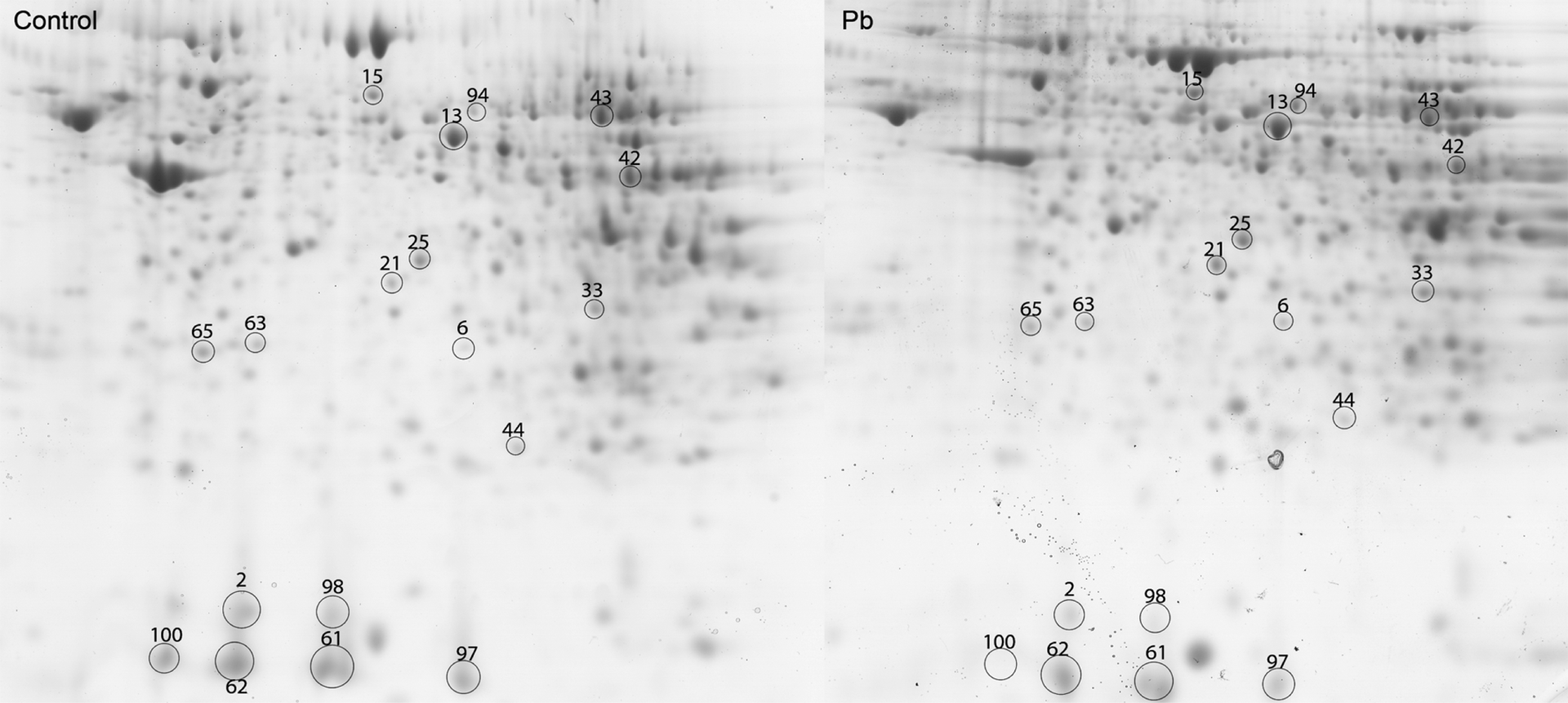
2D-SDS PAGE image comparison of kidney proteome profiles from Fisher 344 rats exposed to 1500 ppm Pb for 5 weeks (blood lead ~50 µg/dL). Protein spots which were significantly up or down in abundance as compared to the control animals (p < 0.05) are circled in black (statistical analysis in Table 2).
Proteins identified as up/down regulated in kidney following Pb exposure for 5 weeks a

a Related to Figure 1.
Discussion
In the current study, Pb-exposed Fisher 344 rats possessed blood Pb levels analogous to CDC Class IV. This blood Pb-level has been observed in children from large, poor urban centers such as Chicago, Illinois and St. Louis, Missouri, as well as children from other industrialized countries, specifically Mexico and China.25–28 This level in adults (e.g. Pb-miners, smelter workers) triggers consultation with physicians and possible chelation therapy. In children, similar blood Pb-levels trigger increased screening of the general population in the affected area, referral to medical personnel, and initiation of chelation therapy. 29
Previous studies have identified oxidative imbalances in the kidney following sub-chronic and chronic Pb-exposure.6,12,13,17 The current study identified significant alterations in the levels of specific proteins following sub-chronic Pb-exposure in a rodent model. Specifically, Pb-exposure induced alterations in abundances of proteins involved in cellular energy metabolism, amino acid metabolism, redox status, chaperone activity, and toxin clearance though not all proteins in a specific functional class were impacted. The present study did not examine whether Pb-exposure, kidney function, and specific protein levels are linked. Candidate biomarkers related to prior reports of kidney functional impairment were identified.
The kidney contains high-affinity metal binding proteins including α-2-microglobulin. 30 In the present study, α-2-microglobulin (Spots 2, 61, 62, 97, and 98) abundance was significantly lowered in the Pb group (58%−74% decrease in abundance except Spot 101). The relatively high %CV of protein abundance for these spots in the Pb group is indicative of varying degrees of excretion of this Pb-binding protein. Our data implies, but does not directly prove, specific effects upon the efficiency of excretion of protein-bound Pb that is most likely due to impact on glomerular filtration rate.
Relative levels of proteins indicative of mitochondrial stress were also altered with Pb exposure. Decreased protein abundances of prohibitin-1 (Spot 63), which regulates mitochondrial function, 31 α-electron transport flavoprotein (Spot 33) a member of the mitochondrial electron transport chain, 32 and ATP synthase H+ transporting mitochondrial F1 complex alpha subunit isoform 1 (Spot 44) was observed in the current study. Thus, sub-chronic Pb-exposure acts as a mitochondrial toxin.
Amino acid metabolism was also negatively impacted by Pb exposure. Decreased abundance of argininosuccinate synthetase which is involved in the arginine synthesis and is particularly abundant in proximal tubules in the cortex, 33 homogentisate 1,2-dioxygenase, which is involved in tyrosine catabolism, 34 and glutamate cysteine ligase, which is the rate-limiting enzyme in GSH synthesis, was observed. 35 The decreases in amino acid catabolism may be a direct result of proximal tubule damage and reflect a switch in use of amino acids from other metabolic pathways to protein synthesis due to the high urinary excretion of the Pb-binding protein, α-2 μ globulin, and continuing protein synthesis.
Increases levels of the endoplasmic reticulum chaperone GRP 78 and lambda crystallin indicates significant tissue stress in response to Pb exposure.36,37 Lambda crystallin is a protein involved in salt-tolerance in the kidney and is also a lenticular enzyme crystallin which shares significant sequence similarity to dehydroascorbate reductase. A change in abundance of between 20% and 40% for enzymatic and chaperone proteins may negatively impact cellular function.
The majority of proteins visible and identified from the gels did not have altered relative abundances including proteins involved with redox homeostasis and repair of oxidative damage.35,38,39 Specifically, peroxiredoxin 1, peroxiredoxin 5, peroxiredoxin 6, glutathione synthetase, and glutathione-S-transferase (Spots 58, 104, 5, 82, and 23, respectively) were present on the gels. GRP75, HSP 65, and GRP90, which are chaperone proteins, were also present as Spots 20, 84, and 78. Mitochondrial fatty acid/amino acid catabolism and detoxification enzymes including isovaleryl CoA dehydrogenase, L-3-hydroxyacyl-CoA dehydrogenase, and aldehyde dehydrogenase precursor (Spots 10, 55, 86 respectively) were also identified.40,41
The decreased abundance of glutamate cysteine ligase, and our prior findings of decreased GSH/GSSG ratios in kidney of Pb-exposed animals, 18 supports an imbalance in redox potential though a general shift to increased production of antioxidant enzymes was not observed. Due to the high energetic demands for continued production of Pb-binding proteins prior to excretion, coupled with the observed decrease in amino acid metabolism, it is possible that the available synthetic capacity has been primarily used to generate proteins dedicated to clearance of the toxin. The present study did not measure enzymatic activity though it is likely that oxidative modification of these proteins is occurring resulting in decreased enzymatic activity and further oxidative stress of the tissue.
In summary, kidney protein profiles are significantly altered in a rodent model of sub-chronic Pb-exposure (BLL ~50 μg/dL). Of note in the present study is the decreased abundances of proteins involved in amino acid catabolism and metal-binding/clearance.
Footnotes
Research described in this article was supported in part by PHS NIH P20 RR-17702 from the COBRE Program of the National Center for Research Resources and PHS NIH P30ES014443 from NIEHS.