
Abstract
This study delves into the synergistic effect of bi-nanofillers, 0-D titanium dioxide (TiO2), and 2-D graphene nanoparticles (GNP) on the microstructure, sorption, anti-microbial, static, and dynamic mechanical behaviour of polylactic acid (PLA). The tensile strength, critical stress intensity factor, and storage modulus (at the optimum filler content of 1wt% GNP and 0.5 wt% TiO2) of P/1 G/0.5T sample at room temperature exhibited an increase of 52%, 42%, and 40% respectively with respect to PLA. This increment is accredited to the physical entanglement of dual nanofillers with PLA chains. The hybrid system revealed a higher surface roughness in atomic force microscopy (AFM) which accounts for the hydrophilic nature of contact angle measurements. The heterogeneous nucleation effect of dual nanofillers improved the percentage of crystallinity by 54% as obtained from differential scanning calorimetry (DSC). The X-ray diffraction (XRD) analysis showed the formation of large number of small crystals in the P/1G/0.5T sample. The amphiphilicity of the hybrid sample reduced the diameter of incubation halos with Staphylococcus aureus in the presence of biocide, ciprofloxacin as it creates a hostile environment for bacterial growth.
Introduction
Plastics have become an integral component of human lives in all aspects, including automobiles, buildings, food packaging, healthcare sectors, and so on. Single-use plastics, such as polybags and packaging materials, pollute the environment through landfills, the ocean, or open dumping, and account for around 5%–12% of total trash creation. Bio-polymers have garnered interest in recent times due to their biocompatibility and biodegradability. 1 Studies revealed that bioplastic production may increase from 2.4 million in 2021 to 7.5 million in 2026. 2 Polylactic acid (PLA) is a very popular biopolymer synthesized from agricultural commodities like sugar beets, corn, cassava, sugar cane, etc. 3 PLA is used in numerous commercial applications spreading from food packaging and textiles to cosmetics and pharmaceuticals. 4 PLA constitutes around 14% of the total commercial biopolymers. 2 PLA has massive potential to be a substitute for conventional fossil-based polymers but is restricted due to their low thermal, mechanical, flame, and barrier properties.5,6 Various studies suggest that the incorporation of nanofillers into the neat PLA matrix will enhance its intrinsic properties.7–9 Diverse inorganic nanofillers like zinc oxide (ZnO), silicon nitride, alumina, nanoclay, and titanium dioxide (TiO2) show the potential to improve the matrix’s properties.10,11 TiO2 nanoparticles have drawn the attention of many researchers owing to their UV resistance, photo-resistance, thermal and chemical stability, and superior refractive index.12,13 Among carbon-based nanofillers, graphene nanoplatelets (GNPs) have a reputation for manifesting high electrical and thermal conductivity, mechanical strength, and barrier properties.14–16 Adding GNP into thermoplastic matrices increases the crystallization temperatures and accelerates the crystallization kinetics due to the heterogeneous nucleation effect, which in turn has a positive influence on the static mechanical properties. 17 Nadaraira and co-authors 18 studied TiO2/GNP composite by sintering method and observed a uniform dispersion of 1 wt% GNPs in the TiO2 matrix resulting in an enhanced fracture toughness compared to the neat TiO2 matrix along with appreciable improvement in electrical properties. With the incorporation of GNPs the grain size showed a drastic reduction which helps in crack bridging or branching. Ghasemi et al. 19 reinforced PLA with cellulose nanofiber (CNF) and maleated PLA (MPLA) as a compatibilizer. The sample with 5 wt% of CNF and MPLA improved the impact strength and tensile strength by 131% and 169%, respectively compared to neat PLA. This can be credited to the superior interfacial adhesion provided by the presence of MPLA and homogenous dispersion of CNF in the PLA as observed from scanning electron microscopy (SEM).
Valapa et al. 20 added graphene (G) into PLA by melt mixing method, and observed that 0.1 wt% of nanofiller exhibited a higher percentage of crystallinity (Xc) owing to the prominent nucleation effect of G. Thermogravimetric analysis (TGA) revealed 0.5 wt% of nanofiller in PLA improved thermal stability. Kissinger’s method revealed that activation energy (Ea) increased from 177 kJ/mol for neat PLA to 184 kJ/mol with the addition of 0.5 wt% of G. Wu et al. 21 incorporated a total of 5 wt% multiwalled carbon nanotubes (MWCNTs) and TiO2 into the PLA matrix, which exhibited enhanced Xc, as verified by X-ray diffraction (XRD) and differential scanning calorimetry (DSC). The storage modulus (E′) of the hybrid composite increased by 72% compared to the neat matrix. The hybrid nanocomposite with a total loading of 9 wt% MWCNT and TiO2 demonstrated better thermal resistance by reducing volatilization, contributing to their superior thermal barrier properties. Piekarska et al. 22 noted that the presence of 3 wt% MMT and 15 wt% cellulose fiber in PLA matrix, raised the E′ value by 50% due to the exfoliation of montmorillonite (MMT) layers as observed from transmission electron microscope (TEM) images. Another group of researchers 23 investigated the synergistic effect of alkylated maleic anhydride grafted graphene oxide (AGO) and silanated nano-TiO2 (ST) in the PLA-starch composite. The Fourier transform infrared spectroscopy (FTIR) and Raman analysis revealed the formation of a strong hydrogen bond between modified nanofillers and the PLA-starch matrix. With the addition of 1 wt% ST and 0.2 wt% of AGO, the contact angle (CA) values were found to be the highest (98.5°), amongst all nanocomposites due to the surface hydrophobicity of uniformly dispersed interpenetrated network, which was confirmed by SEM micrographs. XRD and DSC analysis revealed heterogeneous nucleation and sphero-crystal growth of PLA chains. Another study 24 found that, with the addition of 8 wt% of TiO2, the bacterial colonies (Escherichia coli) reduced by 82% without UV irradiation in comparison with virgin PLA, making it a suitable candidate for biomedical and food packaging applications. Ahmed et al. 25 investigated the surface topography of PLA/polyethylene-glycol (PEG)/graphene oxide (GO) by atomic force microscopy (AFM). At 1 wt% and 2 wt% of GO in PLA/PEG, the surface roughness (root mean square) values were found to be 30.4 and 21.6 μm, respectively due to the decrement in hills and valleys. The tensile strength of the same composites showed an increment of 5.56 and 7.27 MPa, respectively compared to the PLA/PEG system due to their strong interfacial interaction and reduced free volume. Shi et al. 26 incorporated GNPs and CNTs in the PLA matrix fabricated by the 3D printing method. With the addition of 2 wt% of GNPs and 4 wt% of CNTs, Young’s modulus and tensile strength increased by 25.5% and 16.2%, respectively, compared to the neat matrix. The authors also revealed that the hybrid sample showed excellent electromagnetic interference (EMI) shielding in the X-band region. The increase in overall characteristics was accredited to an entangled network formed between 1-D and 2-D nanofiller in the PLA matrix. Bai et al. 27 found that 1 wt% CNT and 2 wt% MMT in PLA provided high fracture toughness owing to a three-dimensional sandwich-like structure formed by multi-dimensional nanofillers.
The present study focuses on the collaborative effect of GNP and TiO2 in the PLA matrix. An effort has been made to comprehend the static and dynamic mechanical properties of the samples. The crystalline nature of the samples is analysed using XRD and DSC analysis. The dispersion of nanofillers is assessed using SEM and TEM, the surface topography, and the roughness of the samples being examined using AFM. Escherichia Coli and Staphylococcus aureus bacteria are being used to evaluate the antimicrobial properties of PLA nanocomposites. The current study also investigates the molecular interaction of fillers and PLA chains by Raman analysis. The transport properties using hexane solvent are being analysed. The production of PLA based hybrid nanocomposites significantly lowers carbon foot print, minimize environmental impacts, decompose in industrial composting conditions and makes it more sustainable over its life cycle. The current work will be a standalone reference which portrays a unique combination of GNP/TiO2 nanofillers in PLA matrix which is expected to enhance static and dynamic mechanical properties, thermal stability, and antimicrobial efficacy. To the best of our knowledge in-depth analysis of the dispersion of bi-nanofillers in PLA, its interfacial bonding, optimization of composite formulations in this unique combination of TiO2/GNP fillers, and its synergisms on various properties are yet to be reported.
Materials and Methods
Materials
Polylactic acid procured from Augment 3Di, Coimbatore, India was used as the base matrix in this study. The nanofillers used are industrial GNPs (supplied by Platonic Nanotech Private Limited, Jharkhand, India) with purity of 99%; thickness 5-10 nm; specific surface area: 200-210 m2/g, as well as industrial grade TiO2 (Platonic Nanotech Private limited, Jharkhand, India) with purity of 99.9% with avg. diameter of 25 nm and specific surface area of 150-170 m2/g.
Experimental Methods
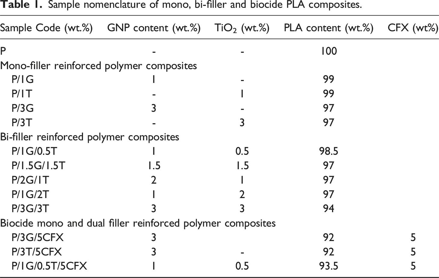
Sample nomenclature of mono, bi-filler and biocide PLA composites.
Characterization Methods
The dispersion of the nanofillers in the prepared samples was analysed using a Field Emission Scanning Electron Microscope (FESEM) of the Carl Zeiss model, Gemini 300. The surfaces of the samples were gold sputtered to a thickness of 2 nm prior to analysis. JEOL/JEM-2100 high–resolution transmission electron microscope (HR-TEM) was used to evaluate dispersion of nanofillers. An Atomic Force Microscope (PARK XE-70), operating at 360 kHz, was employed to analyse the surface topography of the samples. XRD analysis was conducted using Rigaku Ultima IV (40 kV/30 mA) with Cu as an X-ray source. Continuous scanning mode was followed to measure 2θ from 5° to 90° with a step size of 0.02° at a scanning rate of 2°/min. Renishaw Metrological Systems (UK) Invia Reflex Raman Microscope with a spectrometer (514 nm excitation laser), was used for conducting Raman analysis. Tensile tests were carried out using a Universal Testing Machine (UTM), Tinius Olsen (UK) model H25 KT, with a test speed of 50 mm/min at room temperature. The average values obtained after testing 5 samples are reported. The fracture toughness properties of the samples were evaluated by single end notch bending test (SENB) using UTE-40 make-FIE universal testing machine (UTM) at a crosshead speed of 1.25 mm/min (ASTM D5045). DSC analyses were performed with the help of TA instruments Q20 V24.10 Build 122 at non-isothermal conditions. The samples were heated up to 200°C at a heating rate of 10°C/min, maintained at 200°C for 5 min, and then cooled back to room temperature at the same rate for cooling. Dynamic mechanical properties were studied using a DMA850 (TA instruments) in a three-point bending mode from 35°C to 150°C at a heating rate of 5°C/min (frequency of 1 Hz).
The Gram-negative bacteria Escherichia Coli (E.coli) and Gram-positive bacteria Staphylococcus (S.aureus) were inoculated in an agar plate to evaluate the antimicrobial activity of the nanocomposites. The biocide (CFX) was incorporated into PLA/GNP, PLA/TiO2, and PLA/GNP/TiO2 specimens during melt compounding. The CFX diffuses from the circular sample of diameter 2 cm into the agar and inhibits germination and growth of the test microorganisms under ultraviolet (UV) light, and then the diameters of the inhibition zones were measured. Sorption studies were conducted at room temperature using hexane as the solvent. Circular-shaped samples of 2 cm diameter and 3 mm thickness were used. The initial weights of the samples were measured. The samples were immersed in hexane, a non-polar solvent, and the increase in weight of the samples was noted at successive time intervals. The process continued until samples reached the equilibrium swelling stage. Contact angle measurements of samples with water droplets were carried out using a Sony XCD-X710 camera, and images were estimated using Image J software.
Results and Discussion
Scanning Electron Microscopy (SEM) and Transmission Electron Microscopy (TEM)
The homogeneous dispersion of nanoparticles and a well-defined interface between the fillers and the matrix play a significant role in the enhancement of mechanical, thermal, and barrier properties.
28
SEM and TEM analysis was performed to analyse the microstructure and dispersion of nanofillers in PLA. From Figure 1(a), it was evidenced that the PLA manifested a smooth surface compared to all composites. From Figure 1(b) and (c), 0-D TiO2 nanofillers, are visible as white spheres, throughout the matrix indicating homogeneous dispersion. GNP nanoparticles were observed as flake-like structures, as evidenced by Figure 1(d) and (e), due to their multi-layered platelet structures with certain cavities and irregularities. From Figure S1(a) (Supplemental Section S1), the uniform distribution of the dual nanofillers in the matrix was observed. SEM images of (a) PLA, (b), (c) P/3T, at 10K and 20K magnification respectively, (d), (e) P/3G at 10K and 20K magnification respectively, (f) TEM images of P/1 G/0.5 T showing dispersion of nanofillers.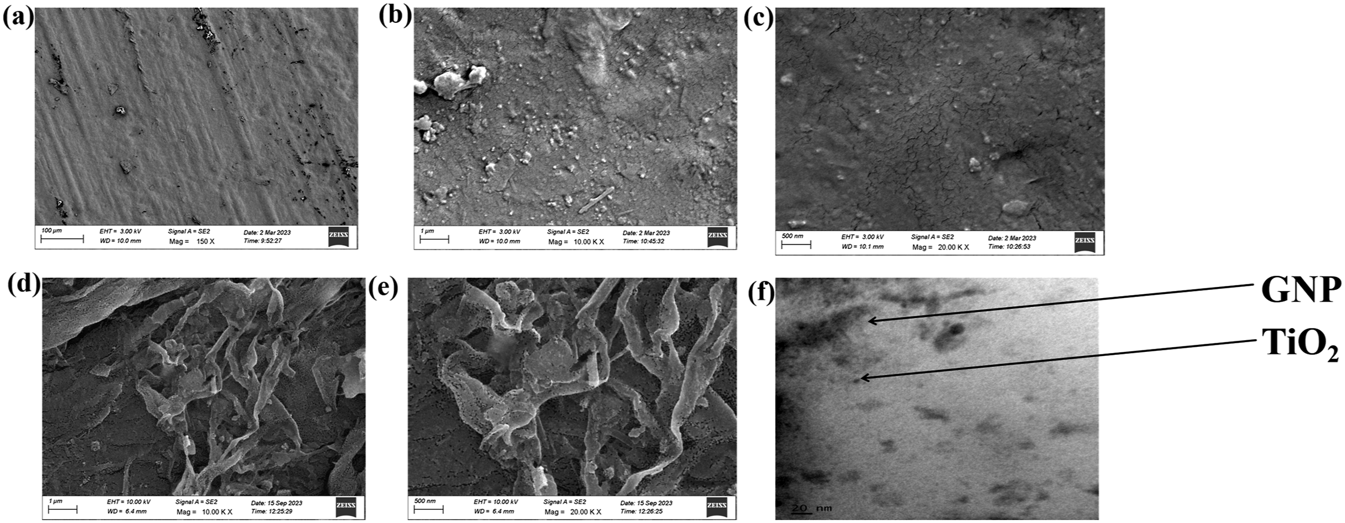
Energy dispersive spectroscopy (EDS) images (Figure S1(c)) reveal the spatial distribution of fillers in the PLA matrix, where the TiO2 nanoparticles are distributed uniformly over large surfaces of the platelets. The TEM micrograph, (Figure 1(f)) depicted the uniform dispersion of GNP and TiO2 in the PLA matrix. From Figure S1(b) and (d)), the aggregation of nanoparticles, as confirmed by the presence of excessive green and blue spots corresponding to the elements O and Ti, respectively, were ascertained. These aggregates act as stress concentration points and deteriorate the properties of the samples.
Raman Spectroscopy
Raman spectroscopy is a powerful characterization technique for polymer nanocomposites, providing critical insights into their molecular structure, composition, and interactions at the nanoscale level.
29
It enables the identification of chemical bonds, functional groups, and polymer-filler interactions, providing detailed information on the composite’s composition and phase distribution. Figure 2 shows the Raman spectra of PLA, P/3G, P/3T, and P/1 G/ 0.5T nanocomposites. Raman spectra (a) C-COO symmetrical stretching, (b) G bands for GNPs, (c) C-O-C stretching, (d) C-H stretching, and (e) over the range of 400 to 3200 cm−1 for P, P/3G, P/3T, and P/1 G/0.5 T.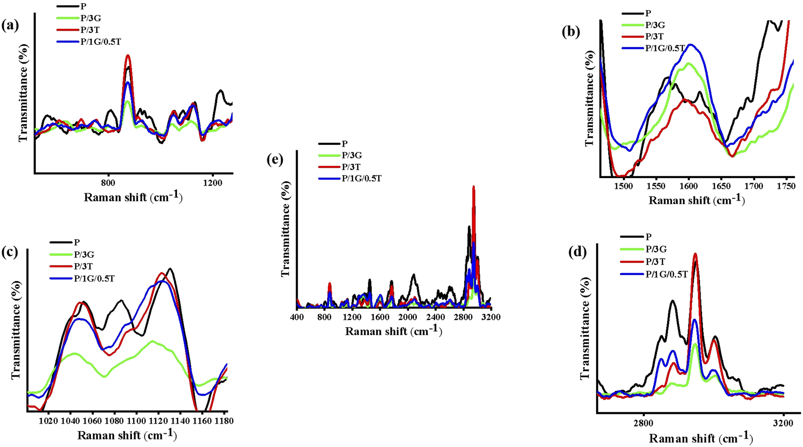
The addition of nano ceramic fillers and GNPs appears to interact with the PLA backbone structure. In the PLA matrix, the peak in the vicinity of 850 cm−1 corresponds to the C-COO symmetrical stretching in the polymer backbone, confirming the presence of ester linkages. 30 For the P/3T sample, the peak’s intensity increases, suggesting enhanced crystallinity and a highly ordered C-COO structure, which contributes to a stronger signal. In contrast, the presence of GNPs in P/3G disrupts the polymer structure due to their high aspect ratio and rigidity, leading to a reduction in peak intensity. In P/1 G/0.5T, the intensity is higher compared to P/3G due to the addition of TiO2 but remains lower than that of neat PLA due to the presence of GNPs. The peaks manifested at around 1180 cm−1 is apparently owing to the stretching of C-O-C within ester groups for neat PLA, 31 which originally display three characteristic and distinct peaks. However, the introduction of nanofillers caused a rearrangement or phase transition in the structure, resulting in structural symmetry and a consequent reduction or merging in the number of observed peaks. The D bands and G bands of GNPs, as seen in Figure 2 and Figure S2 (Supplemental Section S2) , observed at ∼1350 cm−1 and ∼1590 cm−1, respectively32,33 which is absent for neat PLA and P/3T but visible in the P/3G and hybrid nanocomposites.
The peak formed at 1745 cm−1 indicates the C = O stretching of ester linkages in the neat PLA matrix. 30 This peak is typically pronounced for carboxyl groups in the amorphous region, where the polymer chains are in disordered state. As crystallinity increases, the polymer chains become more organized, reducing molecular movement and, consequently, raising the intensity of the peak. The C-H stretching vibrations of aliphatic groups (CH and CH3) are manifested at ∼2945 cm−1. 30 The increased intensity for P/3T can be attributed to the enhanced crystallinity and the highly ordered domains of polymer chains compared to neat matrix. In contrast, the decrease in intensity for hybrid nanocomposites and P/3G is due to the restricted movement of polymer chains caused by the presence of higher aspect ratio nanofiller (GNP). The hybrid nanocomposite shows higher intensity than P/3G because of the lower concentration of GNP and the presence of TiO2, contributing to improved C-H stretching.
X-Ray Diffraction (XRD) Analysis
The crystalline structure and crystal lattices of PLA and their nanocomposites were studied using XRD analysis, and the diffractograms are presented in Figure 3. The growth of crystallites in specific crystal planes due to the influence of nanofillers can be assessed using XRD. In PLA, the reported α peaks at 14.7°, 16.5°, 18.8°, and 22.2° in XRD patterns correspond to (010), (110) or (200), (203) and (105) specific crystal lattice planes, respectively.34,35 X-ray diffractograms of PLA, P/3T, P/3G, and P/1 G/0.5T.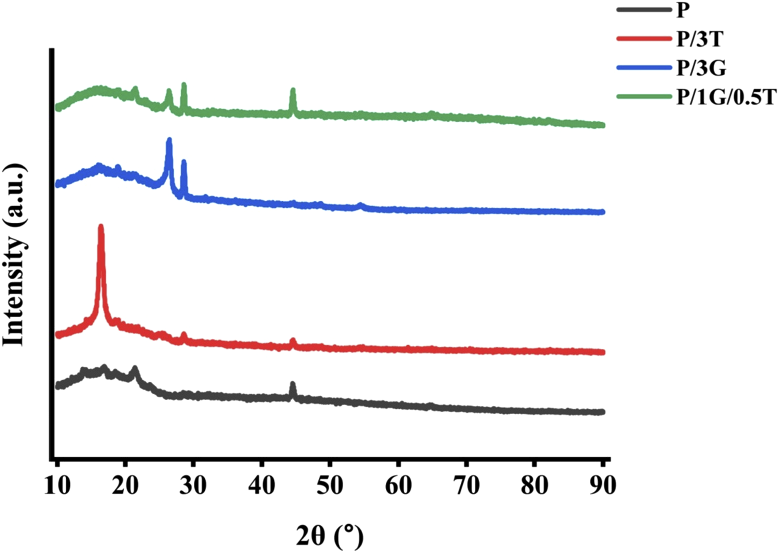
In Figure 3, the presence of an α peak at 21.56°, with a crystallite size of 2.34 nm, along with an additional peak at 44.5°, indicates the passive crystalline regions in PLA. 36 With the addition of TiO2, the additional peaks were observed at 16.4° (110), 18.75° (110), 25.34° (121), and 29.59° (220) with the crystallite sizes of 11.9 nm, 22.9 nm, and 22.7 nm respectively.37,38 The high peak intensity at 16.4° is due to the presence of crystalline regions formed due to the heterogeneous nucleation effect of TiO2. 39 With the incorporation of GNP into the PLA matrix, the average crystallite sizes reduced to 4.4 nm at 16.4° and to 14.2 nm at 18.75°, respectively. The peaks manifested at 21.4° (105), 26.4° (002), and 29.5° (220) resulted in the crystallite sizes of 18 nm, 16 nm, and 26.4 nm, respectively.40,41 The availability of numerous smaller crystals in the P/3G sample is due to the high surface area of GNPs as well as the presence of graphitic planes observed at 26.4°. In P/1G/0.5 T, peaks were predominant at 21.4° (105), 26.4° (002) and 29.3° (220) with crystallites of 9 nm, 14 nm and 24.3 nm size, respectively. The hybrid sample possessed even smaller crystals compared to P/3G and P/3T and is attributed to the synergistic effect of GNP and TiO2. The synergism between the dual nanofillers was further supported by their dispersion, as observed from SEM and TEM.
Atomic Force Microscopy (AFM)
AFM is a high-resolution image technique used to analyse surface topography and 3D surface mapping of materials. The inclusion of nanofillers is expected to impart surface roughness on the nanocomposites. Figure 4 shows 3D images of the topography of PLA and nanocomposites and the roughness values are tabulated in Table T1 (Supplemental Section S3). The root mean square Rq (nm) value of virgin PLA was 9.2 nm and the surface roughness values increased by 78 % and 130% with the inclusion of TiO2 and GNP, respectively. AFM 3D surface images of (a) neat PLA (b) P/3T (c) P/3G (d) P/1 G/0.5T nanocomposites.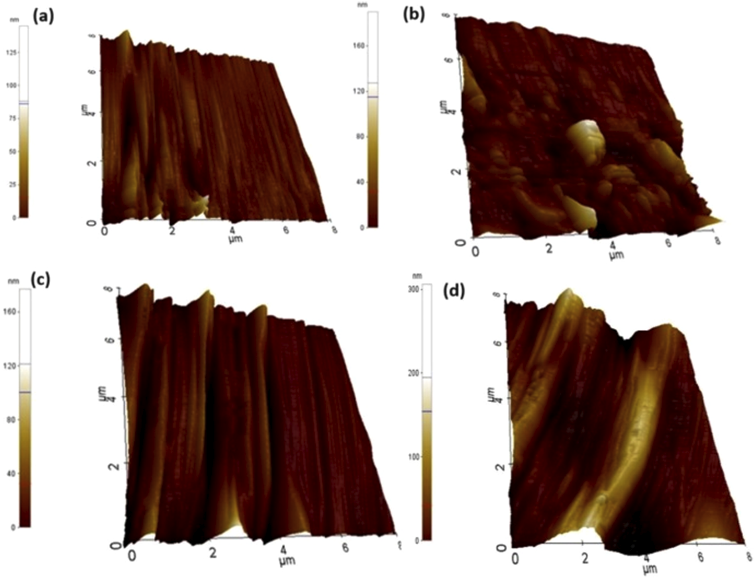
The coexistence of 0D TiO2 and 2D stacks of GNP in hybrid specimens (P/1G/0.5 T) has raised the Rq values by 506.5% with regard to virgin PLA. It is reported that surface roughness has close agreement with filler geometry, and as the particle size increases the nanofillers may protrude from the surface, providing higher roughness. 42 The presence of flaky regions were evident from SEM micrographs of P/1G/0.5 T.
Contact Angle Measurements
The contact angle (CA) measurements of the samples were carried out to understand the wetting behaviour of PLA and its nanocomposites with distilled water using a goniometer are reported in Table T2 (Supplemental Section S4). The improvement in surface roughness due to the presence of nanofillers is expected to raise the surface energy of the nanocomposites. PLA exhibits a contact angle of 69.5° due to the presence of methyl groups and the steric hindrance effect of PLA and results in hydrophobic nature. With the addition of nanofillers, P/G and P/T samples showed a reduction in contact angle indicating the hydrophilic nature of the surface (Figure 5). Literature findings43,44 also agree well with the experimental results showing that contact angle decreases and augments the wetting and hydrophilic nature of the PLA/TiO2 composite specimens. The improved surface roughness of the nanocomposites facilitates better interaction with water molecules, leading to improved adhesion and spreading of droplets. The surface roughness of the hybrid composite, P/1 G/0.5T is high which was vivid in AFM images owing to the increase in filler particle size and difference in the shape of the fillers in dual hybrid composites resulting in CA of 69.5°. At higher hybrid filler concentrations the aggregates of GNP and TiO2 prevents the water droplet from spreading over the surface, enhancing the hydrophobicity as observed from Figure 5.45,46 Average contact angle of samples.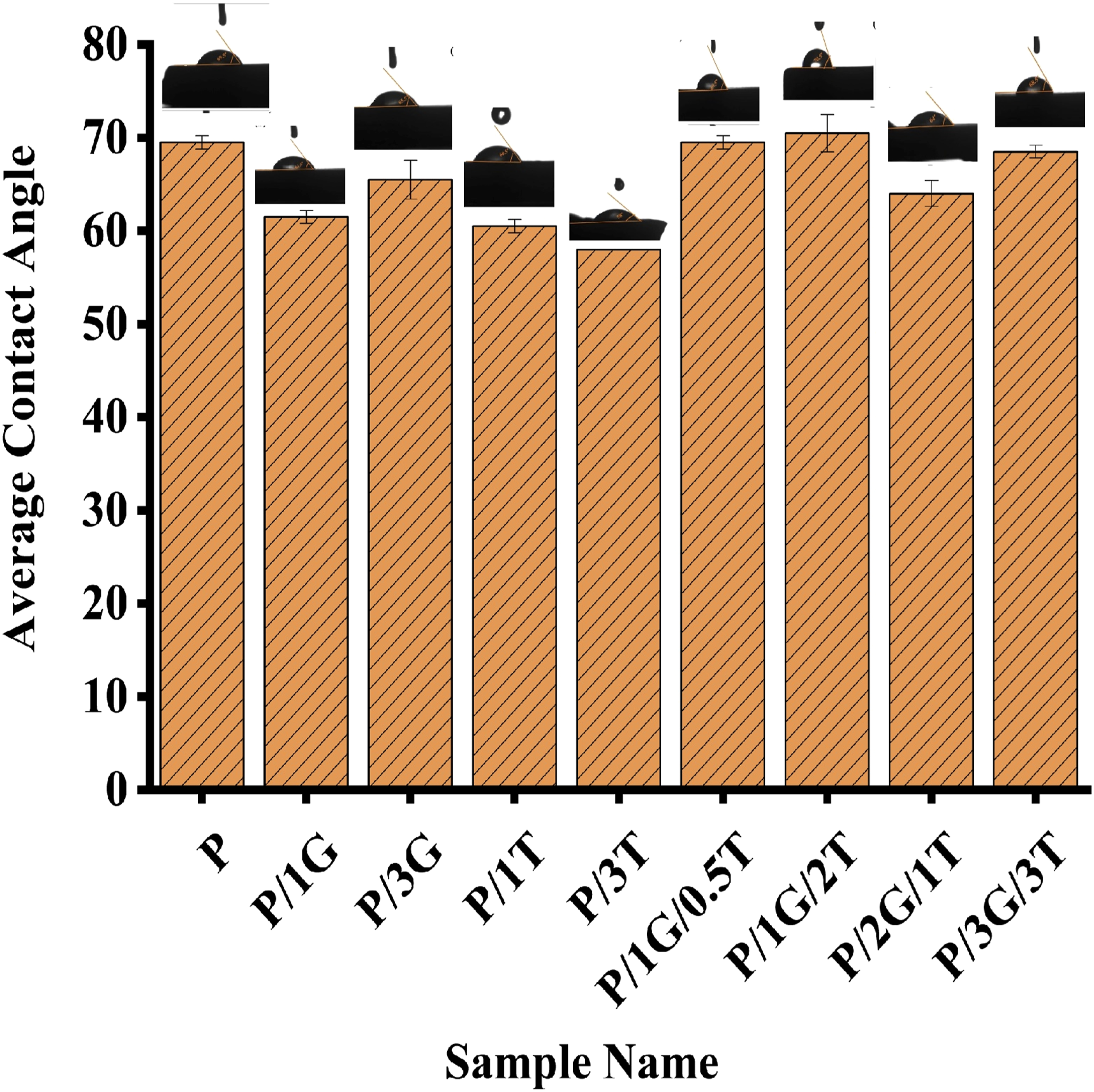
Tensile Test
The tensile test is performed to understand how the material behaves under the tension load. The stress-strain curves of neat PLA, mono, and bi-filler reinforced polymer composites are presented in Figure 6. Neat PLA exhibits a tensile strength of 38 MPa, and its inherent poor ductile behaviour was manifested with low strain at break of 2.8% as tabulated in Table 2. With the inclusion of 1 wt% of 0D TiO2, the tensile strength, and Young’s modulus enhanced by 28 %, and 34% respectively compared to neat PLA. The elongation at break increased by 33% compared to neat PLA. Interestingly, the inclusion of 1 wt% of GNP in PLA has raised the tensile strength to 52.3 MPa, an improvement of 37.6% in comparison with PLA. Furthermore, the modulus of elasticity and elongation at break have also been enhanced by 54% and 34%, respectively in comparison with neat matrix owing to the superior properties of 2D planar nanofillers.
47
The high aspect ratio of GNP augments the mechanical properties of the nanocomposites even at the low composition of fillers. With the increase in loading of GNP in PLA, from 1 to 3 wt%, the tensile strength and modulus increased from 37 % to 50% and 54% to 73%, respectively. It could be inferred that at higher concentrations of TiO2, tensile properties showed decrement compared to the same GNP composition. The planar structure of GNP acts as a bridge and transfers the applied load across a wide area of the PLA matrix. The high strain at the break of GNP composites is evident from the stress-strain curves shown in Figure 6. This verifies the higher energy-absorbing capability of P/G composites compared to P/T composites on the application of external loads. The brittle behaviour of P/3T with higher tensile strength is due to the physical pinning effect of quasi spherical TiO2.
48
Tensile stress-strain curves of (a) Individual nanocomposites (b) Hybrid nanocomposites (c) individual and hybrid nanocomposites. Tensile properties of PLA and nanocomposites.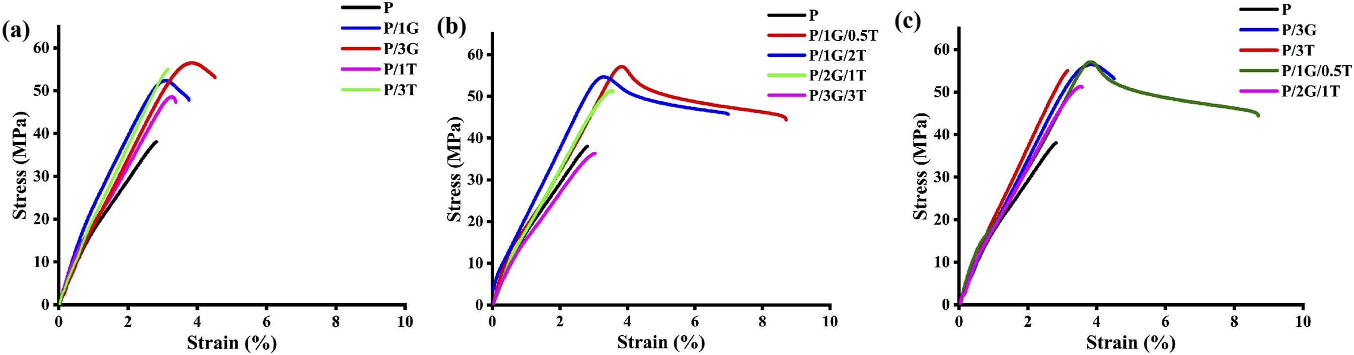
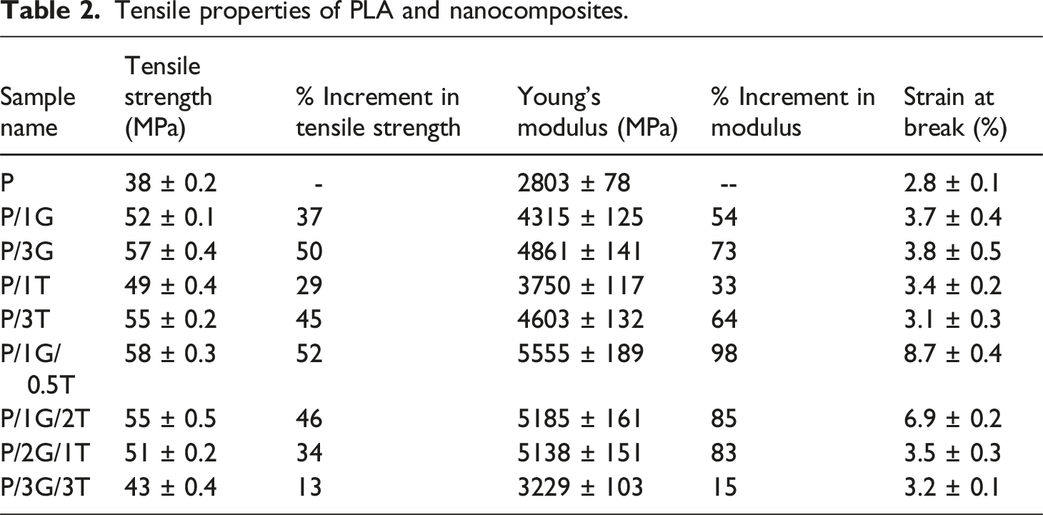
It could be observed that the P/1G/0.5T hybrid specimen revealed a tensile strength of 58 MPa which is 52% enhancement with regard to neat PLA. In addition, there is a drastic improvement in tensile modulus by 98% and strain at break by 210%. These significant improvements are noticed at the minimal content of 0.5 wt% TiO2 and 1 wt% GNP. When comparing the properties of single filler reinforced (both GNP and TiO2) composites at 1 and 3 wt%, with the hybrid specimen P/1G/0.5T, the hybrid sample presented better mechanical properties at low content of filler loading. The synergism of 0D and 1D nanofillers in PLA has played a major role in the escalation of tensile properties. For the P/1G/2T hybrid specimen, tensile strength slightly decreased and maintained ductile behaviour. At a higher content of 6 wt% (P/3G/3T), tensile strength and modulus are drastically reduced (43.2 MPa and 3229 MPa, respectively) due to the presence of agglomerations.
Fracture Toughness
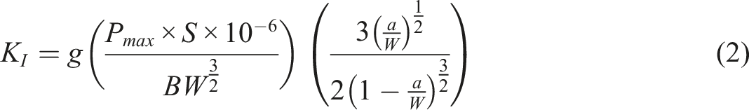
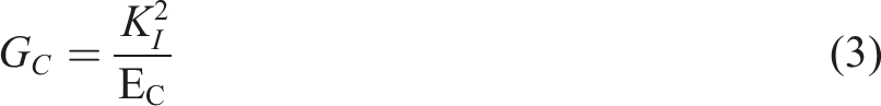
The fracture toughness behaviour of neat PLA and its nanocomposites was examined in a three-point bending mode to investigate the ability of the sample to resist the crack propagation. The strain energy release rate (Gc) and critical stress intensity factor (KI) values were calculated using the following Equations (1) to (2).49,50 KI of a linear fracture can be found from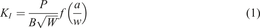
Fracture Toughness parameters obtained for neat PLA, and nanocomposites.
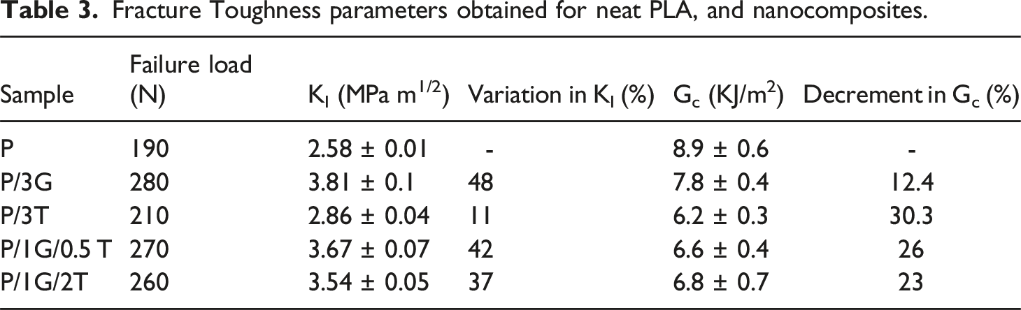
Including 1wt% of TiO2 increased the KI value by 10.85% compared to neat PLA. The increment in the KI value of P/1T nanocomposite was not as appreciable as that of P/3G composites, owing to the 0D geometry of fillers. It could easily promote micro-void development and has little resistance against crack propagation compared with a high aspect ratio GNP. This could lead to interfacial debonding and reduction of fracture toughness properties.
53
In the case of P/1G/0.5T and P/1G/2T hybrids, the failure load increased by 80 N and 70 N, respectively compared to PLA. The bridging effect of GNPs and the pinning effect of TiO2 help to extend the degree of entanglement between the polymer chains and the fillers. The dual nanofillers embedded in the hybrid system act as crack deflection points and change the direction of the crack front. The pinning and bridging effect impedes the loosening of polymer chains under load, thereby decreasing the size of the plastic deformation zone near the crack tip. During crack extension debonding, stretching, and pullouts are observed due to the bridging effect of GNPs whereas due to the presence of 0-D TiO2, the crack deflects and splits into smaller branches.
48
In hybrid nanocomposites, a large amount of energy is required for crack propagation, which reduces the crack growth rate and contributes to higher fracture toughness parameters. The schematic representation of the bridging and pinning effect is delineated in Figure 7. The Gc value of all the nanocomposites showed a decrement due to the high Young’s modulus and the brittle nature of the sample. Schematic representation of (a) Bridging effect of GNPs in P/3G, (b) Pinning effect of TiO2 in P/3T and ( c) Combined bridging and pinning effect in P/1 G/0.5 T.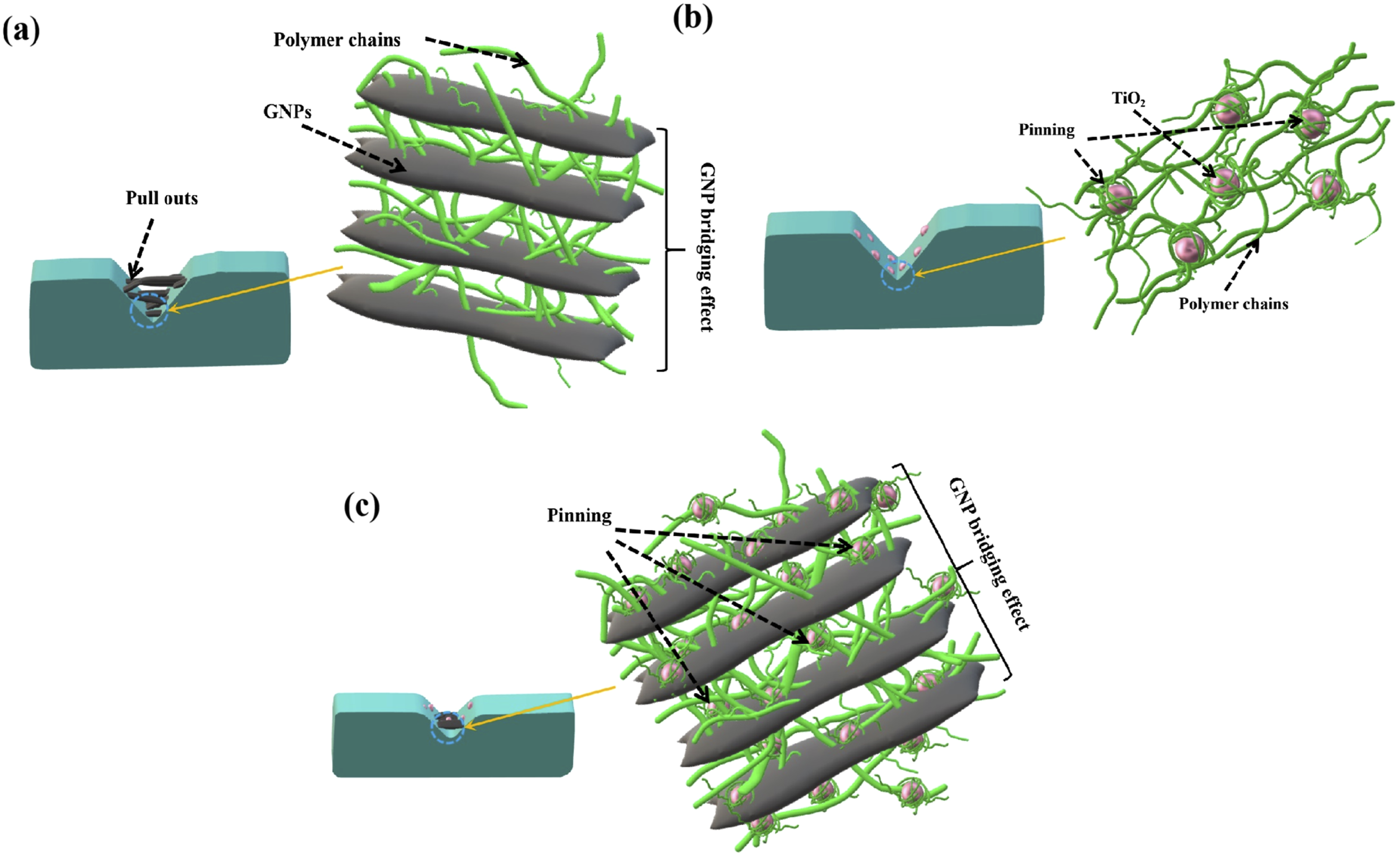
Dynamic Mechanical Analysis (DMA)
The viscoelastic properties of PLA and nanocomposites were examined using DMA. The estimate of the mechanical energy stored in the sample or storage modulus (E′), the amount of energy dissipated from the materials or loss modulus (E″) and the material’s damping or tan δ are discussed in the following sections.
Storage Modulus (E′)
The storage modulus (E′) is an indication of the ability of a viscoelastic material to resist deformation when stress is applied. A material with a higher E′ is more rigid, whereas, the one with lower E′ is relatively soft and flexible. The E′ values of PLA and various nanocomposites are given in Table T3 (Supplementary Section S5) and Figure 8(a). The samples exhibited a glassy phase at low temperatures (35 to 75°C) where the E′ value decreased gradually. From 75°C to 100°C due to the cold crystallization of PLA matrix,
52
the E′ value increased, followed by the decrease in E′ due to pre-melting.
54
The E′ value of P/3T showed an 8% increment compared to neat PLA at 40°C. The spherical TiO2 arrested the molecular chain movements and promoted crystal growth due to the heterogeneous nucleation effect at around 100°C.
54
In P/3G the GNP network in the PLA matrix restricts the macromolecular chain slippage, and results in higher E′ as seen in Table T3. The uniform dispersion and good interfacial interaction between the polymer and GNP propelled the stress transfer in the PLA matrix.54,55 An appreciable E′ value enhancement was also observed in P/1G/0.5T due to the synergistic effect of dual nanofillers. The presence of bi-fillers made the PLA matrix stiffer by providing a generous surface area for spherulite growth; this was evidenced by the higher E′ value around 100°C (Figure 8 (a)). However, in P/2G/1T a slight reduction in E′ with an increase in temperature was observed due to the presence of agglomerates in the PLA matrix.
56
At lower temperatures, the composite system was found to be stiffer, whereas with the increase in temperature, the matrix became flexible due to higher segmental mobility as it is easier for polymer chains to slip through clusters of nanofillers. Dynamic mechanical properties (a) Storage Modulus (b) Loss modulus and ( c) tan δ. (a) Melting and (b) Non-isothermal crystallization DSC thermograms of PLA and its nanocomposites.
Loss Modulus (E″)
Loss modulus is the estimate of energy dissipated by the material when it becomes viscous on the application of heat and shear forces. 57 PLA shows γ, β, and α relaxations around −60°C, 60°C, and 100°C respectively. 58 At α relaxation, the mechanical loss will increase due to cold crystallization. 59 From Figure 8(b) and Table T4 (Supplemental Section S5) it is revealed that neat PLA has the highest loss modulus with the lowest Tα and Tβ indicating that neat PLA exhibits a predominantly viscous nature compared to nanocomposites.
With the incorporation of nanofillers, the viscosity is reduced, this is evident from Figure 8(b). The spherical nature of TiO2 offers a high degree of chain slippage compared to 2D GNP. The larger surface area of GNP particles enhances the interfacial interaction in P/3G compared to neat PLA and P/3T. In P/1G/0.5T the homogeneously dispersed dual nanofillers present in the PLA matrix reduce internal friction by contributing to minimal energy loss. It is evident from Figure 8(a), that P/1G/0.5T has a more elastic behaviour amongst all samples due to limited polymer chain movement by the entanglement of dual nanofillers with PLA chains. In P/2G/1T, the presence of agglomerates might have reduced the effective stress transfer area, diminished their reinforcing efficiency, and led to a reduction in E′ and enhancement of E”. The E″ increased around the Tα region for all the samples due to the cold crystallization effect of the PLA matrix.
Damping Factor (Tan δ)
Tan δ is the ratio of loss modulus to storage modulus and it gives the ability of a material to dissipate energy in the form of heat under the cyclic loading. Table T5 and Figure 8(c) show that incorporating nanofillers improved Tg and Tα compared to neat PLA. The uniform dispersion of TiO2 and the pinning effect of spherical nanoparticles reduced the polymer chain movement and enhanced the Tg value by 3%. The high surface area of GNP limits the chain mobility in P/3G and results in enhancement of Tg and Tα. Among the hybrid samples, the elastic modulus was higher for P/1G/0.5T due to the presence of orderly arranged crystalline regions, similar trend was observed for E′ value as well. The sample exhibited lowering and broadening of the tan δ peak as the nanofillers were effectively intertwining with the polymer macromolecules thus impeding their slippage. On the contrary in P/2G/1T the clusters of nanofillers acted as stress concentration points and reduced the elastic modulus of the sample.
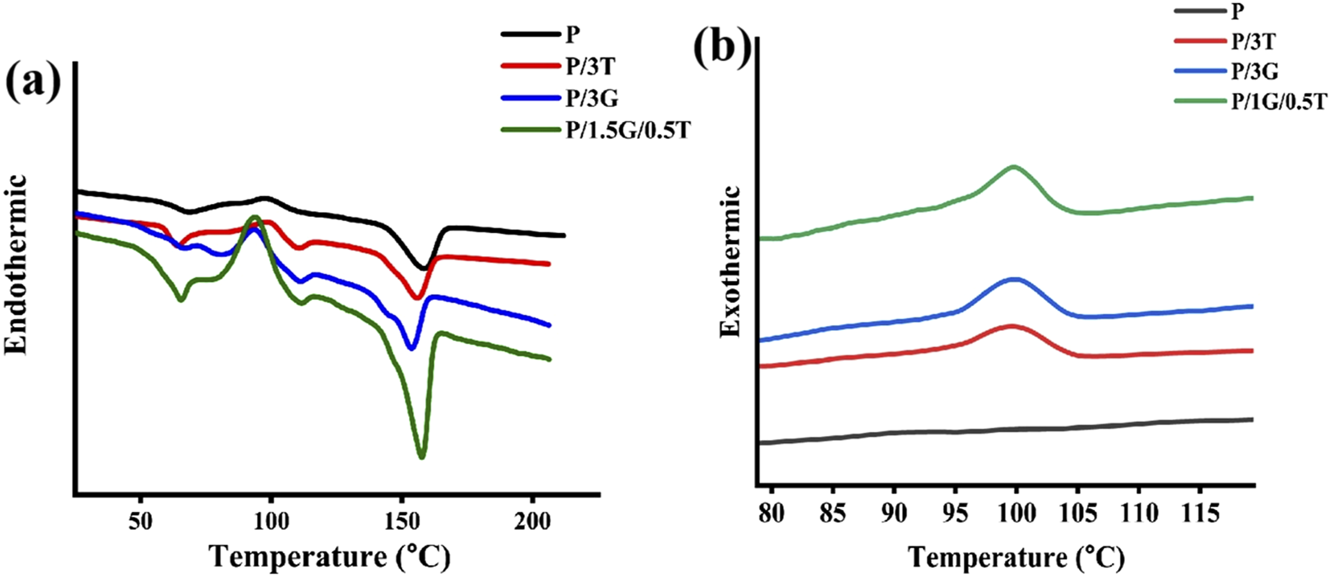
Differential Scanning Calorimetry (DSC)
The non-isothermal crystallization studies of PLA and its nanocomposites were performed using DSC to understand the melting and cooling characteristics of the sample. The incorporation of nanofillers has a telling effect on the crystallization behaviour of semicrystalline polymers.60,61 The percentage of crystallinity (Xc) was calculated using equation (4)62,63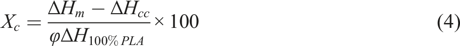
ΔHm and ΔHcc are the enthalpies of melting, and cold crystallization respectively. ΔH100%PLA is the enthalpy of melting of 100% crystalline PLA (93 J/g) and
Were found and tabulated in Table T6 (Supplemental Section S6).
64
Neat PLA samples exhibited a 10% Xc value with Tg of 61°C and Tcc of 91°C. From Figure 9(b) the cooling thermogram of PLA doesn’t show any characteristic crystallization peaks, indicating that PLA exhibits an amorphous nature due to cold crystallization. With the addition of TiO2, the polymer chain spacing was reduced and the Xc value showed a slight increment owing to the dense packing of the polymer chains.
65
The P/3G sample exhibited Xc of 40% with Tc at 93°C, crediting to the presence of 2D GNP, which induces a large number of crystallites due to heterogeneous nucleation and spatial effects.
66
Various studies66–68 have suggested that the incorporation of 2-D nanofillers creates more nucleating sites, attributing to their large surface area to volume ratio. The nanofillers act as nucleation sites for the formation of polymer spherulites as presented in Figure 10. These spherulites grow freely until they impinge upon each other. The high surface area of graphene results in strong interfacial interactions and restricts chain mobility which promotes chain stacking. In P/1G/0.5T the Xc was found to be highest amongst all the samples due to the synergistic effect of TiO2 and GNP. The Tcc of hybrid was found lower than neat PLA indicating the positive effect of the inclusion of dual nanofillers, by reducing the nucleation barrier.
69
The amorphous region in the sample gains energy and rearranges themselves in a much more orderly manner at lower temperatures than PLA, which enhances the stiffness and strength of the nanocomposite.
70
The dual nanofillers present in the matrix provide an enormous surface area for heterogeneous nucleation.71,72 The Tm recorded a slight increase for this sample with regard to other nanocomposites. The uniform dispersion of dual nanofillers restricts the macromolecular chain mobility of PLA even with the increase in temperature. Schematic representation of nucleating sites offered by (a) GNP (b) TiO2 nanoparticle for PLA crystallization.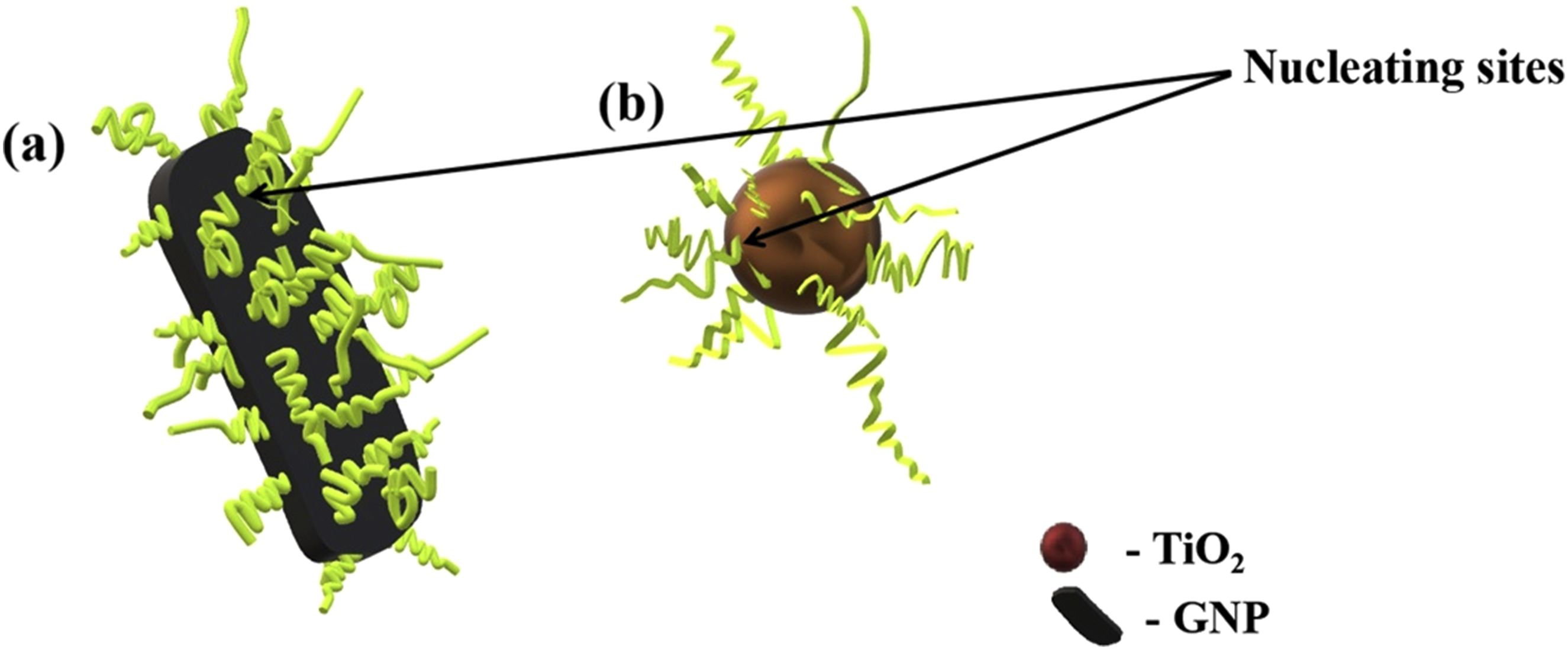
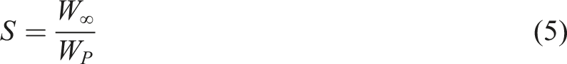
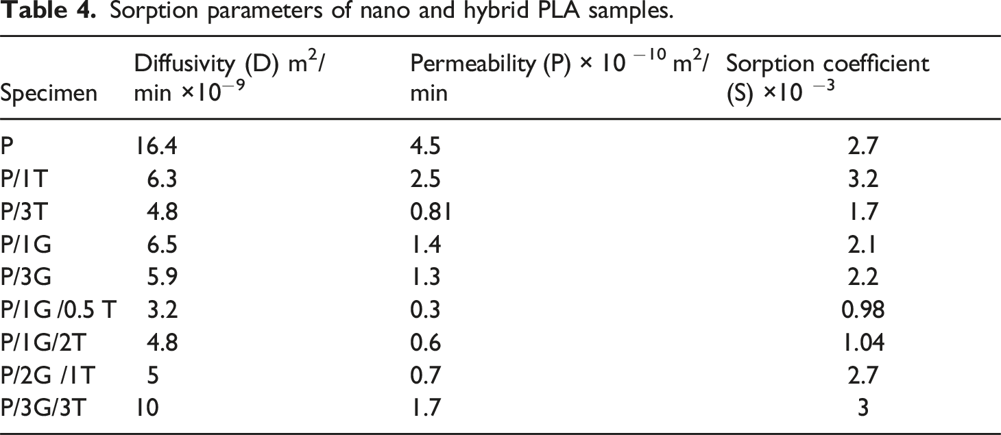
Sorption Behaviour
Sorption parameters of nano and hybrid PLA samples.
W∞ is the solvent mass absorbed at equilibrium uptake and WP is the initial weight of the composite. Fick’s law was used to evaluate the diffusion (D) of solvent molecules through the samples as in equation (6).
75
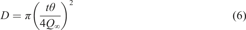
Q∞ is the mass percentage of the solvent at the equilibrium swell. θ is the slope of the initial portion of the sorption curve and t is the thickness of the sample. The permeability can be evaluated from diffusivity and sorption coefficient as follows (equation (7))
75

PLA is strongly hydrophobic and insoluble in water, alcohol, and linear hydrocarbon solvents like hexane. During the diffusion of molecules, the ester bonds will break leading to the disintegration of PLA macromolecular chains.
47
The chain scission increases free volume and the formation of wider paths for the diffusion of solvent molecules resulting in higher permeability (4.5 × 10⁻¹⁰ m2/min) and diffusivity (16.4 × 10⁻⁹ m2/min) amongst all samples (Figures S3(a) and (b) in (Supplemental Section S7), and Figure 11(a)). Schematic representation of sorption pathways of (a) PLA, (b) P/3T, (c) P/3G and (d) P/1G/0.5T.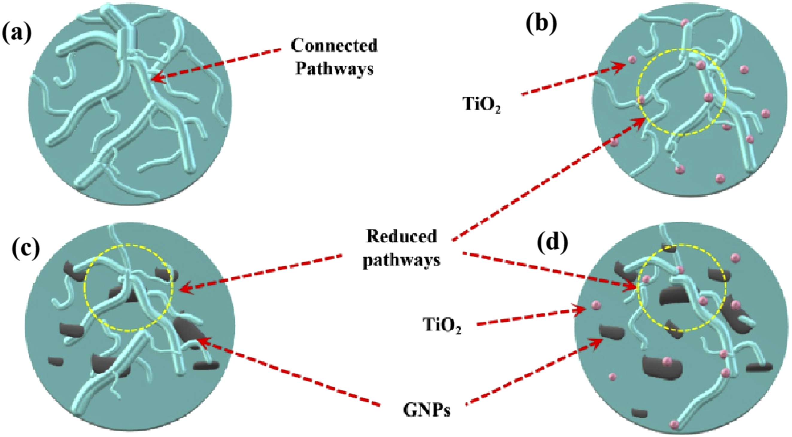
With the addition of 1 wt% and 3 wt% of TiO2, the diffusivity dropped significantly by 61% and 71%, respectively, compared to the neat matrix owing to the hindrance offered by the 0-D ceramic nanofiller. P/3G nanocomposites exhibited a decrease in diffusivity by 60% and in permeability by 69%. GNP nanocomposites offer tortuous pathways for solvent than TiO2 nanocomposites, crediting to the 2D planar structure of GNPs and their impediments (Refer Figure 11(b) and (c)). The lowest diffusivity and permeability were observed for P/1G/0.5T amongst all the samples due to the synergism of hybrid fillers in the PLA matrix. At optimum filler content, more blockages were offered to the solvent molecule movement owing to the uniform distribution of fillers. The voids existing between the well-distributed GNP platelets in the PLA are filled by the 0D nanofiller (as represented in Figure 11(d)) resulting in the decline of the sorption coefficient by 63% compared to the neat matrix. In higher hybrid compositions, the agglomeration of GNPs and TiO2 resulting from Van der Waal’s force of attraction forms more free channels for the solvent flow.
Antimicrobial Activity
Antibacterial parameters obtained via Agar diffusion method for PLA, P/T, P/G and P/T/G nanoplatelets against E. coli and S. aureus strains.
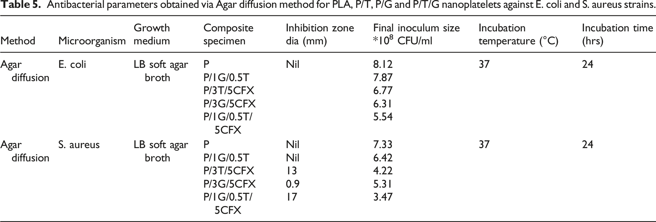
CFX biocide from the sample. In the case of the 0D TiO2 nanoparticles, the biocide is readily released, resulting in a larger inhibition zone. Conversely, the 2D GNPs might be restricting the release of CFX, leading to a much smaller inhibition zone. The presence of 2D nanofiller in the PLA matrix creates a torturous pathway for the release of CFX, resulting in prolonged drug release.76,77 In the case of E. coli, due to the presence of outer membrane, the inhibition zone was absent in P/3G/5CFX, which acts as a limiting barrier for the entry of CFX. In contrast, CFX was able to enter S. aureus, a Gram-positive bacterium, due to the absence of an outer membrane, which caused cell wall destruction and ultimately bacterial death. 78 Further, distinct inhibition zones were created in those nanocomposites. In P/3T/5CFX, the TiO2 nanoparticles generated electron-hole pairs owing to its photocatalytic effect, inducing redox reactions with S. aureus causing the formation of a large inhibition zone. TiO2 destructs the microbes by producing reactive oxygen species (ROS). 79 From the UV light received, the electrons (e−) are excited from the valence band (VB) to the conduction band (CB) leaving behind holes (h+). 80 The electron combines with oxygen to form superoxide anion radical, which is further converted to hydrogen peroxide and hydroxyl radical, and these radicals are toxic to bacteria. Highly reactive hydroxyl radicals are formed by the reaction of h+ and water, this attacks bacterial cell components. The recombination of e− and h+ reduces the efficiency of attacking the cell wall by decreasing the ROS. The available ROS reacts with lipids in the cell membrane of microbes and results in rupture, leading to denaturing of enzymes and proteins.78,79,81 It could be observed that Gram-negative, E-Coli showed resistance to CFX than Gram-positive S. aureus. In P/1G/0.5T/5CFX, the generation of a tortuous path of GNP along with the photocatalytic activity of TiO2 under UV light resulted in smaller inoculum size. This highlights the role that nanofiller geometry and interactions within the composite play in controlling the release rate and effectiveness of the embedded biocide.
Conclusion
A systematic study was carried out to understand the collaborative effect of TiO2 and GNP nanoparticles in the PLA matrix. The uniform distribution of dual nanofillers in P/1G/0.5T was revealed by SEM and TEM analysis, the sample also showed the presence of a large number of small and uniform-sized crystallites from XRD analysis. Tensile strength and Young’s modulus of the hybrid sample showed a 52% and 98% increase respectively. The sample also exhibited appreciable improvement in fracture toughness due to the pinning effect of 0D-TiO2 and the bridging effect of GNPs. The hybrid sample manifested higher storage and loss modulus along with lowering and broadening of tan δ due to the reduction in chain slippage by the entanglement of the spherical nature of TiO2 and the larger surface area of 2D GNP particles elevating the interfacial interaction between the filler and polymer matrix. The DSC analysis revealed that the crystallinity of hybrid samples increased to 54% along with a slight increase in cold crystallization, melting, and crystallization temperatures due to spherulite growth and heterogeneous nucleation. The hybrid system exhibited higher surface roughness with hydrophilic behavior due to which the adhesion of water molecules improved with the spreading of droplets. At optimum filler content, in P/1G/0.5T, more obstructions were offered to the hexane solvent transport owing to the entanglement by the intertwining of bi-nanofillers and polymer chains, resulting in the lowest sorption coefficient amongst the samples. P/1G/0.5T/5CFX showed a final inoculum size of 3.47 × 10⁸ CFU/ml with Staphylococcus aureus, the lowest bacterial count as CFX will inhibit the production of DNA gyrase and topoisomerase IV in bacteria. Overall, the hybrid sample P/1G/0.5T/5CFX can be considered for potential applications in food and edible product packaging and membranes used in air quality management.
Supplemental Material
Supplemental Material - Multifunctional behaviour of PLA biomacromolecules by GNP/TiO2 hybrid nanofiller integration: Amplifying strength, solvent sorption resistance and antimicrobial properties
Supplemental Material for Multifunctional behaviour of PLA biomacromolecules by GNP/TiO2 hybrid nanofiller integration: Amplifying strength, solvent sorption resistance and antimicrobial properties by Snaha Leena, Sai Gopal Krishna Bhagavatula, Rasana Nanoth, K. P. R. Mohan Ram, A. W. Jerfin, S Phani Abhinay, C.S. Tharun Venkat, K. Jayanarayanan, Krishna Prasad Rajan, Rajesh Theravalappil, Megha S. Kumar and Punathil Vasu Suneesh in Journal of Thermoplastic Composite Materials.
Footnotes
Acknowledgements
The authors thank Mr Vijaya Kumar, Polymer Processing Laboratory, Amrita, Coimbatore, for assisting in processing the samples. The authors would like to express honest gratitude to the Department of Mechanical Engineering and the Department of Civil Engineering, Amrita Vishwa Vidyapeetham, Coimbatore, for providing the facilities for conducting the static mechanical tests. The authors are grateful to the Amrita Biosensor Research Lab., Amrita School of Engineering, Amrita Vishwa Vidyapeetham, Coimbatore, India for assistance in anti-microbial studies.
Author contributions
Snaha L: Data curation, Visualization, Methodology, Formal analysis, Investigation, Writing- Original draft preparation, Bhagavutula Sai Gopal Krishna: Investigation, Methodology, Rasana Nanoth: Validation, Conceptualization, Investigation, Supervision, Reviewing and Editing, Mohan Ram. K.P.R: Methodology, Formal analysis, A.W.Jerfin: Data curation, Investigation, S Phani Abhinay: Visualization, Formal analysis, Tharun Venkat.C.S: Conceptualization, Investigation, Jayanarayanan K: Methodology, Investigation, Supervision Reviewing and Editing, Krishna Prasad Rajan: Investigation, Supervision, Rajesh Theravalappil: Investigation and Visualization, Megha S Kumar: Methodology, Punathil Vasu Suneesh: Methodology.
Declaration of conflicting interests
The author(s) declared no potential conflicts of interest with respect to the research, authorship, and/or publication of this article.
Funding
The author(s) received no financial support for the research, authorship, and/or publication of this article.
ORCID iDs
Supplemental Material
Supplemental Material for this article is available online.
References
Supplementary Material
Please find the following supplemental material available below.
For Open Access articles published under a Creative Commons License, all supplemental material carries the same license as the article it is associated with.
For non-Open Access articles published, all supplemental material carries a non-exclusive license, and permission requests for re-use of supplemental material or any part of supplemental material shall be sent directly to the copyright owner as specified in the copyright notice associated with the article.