
Abstract
Human articular cartilage, a nonlinear, viscoelastic solid-liquid biphasic tissue, has different mechanical properties and undertakes different functions in different layer. The compression deformation and fatigue damage mechanism of each layer of cartilage is also different when in injury. Compression properties of cartilage can predict cartilage integrity and the likelihood of osteoarthritis. Therefore, the confined and unconfined compression of cartilage is taken to systematically study the dynamic mechanical properties of each layer and reveal the relationship between the dynamic mechanical properties of each cartilage layer and the function of the corresponding cartilage layer. Under confined and unconfined compression conditions, the larger the cyclic loading rate, the greater the deformation rate of each layer. In addition, under the same cyclic loading rate, the superficial layer had the highest deformation rate, followed by the middle layer and the deep layer, which indicates that the deep layer mainly assumes the compression load. Furthermore, under the same loading displacement, the loading stress of cartilage and the deformation rate of each layer in confined compression were greater than those in unconfined compression. Simultaneously, with the increase in the number of loading cycles, the deformation rate in different layers increased first and then stabilized.
Keywords
Introduction
Articular cartilage, an important connective tissue in human joints, has low elastic modulus, high deformability, excellent lubricity, wear resistance properties and can buffer friction and transmit loads in joint activities by relying on the special structural and composition.1-3 Due to the complex mechanical environment of the physiological parts, cartilage is easily damaged and difficult to repair, which directly affects people’s walking activities. Once damaged, it will bring inconvenience to human life. Osteoarthritis usually occurs, and related studies have shown that no correlation of frequency of osteoarthritis with parameters of bone strength and biomechanical parameters was found, suggesting that bone is only secondarily affected in osteoarthritis and that cartilage is the initial target of the disease. 4 Therefore, biomimetic articular cartilage materials that mimic the structure and mechanical properties of natural articular cartilage have been extensively developed and studied. And there has been great progress in seed cells, 5 scaffold materials, 6 etc. Gelatin-polyvinyl alcohol polymer films with good biocompatibility, cell adhesion and growth activity have made it unique in artificial cartilage preparation. 7 Polyelectrolyte hydrogels with a wide range of compressive moduli demonstrate excellent swelling and mechanical properties as potential synthetic cartilage substitute materials. 8 The FDP-based polylactic acid (PLA) structures embedded with biologically treated natural fibers reveal good sustained scaffold stability, cell ingrowth and viability, which can be used for many biomedical applications like cartilage, intervertebral disc and bone, in a combination of hydrogels. 9 Therefore, the research on the mechanical properties of articular cartilage is of great significance.
Mature articular cartilage has heterogeneous components that depend on depth. It is usually divided into three regions: superficial layer (10%–20%), middle layer (40%–60%) and deep layer (30%). The collagen fibers in the superficial zone are arranged parallel to the articular surface, the network structure is dense, with a good lubrication.10,11 The collagen fibers in the middle zone are oriented more randomly and the collagen fibers in the deep zone are arranged perpendicular to the articular surface. 12 Both the middle and deep zone have good performance against compression, playing the role of maintaining body fluid and cushioning. 13 Therefore, it is of great scientific value to study the dynamic mechanical properties of each layer of articular cartilage and further refine the damage process of layered structure. The axial compressive strain of the surface layer and middle layer of cartilage in the relaxation period increases significantly under dynamic load conditions. 14 The strain-dependent change of ultrasonic velocity during dynamic compression shows that the reorientation of collagen matrix can lower the ultrasonic velocity, 15 and the amplitude and waveform of load can affect the transport of the articular cartilage matrix. 16 In addition, with the help of hydrodynamic analysis, there is an interactive relationship between confined compression load and cartilage fiber hardening, which is large in contract, the faster the fiber hardens. 17 The distribution direction of the matrix in cartilage has an important impact on the properties of materials and material transportation.
To establish the biomechanical theory and anisotropic model of cartilage, the mechanical anisotropy of cartilage under confined and unconfined compression is studied, which shows that the compressive equilibrium modulus in both directions is lower than the tensile equilibrium modulus. 18 In addition, the compressive Young’s modulus and Poisson’s ratio of bovine cartilage in multiple directions were explored, and a deeper understanding of the symmetry of cartilage materials was obtained. 19 The test of bovine cartilage under finite compressive stress relaxation and cyclic load shows that the viscoelasticity of fluid dominates the compressive response of cartilage, while the inherent viscoelasticity of the solid matrix dominates the tensile response. 20 The variable frequency limited dynamic compression test shows that the interstitial cartilage fluid is under different degrees of pressure, which significantly increases the dynamic stiffness of cartilage. 21
At present, the research on the dynamic mechanical properties of natural articular cartilage mainly focuses on the macroscopic integral mechanical behavior. Few researchers have studied the mechanical properties of each layer of articular cartilage and the role of its corresponding layers. Therefore, in this study, bovine knee articular cartilage was used as the research object, and the dynamic mechanical properties of each layer of cartilage under cyclic loading were studied systematically. Finally, the relationship between the dynamic mechanical properties of each layer and the function of the corresponding layer was also revealed, which can help to understand the degeneration process of cartilage, has theoretical guiding significance for the preparation of cartilage repair materials, and also contributes to our understanding, prevention and treatment of osteoarthritis.
Experiment
Materials
Natural articular cartilage samples were taken from 18-month-old adult achyranthes femur, obtained within 4 h after slaughter, stored in normal saline within 36 h, and frozen at −20°C for later use
22
The cartilage pin sample (φ6 mm) is shown in Figure 1. Each group of experiments was carried out three times to ensure the accuracy and stability of the experimental results. The articular cartilage pin sample.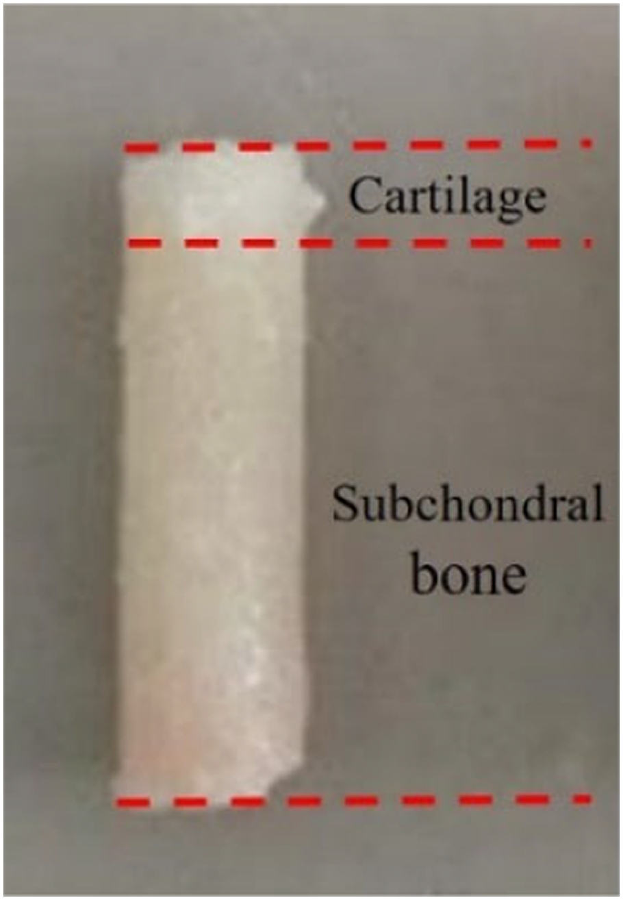
Methods
In-situ dynamic mechanical performance testing system
A self-made in-situ dynamic mechanical performance testing system was developed to study the dynamic changes of articular cartilage in different layers under cyclic loading, as shown in Figure 2, which is composed of a driving device, collection device and in-situ measuring device. In-situ dynamic mechanical properties testing system.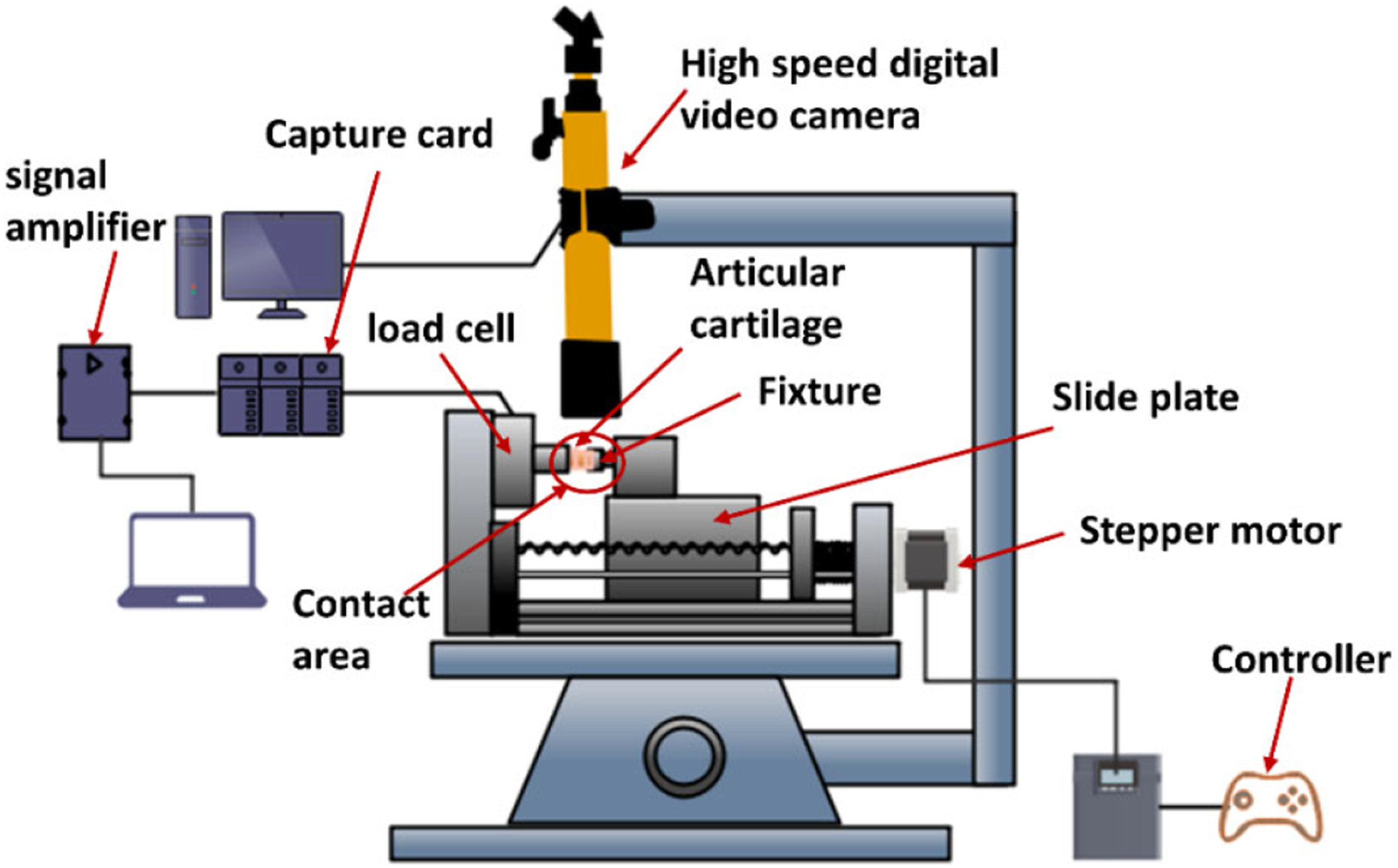
The driving device is driven by a 42FH01-01 stepping motor with a ball screw diameter of 12 mm, lead distance of 4 mm, and effective stroke of 100 mm. The collection device includes a load cell (range 0–100N), acquisition card, signal amplifier, etc., with an output voltage of ±5V. The in-situ measurement device uses a high-speed digital video camera (VW-9000, Keyence) for dynamic acquisition, with a frame rate of 1000fps and a resolution of 640×480 dpi.
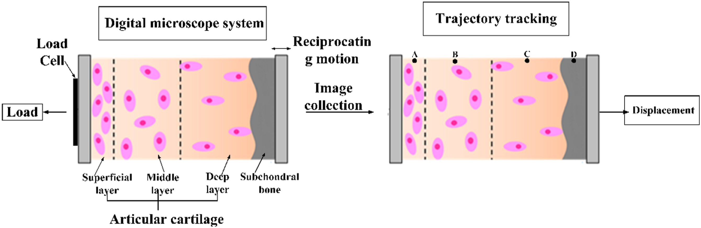
The working principle of this system is shown in Figure 3. It relies on a ball screw to drive and cyclically load cartilage samples. The load cell detects the load, and the VW-9000 high-speed digital camera records the dynamic deformation process of the cartilage sample. In-situ dynamic mechanical properties testing machine.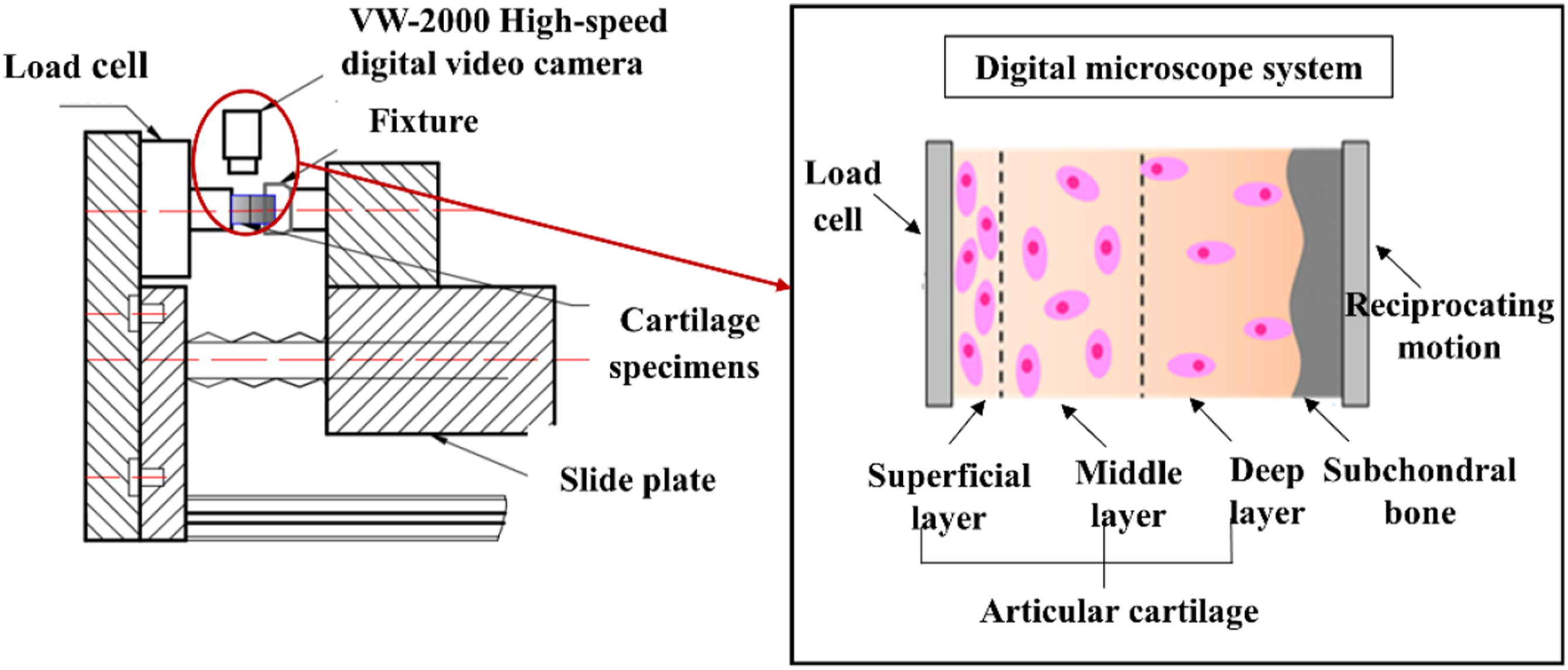
Digital image-related technology was applied to take a tracer point with obvious characteristics in the superficial layer, middle layer, deep layer and subchondral bone respectively, which were respectively marked as A, B, C, and D. The cartilage is divided into three layers by referring to the thickness ratio of each layer (superficial layer (10%–20%), middle layer (40%–60%) and deep layer (30%)) and combining the interlayer tide lines in the optical topography, as shown in Figure 4. Micro-tracking technology is used to track the trajectory of the feature points, and observe the position of the feature points relative to the coordinate axis, calculate the displacement of each layer of cartilage. The in situ compression mechanism of cartilage is shown in Figure 5. Optical morphology of the bovine knee articular cartilage. The dynamic mechanical characteristics of bovine knee articular cartilage.
Articular cartilage compression
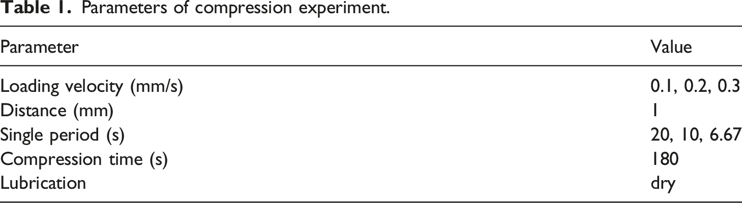
The articular cartilage compression includes unconfined compression and confined compression, as shown in Figure 6. Specifically, unconfined compression refers to the axial load of the cartilage without radial restriction, and confined compression refers to the radial constraint of the cartilage with no sliding, only in the axial direction of the load. Each test is performed three times. The parameters of the compression experiment are shown in Table 1 and the parameters of the in-situ measuring device are shown in Table 2. (a) Unconfined compression, (b) confined compression. Parameters of compression experiment. Parameters of microscope equipment.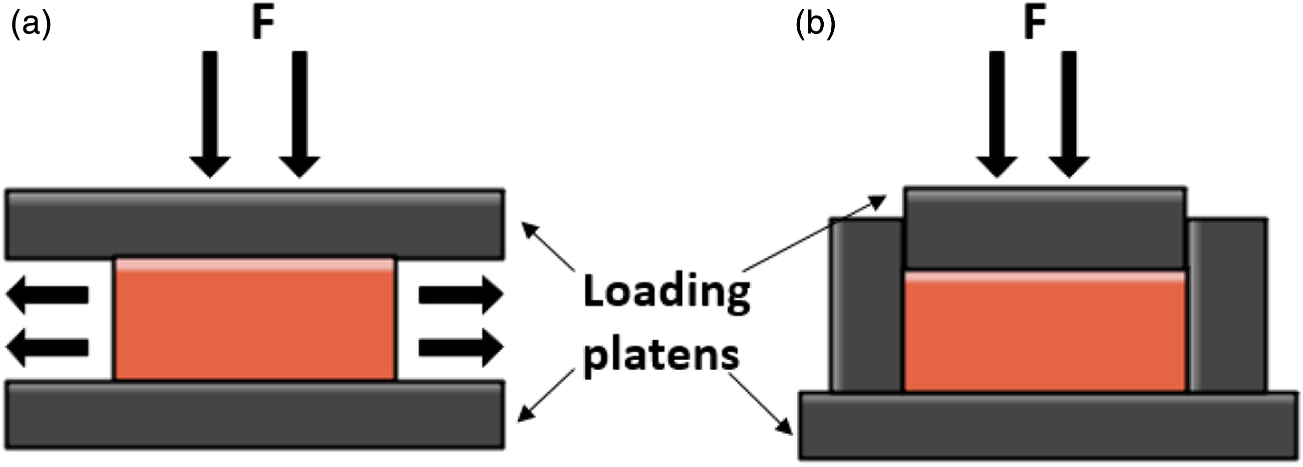

Results
Macroscopic dynamic deformation behaviors of articular cartilage
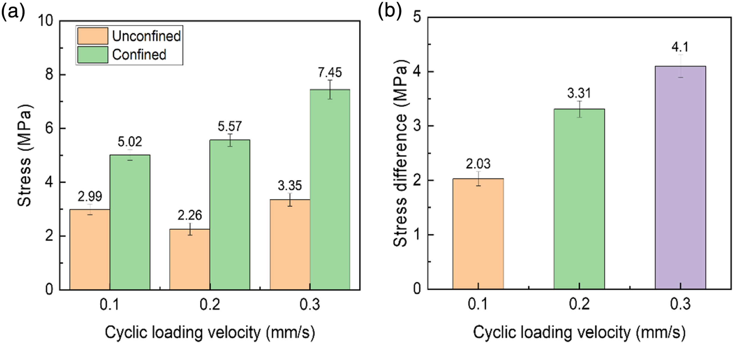
Figure 7 shows the stress waveform of the articular cartilage in confined and unconfined compression, with different loading velocities (0.1 mm/s, 0.2 mm/s, 0.3 mm/s) and the same displacement (1 mm). Under the same cyclic loading velocity, the loading stress of confined compression is significantly higher than that of unconfined compression, and the bigger the cyclic loading velocity, the greater the difference between the two compression states, as shown in Figure 8. The force-loading curves of compression. (a) 0.1 mm/s-unconfined compression, (b)0.2 mm/s-unconfined compression, (c) 0.3 mm/s-unconfined compression, (d) 0.1 mm/s-confined compression, (e) 0.2 mm/s-confined compression, (f) 0.3 mm/s-confined compression. The compression stress under different force-loading velocity.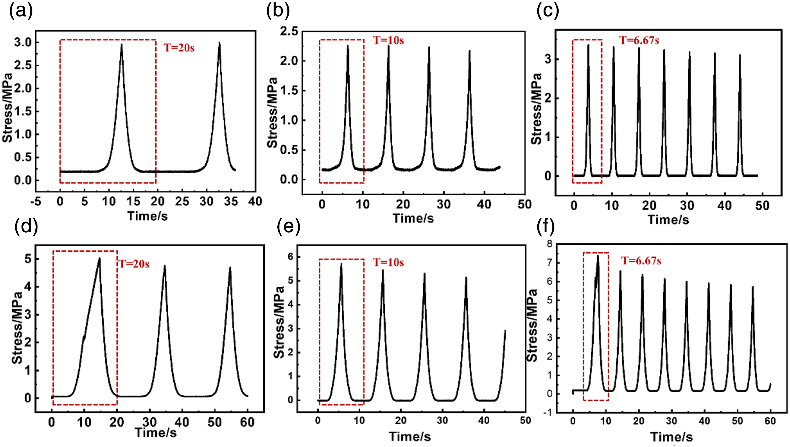
Dynamic deformation behaviors of different layers of articular cartilage
Figure 9 shows the movement displacement of different layers of articular cartilage under different compression states and loading velocities. It can be seen that the movement displacement change curve can be divided into the slow stage and the rapid stage. The slow stage lasts for a short time, the three layers of cartilage begin to deform at the same time, and the movement displacement curves almost overlap. However, in the rapid stage, the strain growth rate gradually increases, and the cartilage layers deform at different times until the end of the loading process. Under a constant cyclic loading velocity, the superficial layer has the strongest sensitivity to the increase of loading, followed by the middle layer and the deep layer. Simultaneously, the largest movement deformations are mainly accumulated in the superficial layer and the middle layer, and the deep layer has the smallest deformation. During the unloading rebound process, the spring back strain of the superficial layer decreases the most, followed by the middle layer and the deep layer. With the extension of the unloading rebound time, the strains of different layers showed a gradually decreasing trend and finally tended to the state before compression. As the cyclic loading rate increases and the number of cyclic loadings per unit time increases, the compressed and deformed cartilage becomes more difficult to return to the original state, and the deformation rate of each layer of cartilage also increases. Displacement of each layer of articular cartilage at different loading velocity. (a) 0.1 mm/s-unconfined compression, (b) 0.2 mm/s-unconfined compression, (c) 0.3 mm/s-unconfined compression, (d) 0.1 mm/s-confined compression, (e) 0.2 mm/s-confined compression, (f) 0.3 mm/s-confined compression.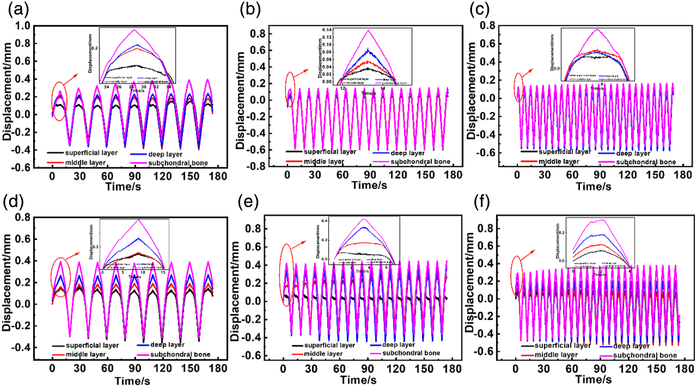
Figure 10 shows the deformation ratios of different layers of cartilage after the first cycle of loading under different compression states and different loading velocities. Compared with unconfined cyclic compression, the displacements of each layer are more evenly spaced under confined cyclic compression. Deformation ratios of different layers of articular cartilage under confined and unconfined compression in different loading velocities at the first period.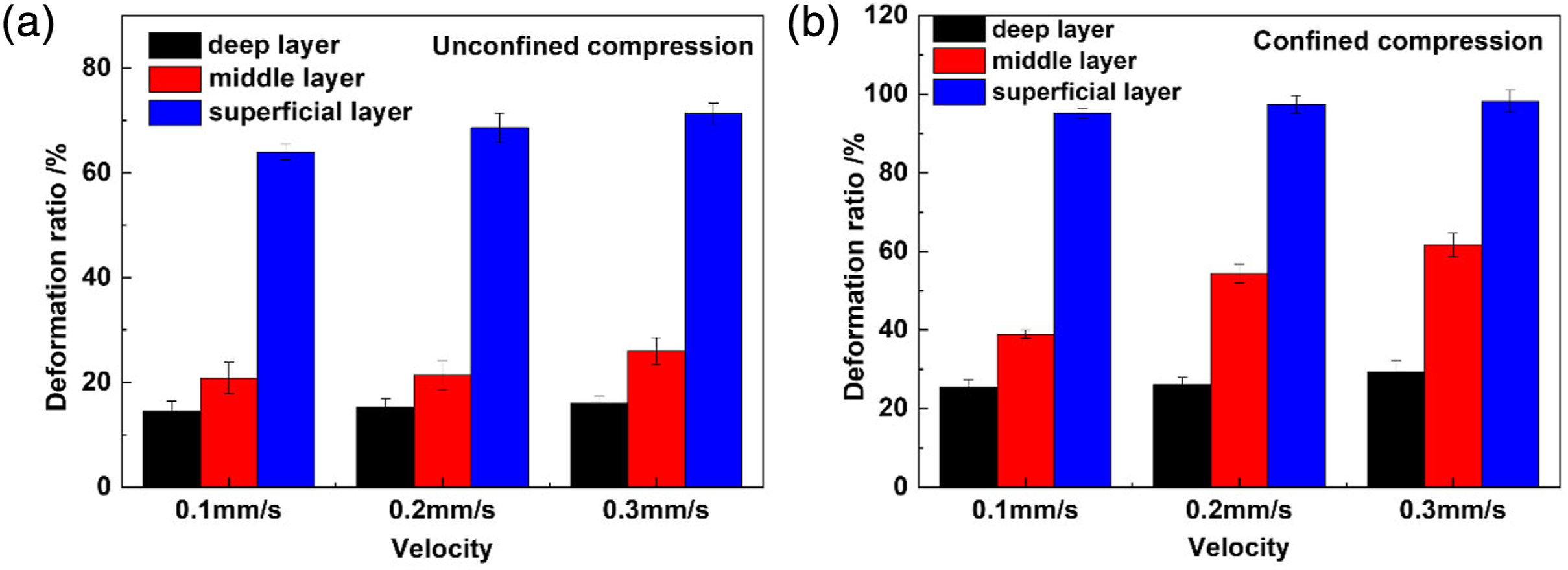
Figure 11 shows the dynamic deformation of the articular cartilage in the unconfined and confined loading process under different cyclic loading velocities. It can be seen that the initial position of the feature points (A, B, C, D) are distinct from the end position of the feature points after the 180s compression tests. Table 3 shows the compression ratio of each layer of articular cartilage after unconfined and confined compression under different cyclic loading velocities. The analysis shows that, under both compression conditions, when the loading displacement is the same, the greater the cyclic loading velocity, the greater the compression ratio of the superficial layer. When the velocity is 0.3 mm/s, the compression ratio of the superficial layer is 56% (confined compression) and 57.1% (unconfined compression), which are higher than that of the lower velocity. When the cyclic loading velocity is the same, the compression ratio of the superficial layer is the highest, followed by the middle layer and the deep layer. Furthermore, compared with unconfined cyclic compression, the compression ratio of each layer of cartilage under confined compression is larger. Dynamic deformation of articular cartilage at different loading velocity (a)0.1 mm/s-unconfined compression; (b)0.2 mm/s-unconfined compression; (c)0.3 mm/s-unconfined compression; (d)0.1 mm/s-confined compression; (e)0.2 mm/s-confined compression; (f)0.3 mm/s-confined compression. Compression ratios of different layers of articular cartilage under confined and unconfined conditions at different loading velocity.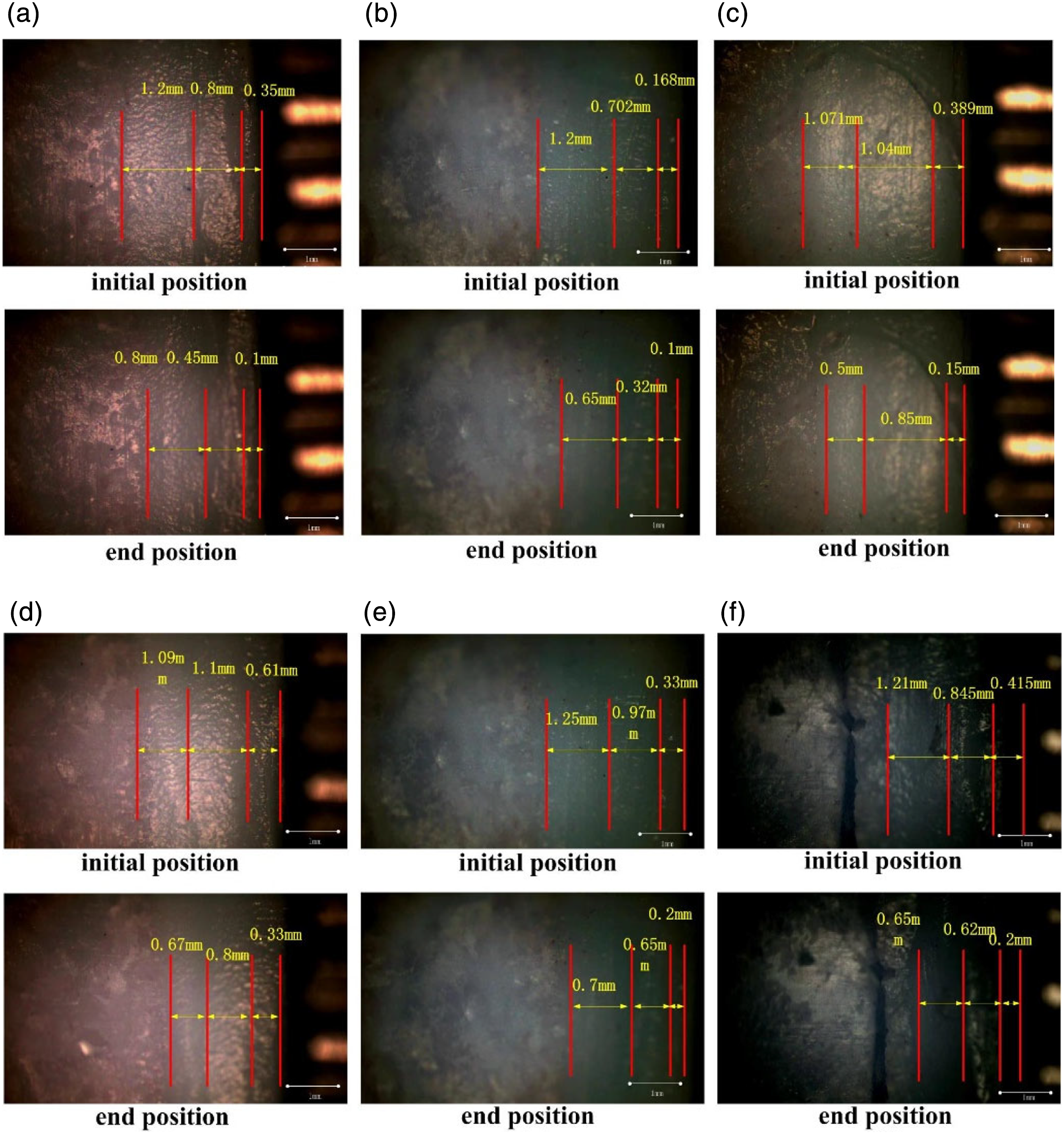
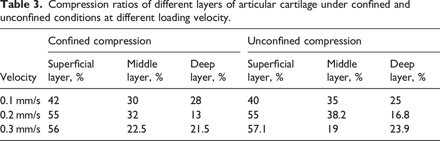
Figure 12 shows the deformation ratio of each layer of articular cartilage in different cycles of compression under confined and unconfined states for different loading rates (0.1 mm/s, 0.2 mm/s, and 0.3 mm/s). It can be seen that with the increase in the number of cycles, the deformation rate of different layers of articular cartilage increases and then tends to be stable. Moreover, under the action of a constant loading rate, the deformation rate of different layers of cartilage do not coincide, which indicates that the mechanical properties of each layer of articular cartilage are different. The deformation rate of the superficial layer of articular cartilage is the largest, the deformation rate of the deep layer is the smallest, and then that of the middle layer. With the increase in cyclic loading rate, the deformed parts of each layer of articular cartilage are too late to recover, and then the next cyclic compression experiment is carried out, resulting in an increase in the deformation rate again. Simultaneously, the increase in loading rate has the greatest effect on the deformation rate of the superficial layer but has little effect on the deep layer. Moreover, compared with the deformation rate of unconfined cyclic compression, under the state of confined compression, the radial deformation of articular cartilage under the condition of a constant loading rate is limited, and the liquid phase can not seep out, which leads to the deformation rate of each layer significantly higher than that of the unconfined compression, and the confined compression testing can better reflect the viscoelasticity of articular cartilage. Deformation ratio of different layers of articular cartilage in different cycles under A) confined and B) unconfined conditions. (a) Superficial layer (b) middle layer (c) deep layer.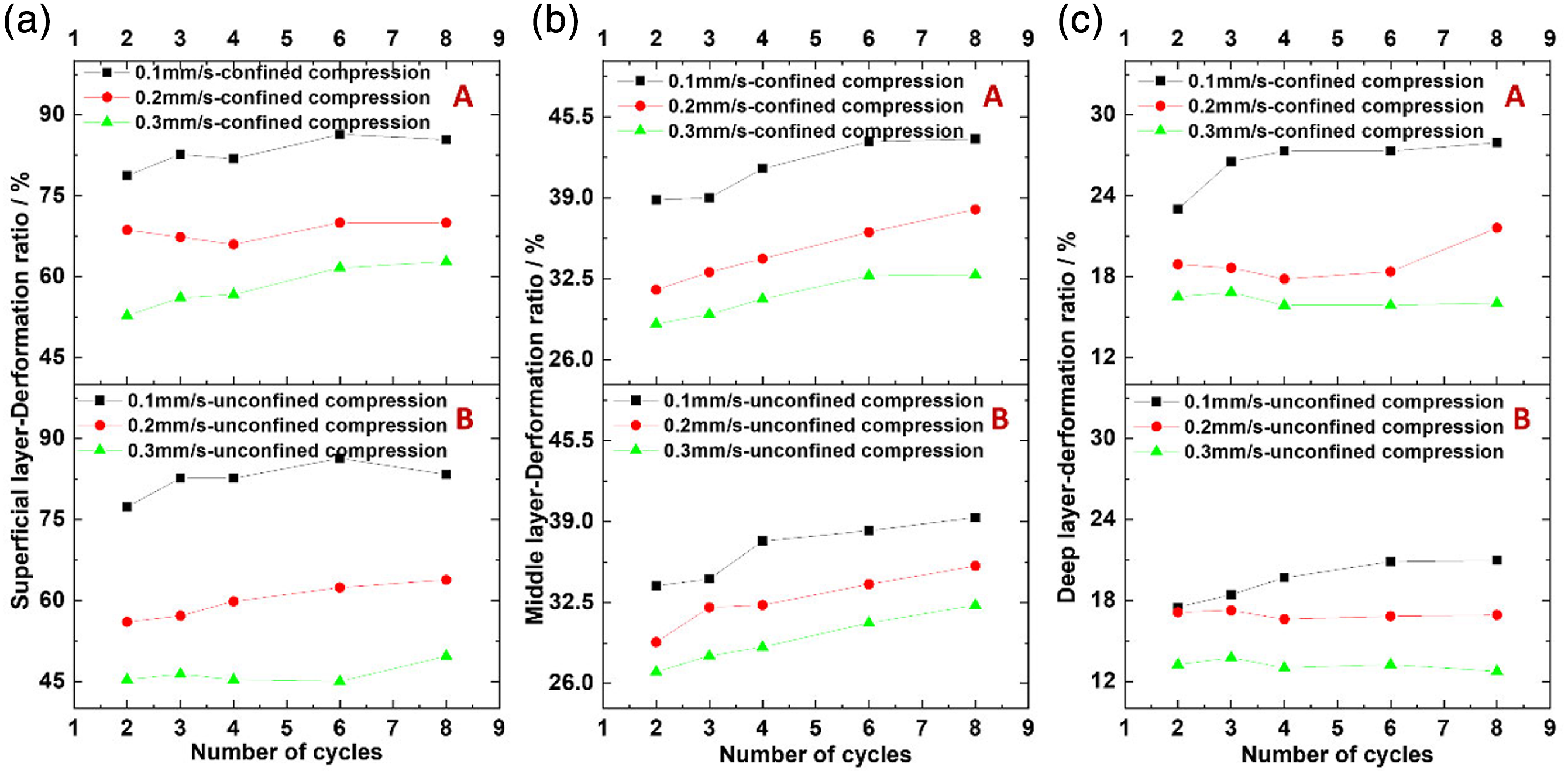
Discussion
The difference in deformation behavior in different layers of articular cartilage is closely related to its composition and microstructure and also determines its different roles. Figure 13 shows the deformation mechanism of different layers of cartilage under unconfined and confined compression. As can be seen, cartilage can be divided into the superficial layer, middle layer and deep layer according to the arrangement of macromolecules and collagen fibers. The collagen fibers in the superficial layer are densely arranged and distributed parallel to the articular surface, with the least proteoglycan and the highest water content. Therefore, the superficial layer is prone to normal deformation and the minimum elastic modulus. The collagen fibers in the middle layer are irregularly arranged, with high proteoglycan and low water content, and can bear part of the compression load to resist deformation. The collagen fibers in the deep layer are distributed perpendicular to the articular surface, with the highest proteoglycan content and the lowest water content. Therefore, the elastic modulus of the deep layer is the largest and the deformation range is the smallest,
23
which is the main area to resist compression deformation and transmit compression load. The deformation mechanism of the different layers of cartilage under unconfined and confined compression conditions.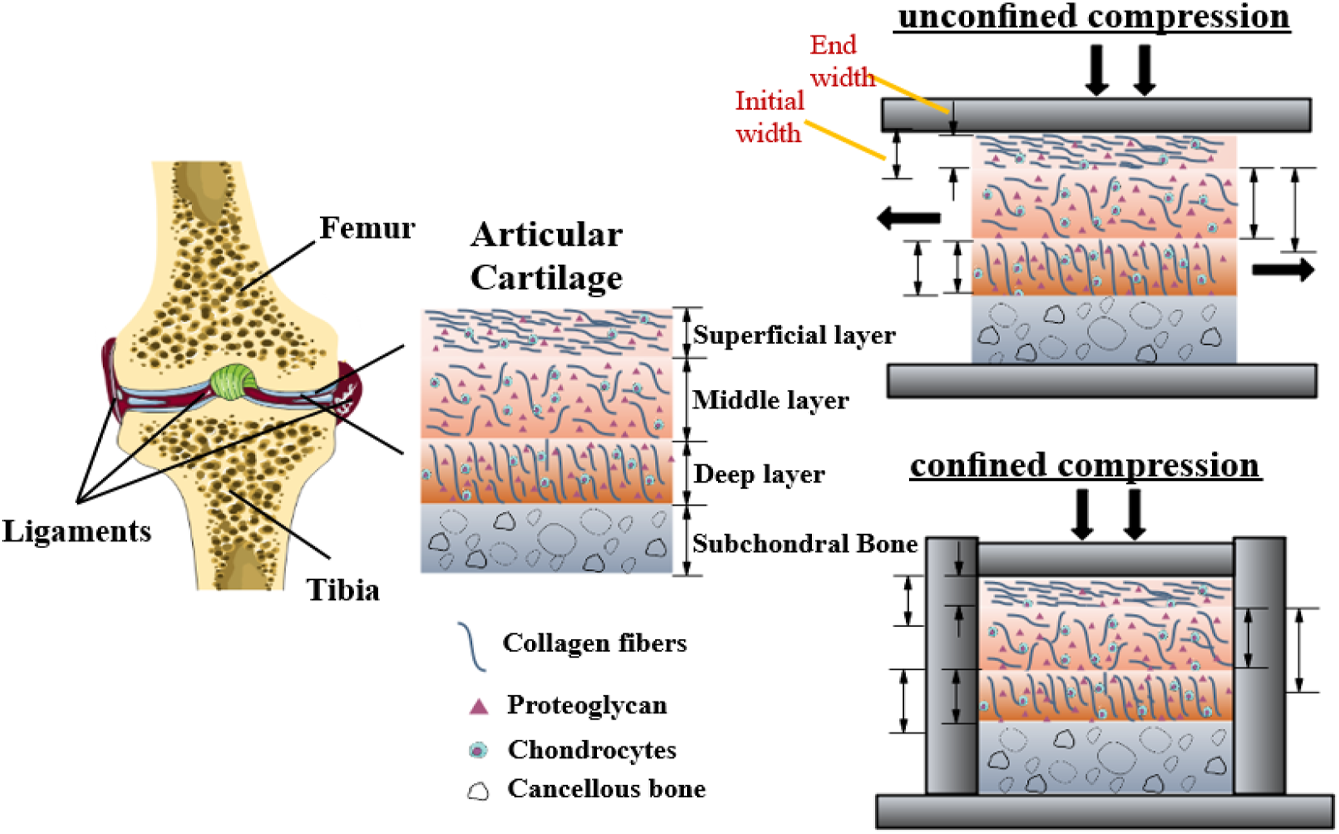
Cartilage is the initial target of the osteoarthritis. 4 The damage of cartilage can be reflected by the mechanical properties, so the confined and unconfined compressive properties of different layers of cartilage should be studied to reveal the evolution and load-bearing laws of cartilage damage. At the initial stage of loading, due to the low permeability, the synovial fluid cannot be flowed out in time, and the interstitial hydraulic pressure is formed in the cartilage. Subsequently, the hydraulic pressure can bear most of the mechanical load in a short time and also plays a good role in lubrication. This stage corresponds to the slow growth stage in movement displacement growth. As the load continues to be applied, the liquid quickly seeps out, and asynchronous deformation occurs. This stage corresponds to the rapid growth stage in movement displacement growth. As the amount of liquid in the pores decreases, the collagen fiber network plays a significant role in supporting the cartilage, and the strain rate gradually slows down. Since the bearing part of the cartilage gradually transforms from the initial pore fluid into the collagen fiber network, the ability of the collagen fiber network to resist deformation is much better than that of the pore fluid. With the rapid exudation of the liquid, the ability of cartilage to resist deformation increases rapidly, and its elastic modulus also increases. When the main load-bearing unit is the collagen fiber network, the compressive elastic modulus of cartilage continues to increase with the increase of collagen fiber deformation, but the increase rate is significantly slower, 24 which is consistent with the finite deformation biphasic material properties of bovine articular cartilage.25,26
Under confined compression, the cartilage samples only deform along the axis due to axial loading and radial constraint. During the process of loading, the water in the cartilage is squeezed out and slowly lost through the gap. Therefore, in the two compression states, when the displacement of loading is the same, the deformation of cartilage under confined compression is larger. However, some limitations should be noted. First, the division of the cartilage layer area is based on the proportion of the thickness of each layer recorded in the reference, and there is a deviation in the selection of the tracking points of each layer. Secondly, the resolution of the in-situ measuring device is only 640×480 dpi and needs to be enhanced for clearer photos and videos.
Conclusion
(1) The dynamic mechanical properties of articular cartilage were evaluated by strain and depth. In the confined and unconfined compression of articular cartilage, the deformation rate of the superficial layer is the largest, followed by that of the middle layer, and that of the deep layer is the smallest. It is the deep layer of cartilage that bears compression load and resists compression deformation. (2) The deformation of articular cartilage is small in the process of a single stress cycle. As the number of loading cycles increases, the deformation also accumulates and increases. When the loading rate is the same, the confined compression deformation rate of each layer of the articular cartilage is significantly higher than that of unconfined compression.
Footnotes
Declaration of conflicting interests
The author(s) declared no potential conflicts of interest with respect to the research, authorship, and/or publication of this article.
Funding
The author(s) disclosed receipt of the following financial support for the research, authorship, and/or publication of this article: This research is supported by National Natural Science Foundation of China (Grant No.51875564, 51705517, 52105231, 52175204), Natural Science Foundation of Jiangsu Province (Grant No. BK20211243), and A Project Funded by the Priority Academic Program Development of Jiangsu Higher Education Institutions (PAPD).