
Abstract
Magnetite (Fe3O4) nanoparticles/polyvinylpyrrolidone (PVP) composite nanofibers (FCNFs) have been fabricated to evaluate the potential of FCNFs as electromagnetic interference (EMI) shielding material in the frequency range of 8.2–12.4 GHz. The scanning electron microscope and viscosity analyses confirmed the presence of good dispersion Fe3O4 nanoparticles encapsulated within the electrospun nanofibers and showed FCNF morphologies with diameters of 150–500 nm. The magnetic properties and electrical conductivity of FCNFs were found to be dependent on Fe3O4 nanoparticles concentration and showed an increase with increasing Fe3O4 nanoparticles loading. The EMI shielding efficiency of FCNFs increased up to approximately 22 dB. The EMI shielding results for FCNFs showed that absorption was the major shielding mechanism and reflection was the secondary shielding mechanism. The present study has shown the possibility of utilizing magnetic FCNFs as EMI shielding/absorption materials.
In recent years, with the development of electronic and electrical instruments with frequency ranges from 300 MHz to 300 GHz, microwave shielding materials are widely known to prevent electromagnetic interference (EMI) and microwave pollution. 1,2 In addition, EMI may induce human diseases such as insomnia, nervousness, headaches, leukemia, miscarriages, and breast cancer. 3,4 Therefore, there is a critical requirement to develop effective and practical EMI shielding materials for microwaves of the EM spectrum in order to make the environment safer and improve the lifetime of electronic instruments and the efficiency of use of frequency sources. 5
Magnetic materials are good potential candidates as microwave shielding due to their combination of dielectric and magnetic loss as well as controllable morphologies. 6 –8 Specially, among the available various magnetic materials, magnetite (Fe3O4) nanoparticles are one of the most promising conduction allotropes, on the basis of their superior electrical and thermal properties comparing to other magnetic materials. 9,10 Fe3O4 nanoparticles have recently attracted great attention because of their valuable properties, including extraordinary magnetic properties, low toxicity, good electrical conductivity, and nearly full spin polarization at room temperature; these properties have great potential for applications in EMI shielding/absorbing materials. 1,6,7,11 At present, Fe3O4 nanoparticles have been intensively examined for their superior properties, which make them an excellent choice to fabricate magnetic polymer composite for EMI shielding at very low loading. 5,7 Various techniques have been developed to fabricate Fe3O4/polymer nanocomposites. 1,7,12 But, among the various methods, electrospinning is recognized as an economical method for manufacturing magnetic composite with nanofibrous structure, when compared with other techniques. 11 –15 Some literatures 10 –15 have reported the electrospinning process of Fe3O4/polymer composite nanofibrous structure, there is almost no report studied on simultaneous morphology controlling and EMI shielding of a Fe3O4/polymer nanofibrous mat.
Polyvinylpyrrolidone (PVP), an important synthetic polymer, is very biocompatible and nontoxic. The PVP has high tensile strength, excellent thermal stability, low chemical toxicity, and good spinnability. 16 –18 Therefore, PVP has been considered as a promising polymer for EMI shielding application. Although very few research 11,19,20 have reported the electrospinning process of Fe3O4/PVP composite with nanofibrous structure, there is almost no report studied on simultaneous morphology controlling and EMI shielding of a Fe3O4/PVP nanofibrous mat. Therefore, during this study, Fe3O4/PVP composite nanofibers (FCNFs) were fabricated by electrospinning procedure. The effect of Fe3O4 nanoparticles on the morphological properties, electrical conductivity, magnetic properties, and EMI shielding of FCNFs in the X-band frequency range is investigated for the first time to the best of our knowledge.
Experimental
Materials
The PVP powders with molecular weight of 360,000 g mol−1 were supplied from Sigma-Aldrich (Saint-Louis, Missouri, USA). The Fe3O4 nanoparticles (diameter = 20–30 nm), with a purity of 95%, were provided by US Research Nanomaterials, Inc (Houston, Texas, USA). The solvent used for dissolving PVP and Fe3O4/PVP dispersion was N,N-dimethylformamide (DMF, Merck, Darmstadt, Germany).
Preparation of Fe3O4/PVP solutions
The different weights of Fe3O4 nanoparticles were dispersed in the DMF by using an ultrasonic homogenizer at 0°C for 20 min. The electrospinning Fe3O4/PVP solutions were prepared by dissolving 13 wt% of PVP with in dispersion solutions by using a magnetic stirrer at room temperature for 24 h.
Fabrication of FCNFs
The electrospinning process was carried out using Electroris (FNM, Iran). The prepared magnetic Fe3O4/PVP solutions were added to a glass syringe with a needle (L = 35 mm, D = 0.7 mm, and L/D = 50) and connected to the power supply, which can generate DC voltages in the range of 0–50 kV. The needle was connected to a positive high-voltage power supply. An aluminum foil was wrapped on the Electroris grounded rotating drum as collector and was located at a distance of 20 cm from the needle. The electrospinning was carried out by applying a high voltage of 15 kV between the needle and the rotating collector. The feed rate of the polymer solutions was 0.25 mL h−1 and a take-up speed of 100 r min−1 was selected to collect the magnetic FCNFs. These composite nanofibers were collected and then dried in oven at 50°C for 4 h. The magnetic FCNF products were categorized as FCNF-0 (0 wt% Fe3O4), FCNF-0.5 (0.5 wt% Fe3O4), FCNF-1 (1 wt% Fe3O4), FCNF-2 (2 wt% Fe3O4), FCNF-3 (3 wt% Fe3O4), FCNF-4 (4 wt% Fe3O4), and FCNF-5 (5 wt% Fe3O4).
Measurement and characterization
The viscosity of magnetic Fe3O4/PVP solutions was measured by a viscometer (DV-II+Programable; Brookfield, Middleborough, Massachusetts, USA). The surface morphology of the electrospun nanofibers was examined by scanning electron microscope (SEM, XL-30; Philips, Netherlands) at an accelerating voltage of 25 kV under magnification of 5000×, and the average fiber diameter was measured with the SEM images using Image J software (National Institute of Health, Bethesda, Maryland, USA) from 100 randomly a selection of FCNFs. The X-ray diffraction (XRD) characterization of the Fe3O4 nanoparticle and FCNFs was carried out by an EQuniox-3000 XRD (Artenay, France) using Copper Kα (λ = 1.5418°A). The XRD patterns were recorded in the scanning angle range of 5–70° with a scanning speed of 1° min−1. The magnetic properties of the electrospun Fe3O4/PVP nanofibers were characterized by vibrating sample magnetometer (VSM) at room temperature.
The electrical conductivity of the electrospun FCNFs was analyzed on the 4-point probe technique. Before measuring the conductivity, the composite nanofibers were cut into 2 × 2 cm2 rectangular shapes (thickness = 1 mm) and conditioned for 24 h in 25 ± 2°C and 35 ± 5% relative humidity. The electrical current (I) was measured with a Keithley 224 electrometer and measurements of voltage (V) were performed with a Keithley 196 (Cleveland, Ohio, USA). The electrical conductivity (σ) was calculated using the equation 21
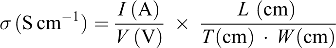
where L, W, and T is length, width, and thickness of the FCNF sample, respectively.
The electromagnetic shielding measurements have been carried out using vector network analyzer (Agilent 8510C, Santa Clara, California, USA) in the X-band region (8.2–12.4 GHz). Rectangular FCNFs samples with dimensions of 22.86 × 10.16 × 1 mm3 were fixed into the waveguide sample holder. At least three specimens were prepared for each kind of composite nanofibers. The final results were averaged from all the tested composites.
Results and discussion
Morphology of the FCNFs
Figure 1 displays the SEM photographs and corresponding fiber diameter distribution of PVP nanofibers and FCNFs at different concentrations of Fe3O4 nanoparticles. As it can be seen, the surface morphology of pure PVP nanofibers is smooth and without beads. The surface morphology of FCNFs is very smooth in the 0.5–2 wt% ranges of Fe3O4 nanoparticles loading. Low surface roughness and disproportion in the FCNF-3 and FCNF-4 samples were observed, whereas still good arrangement of Fe3O4 nanoparticles in the Fe3O4/PVP composite structures can be seen (Figure 1(e) and (f)). However, considerable aggregation and irregularities were observed as the Fe3O4 nanoparticles content in the FCNFs increased to 5 wt%.
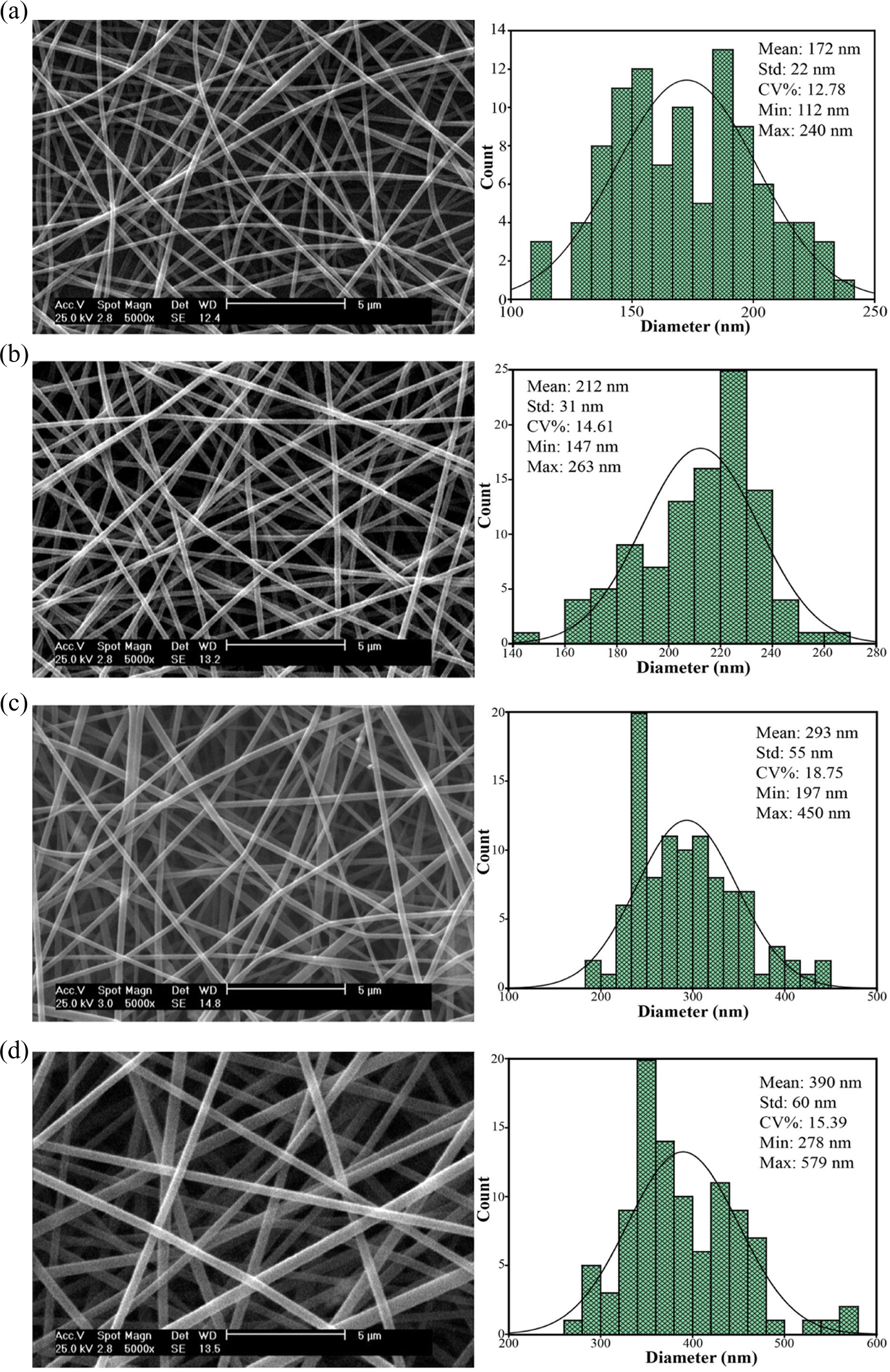
SEM photographs (left) and corresponding fiber diameter distribution (right) of FCNFs samples at magnification of ×5000; (a) FCNF-0, (b) FCNF-0.5, (c) FCNF-1, (d) FCNF-2, (e) FCNF-3, (f) FCNF-4, and (g) FCNF-5 samples. SEM: scanning electron microscope; FCNF: FCNF: magnetite/polyvinylpyrrolidone composite nanofiber.
Changes in the morphology of FCNFs versus the Fe3O4 nanoparticles concentration are shown in Table 1. With increasing the concentration of Fe3O4 nanoparticles in the solution from 0 to 0.5 wt%, the average diameter of electrospun nanofibers moderately increased from 172 ± 22 nm for FCNF-0 to 212 ± 31 nm for FCNF-0.5 sample. Further increase in Fe3O4 nanoparticles concentration from 1 to 4 wt%, the average diameter of FCNFs considerably increased from 293 ± 55 to 482 ± 101 nm. But, when Fe3O4 nanoparticles concentration was increased to 5 wt% the average diameter decreased to 428 ± 106 nm. In the electrospinning method, the viscosity of electrospun solution is very significant, which can affect the electrospun nanofibers’ diameter and surface morphology. As described in other researches, 22,23 mostly with increasing the viscosity of the polymer solution, the average diameter of electrospun nanofibers increased. Variations in the Fe3O4/PVP solution viscosity against the concentrations of Fe3O4 nanoparticles are shown in Table 1. With the addition of Fe3O4 nanoparticles content from 0 to 4 wt% in polymer solution, there was a significant increase in the viscosity. But, when the Fe3O4 nanoparticles concentration was increased to 5 wt%, the viscosity decreased noticeably. These variations can be related to the polymer chain entanglement changes in the solution produced due to adding Fe3O4 nanoparticles. To improve the morphological properties of the prepared FCNFs containing Fe3O4 nanoparticles, the dispersion condition is critical and plays an important role in achieving effective properties of FCNFs.
Effect of Fe3O4 nanoparticles concentration on morphology of FCNFs.
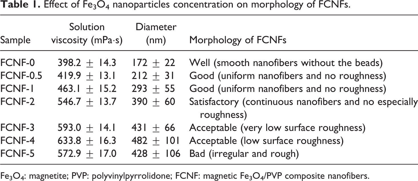
Fe3O4: magnetite; PVP: polyvinylpyrrolidone; FCNF: magnetic Fe3O4/PVP composite nanofibers.
Microstructural properties of FCNFs
The XRD test was undertaken to determine the physical status of Fe3O4 in the composite nanofibers. Figure 2 shows X-ray patterns of the pristine Fe3O4 nanoparticle, pure PVP nanofibers, and FCNFs with 4 wt% Fe3O4. From Figure 2, it can be seen that the diffractogram of Fe3O4 nanoparticle exhibits the typical peaks at 18.2°, 30.0°, 35.4°, 37.0°, 43.0°, 53.4°, 56.9°, and 62.4°, corresponding to the characteristic peaks of pure Fe3O4 (111), (220), (311), (222), (400), (422), (511), and (440) reflections, in good agreement with that of the literatures. 24 –28 The pure PVP nanofiber sample is amorphous with only two broad peaks at 11.5° and 21.0°. Similar peaks have been reported for PVP nanofibers in other works. 29,30 But, the FCNFs shows obvious crystalline diffraction patterns similar to the Fe3O4, which indicates the interaction between Fe3O4 units and PVP molecules and the formation of the regular microstructure composite nanofibers.
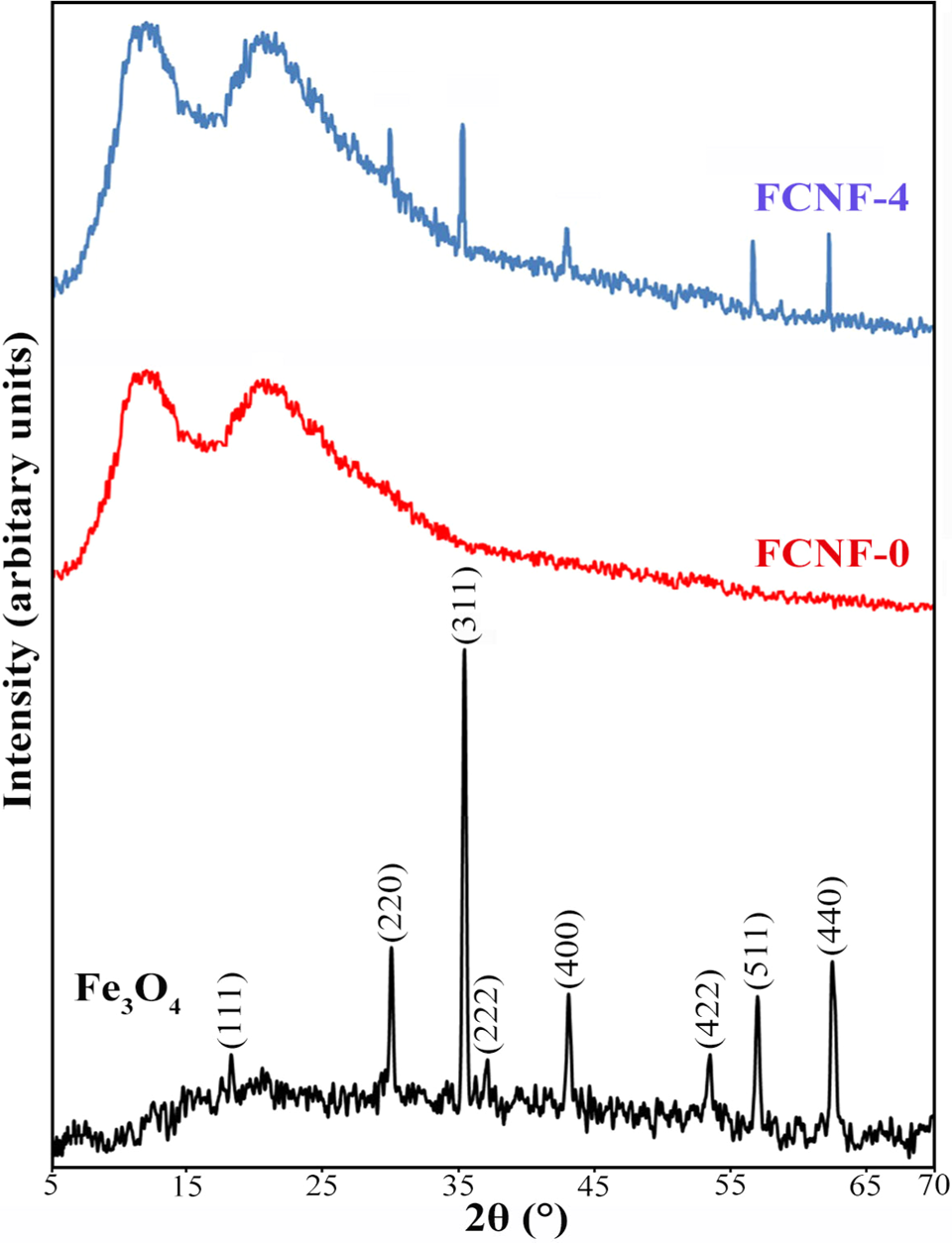
XRD patterns of Fe3O4 nanoparticle, pure PVP nanofibers, and FCNFS. XRD: X-ray diffraction; Fe3O4: magnetite; FCNF: magnetite/polyvinylpyrrolidone composite nanofiber.
Magnetic properties of FCNFs
Static magnetic property is a very useful method for the quality of dispersion characterization of magnetic nanoparticle-based composites. 7 The magnetic hysteresis loop of the Fe3O4 nanoparticles and FCNFs are shown in Figure 3. The saturation magnetization (M s), remanent magnetization (M r), and coercivity (H c) are main technical parameters to characterize the magnetism of a ferromagnetic sample. The Fe3O4 nanoparticle sample exhibits a ferromagnetic behavior with M s, M r, and H c values of 70.2 emu g−1, 8.7 emu g−1, and 91 Oe, respectively. The M s of our magnetic nanoparticles is higher than those of Fe3O4 ferromagnetic particles in other works. 31 –33
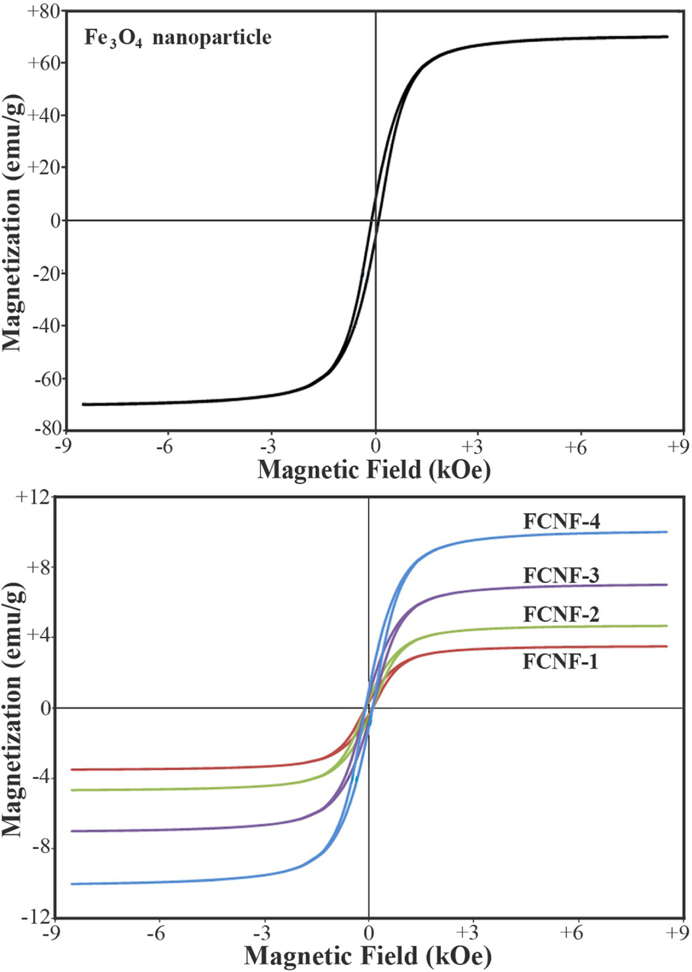
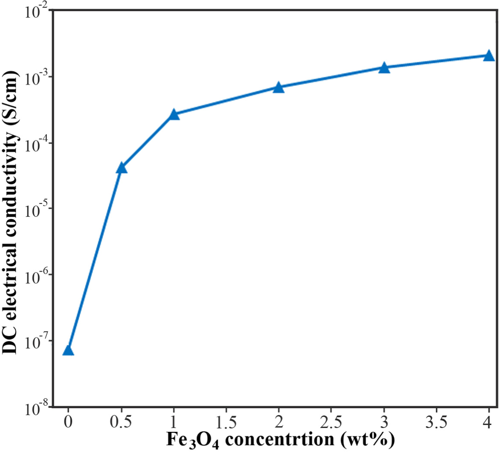
The magnetic hysteresis curves measured at room temperature for Fe3O4 nanoparticles and FCNFs. Fe3O4: magnetite; FCNF: magnetite/polyvinylpyrrolidone composite nanofiber.
The results given in Figure 3 for composite nanofibers reveal that magnetic properties of FCNFs increase to some extent with increase in Fe3O4 nanoparticles loading. The M s of the nanofibrous membranes increased from 3.51 emu g−1 for FCNF-1 and 4.67 emu g−1 for FCNF-2 to 10.02 emu/g for FCNF-4, which basically kept a linear growth model with the increase of Fe3O4 nanoparticles loading. The saturation magnetization shows the ability of a material to store the electromagnetic energy under the influence of a magnetic field. 7 It is observed that saturation magnetization values of nanofibers increase with increasing Fe3O4 nanoparticles content in composite nanofiber structures. This enhancement of magnetic properties by Fe3O4 nanoparticles loading can be due to the barrier effect of individual-dispersed Fe3O4 nanoparticles into the PVP matrix. From the VSM analysis results, it can be concluded that FCNFs with good magnetic properties can be used as high absorbing loss EMI shielding material in the X-band frequency range.
Electrical conductivity of FCNFs
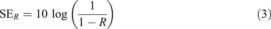
The electrical conductivity of FCNFs as a function of weight fraction of Fe3O4 nanoparticles are shown in Figure 4. The electrical conductivity of a nanofibrous web made of pure PVP is very small (7.41 × 10⁻⁸ S cm−1). The electrical conductivity of the samples increases with the increase of the magnetic nanoparticles content. The highest increase in the electrical conductivity occurs when the Fe3O4 nanoparticles’ concentration is increased from 0 to 0.5 wt%. Therefore, the electrospun FCNFs showed that the insulator to semiconductor transition took place for 0.5 wt% Fe3O4 nanoparticles content in nanofibers structure. The electrical conductivity of these composite nanofibers could be adjusted from approximately 10−8 S cm−1 (insulator) to approximately 10−3 S cm−1 (semiconductor) by choosing the different amounts of Fe3O4 nanoparticles, which is very useful for their EMI shielding application.
Electrical conductivity of electrospun FCNFs samples. FCNF: magnetite/polyvinylpyrrolidone composite nanofiber.
Shielding effectiveness of FCNFs
The total EMI shielding efficiency (SE) is the sum of the reflection from the surface of composite (SER), the absorption loss (SEA), and the multiple internal reflections (SEM) of electromagnetic radiation. 4,34 The scattering parameters (S-parameters), that is, S 11 (or S 22) and S 12 (or S 21) of the two-port network system represent the reflection and transmission coefficient, respectively. According to the analysis of S-parameters, transmittance (T) and reflectance (R) coefficient of the shielding composite can be described as below 3,35


where Er is the electric field of reflected EM wave.
The effect of internal reflections between both interfaces of the composite is negligible when SEA > 10 dB. Therefore, the SER, SEA, and SET of the shielding composite structure are correlated with reflectance and transmittance coefficient by the following equations 36
The SEA and SER of the PVP nanofiber and their composite Fe3O4/PVP nanofibers with various magnetic nanoparticles loadings as a function of X-band frequency are shown in Figure 5(a) and (b), respectively. From the experimental shielding measurement, the average SEA has been found to vary from 2 to 17 dB with increase in the Fe3O4 nanoparticles content whereas the average SER increases from 3 to 5 dB for the same. Therefore, with the increasing Fe3O4 nanoparticles content, the absorption increases greatly, whereas the reflection only increases a very little. This result suggests that the FCNFs should be more absorptive and less reflective to EM radiation, that is, the primary EMI shielding mechanism of the composite nanofibers is dominant by absorption rather than by reflection in the X-band frequency. This result is in agreement with the investigation of the shielding mechanism of Fe3O4/polymer composites. 1,7
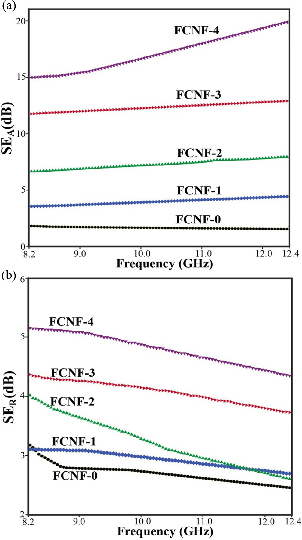
Plot of SEA (a) and SER (b) of FCNFs as a function of the X-band frequency. SER: surface of composite; SEA: the absorption loss; FCNF: magnetite/polyvinylpyrrolidone composite nanofiber.
The total EMI shielding of FCNFs as a function of weight fraction of Fe3O4 nanoparticles for X-band frequency is shown in Figure 6. It can be observed that the total shielding of pure nanofiber (FCNF-0) is about 3 to 4 dB, which is primarily attributed to the slight conductivity of nanofibers’ structure. As expected from the magnetic properties and electrical conductivity data, the EMI shielding increases with increasing content of Fe3O4 nanoparticles in the composite nanofibers and the contribution to the EMI shielding should come from the addition of Fe3O4 nanoparticles. The total EMI shielding of the FCNFs presented in this work was compared with the polymeric composite EMI shielding results reported in the literature and summarized in Table 2. Comparing the summarized results in this table is not straightforward as the EMI shielding values are reported at different thicknesses.
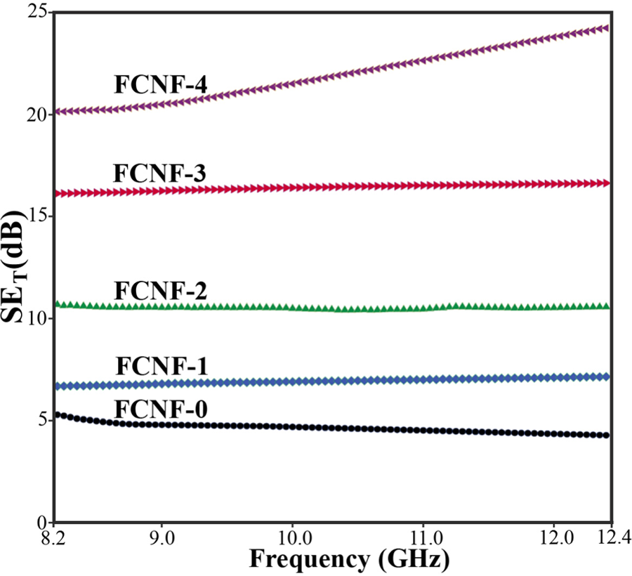
Total EMI shielding of FCNFs as a function of the X-band frequency. EMI: electromagnetic interference; FCNF: magnetite/polyvinylpyrrolidone composite nanofiber.
Comparison between EMI shielding of polymeric composites proposed in the literatures and this work for X-band.
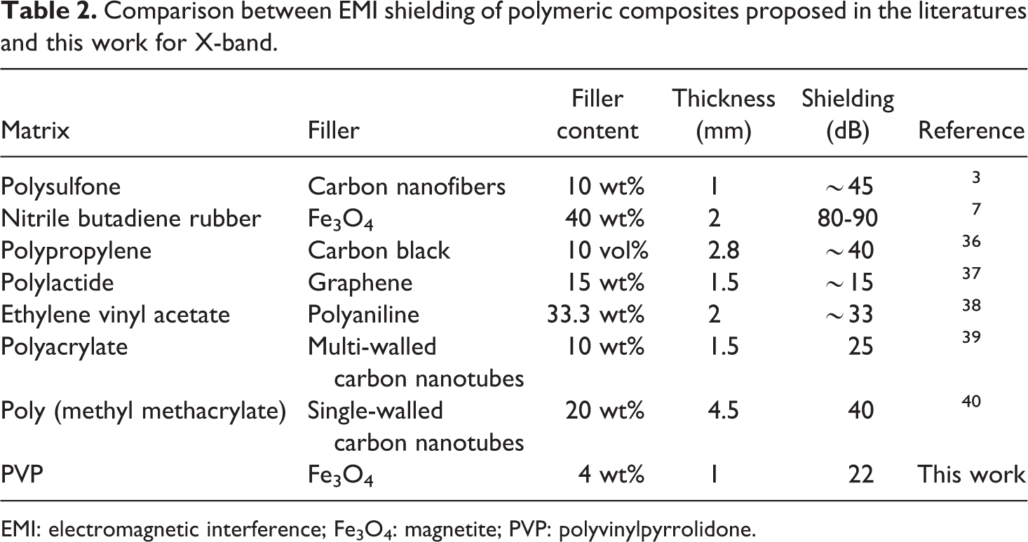
EMI: electromagnetic interference; Fe3O4: magnetite; PVP: polyvinylpyrrolidone.
The total SE achieved for the FCNF is approximately 22 dB (FCNF-4) which is much higher than the pure nanofibers (FCNF-0). The total shielding of 20 dB means that 99% of the EM waves have been attenuated. For magnetic FCNFs containing 4 wt% of Fe3O4 nanoparticles, the total SE shows more than 99% shielding of EM energy over the entire frequency range of measurement. The total SE of 20 dB is considered an adequate level of shielding for many industrial applications. 5 It is evident that the FCNFs with 4 wt% Fe3O4 nanoparticles can be used as appropriate shielding materials for EMI, especially in X-band and can meet the commercial requirements.
Conclusions
Fe3O4 nanoparticles were dispersed in PVP by ultrasonication to prepare FCNFs applied on building interior wall for EMI shielding applications. Well-dispersed FCNFs with varying amounts of Fe3O4 nanoparticles (0.5–4 wt%) were successfully prepared by electrospinning method. With increasing the concentration of Fe3O4 nanoparticles in the solution from 0.5 to 4 wt%, the average FCNFs diameter increased from 172 ± 22 nm to 482 ± 101 nm. Magnetic properties and electrical conductivity were found to improve with increase in Fe3O4 nanoparticles’ concentration. Total EMI shielding of approximately 22 dB was obtained at 4 wt% Fe3O4 nanoparticles loading (FCNF-4) in the frequency range of 8.2–12.4 GHz. The mechanism of EMI shielding of FCNFs has been well explained by comparing the contribution of reflection and absorption to the total EMI shielding and revealed that shielding due to absorption plays a significant role compared to reflection in determining the total EMI shielding. In conclusion, it can be concluded that FCNFs having 4 wt% Fe3O4 nanoparticles can be used as an effective EMI shielding material.
Footnotes
Acknowledgement
We would like to thank to National Elites Foundation of Iran for supporting this study.
Declaration of Conflicting Interests
The author(s) declared no potential conflicts of interest with respect to the research, authorship, and/or publication of this article.
Funding
The author(s) disclosed receipt of the following financial support for the research, authorship, and/or publication of this article: This work was supported by National Elites Foundation of Iran.