
Abstract
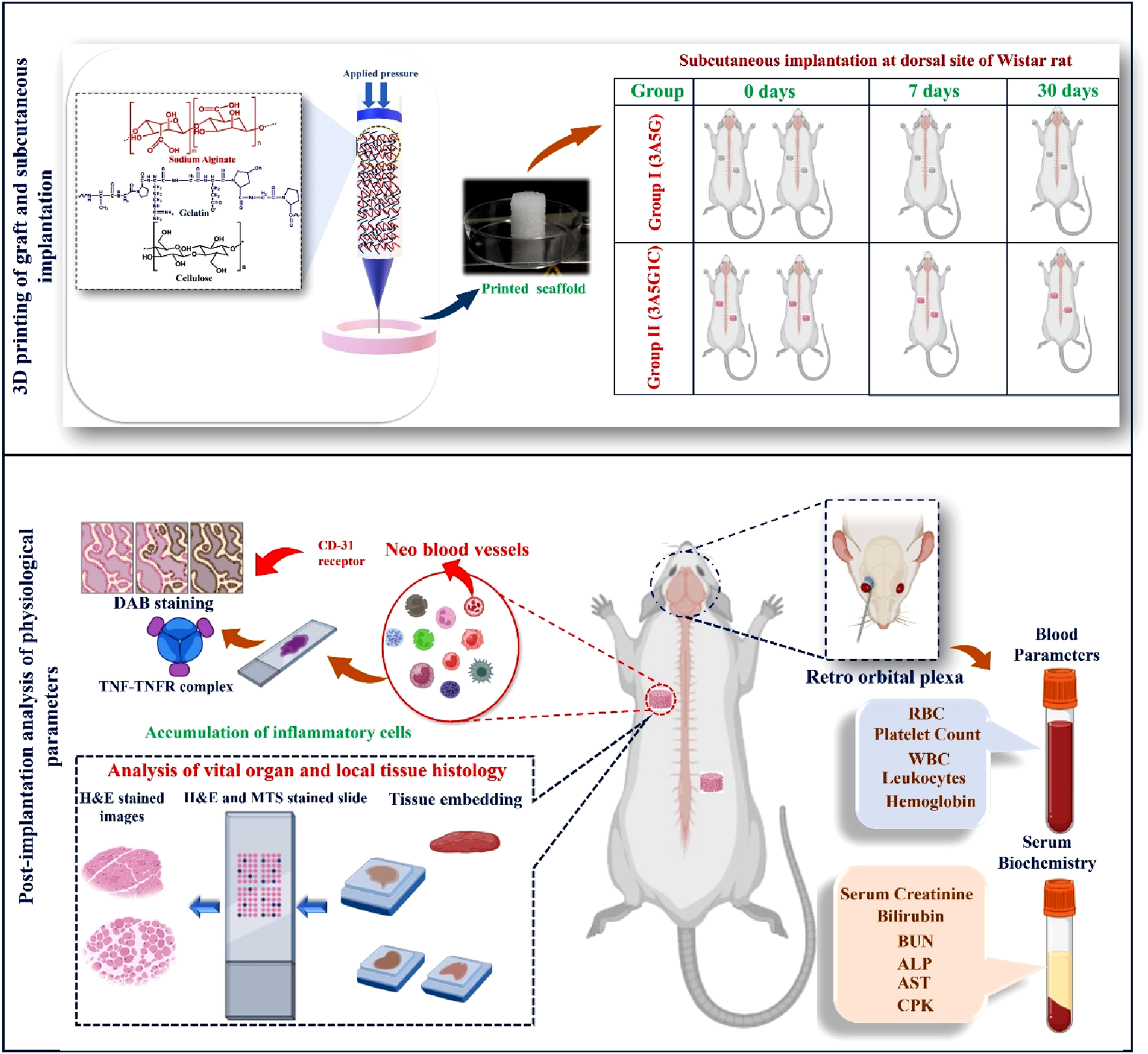
The last two decaes have witnessed significant efforts to develop gelatin/alginate based scaffolds using variants of 3D printing techniques. However, their biocompatibility for regenerating complex soft tissues remains insufficiently explored. Addressing this gap, we fabricated 3D-printed alginate-gelatin (3A5G) and nanocellulose-reinforced (3A5G1C) hydrogel scaffolds with clinically relevant dimensions (15 mm diameter, 5 mm height) and the host tissue responses were critically analyzed. The distinct advantages of nanocellulose in modulating mechanical strength, viscoelasticity, swelling, and degradation characteristics were established in our prior studies. This investigation aimed to comprehensively evaluate the foreign body response of these scaffolds in a rat model. The animals exhibited healthy metabolic activity, evidenced by progressive weight gain, localized tissue healing, and normal mobility over 30 days. Histological analyses could not reveal any adverse immune reaction at 7- or 30-days, post-implantation. Hematological and serum biochemical assessments indicated a progression from acute (7 days) to sub-acute (30 days) inflammation, following subcutaneous implantation, without any signature of systemic toxicity. Immune marker evaluation (TNF-α, CD-8, CD-68, COX-2, IL-6) confirmed the absence of pathological immune responses, even with nanocellulose incorporation. Immunohistochemical analysis using CD31 staining demonstrated enhanced vascularization in nanocellulose-reinforced scaffolds at both 7 and 30 days. The absence of systemic toxicity from scaffold degradation products and the favorable biocompatibility outcomes underline the potential of these hydrogel scaffolds for soft tissue regeneration. The incorporation of nanocellulose further enhanced the scaffolds’ functional performance, particularly in promoting vascularization, positioning them as promising candidates for complex tissue engineering applications.
Get full access to this article
View all access options for this article.