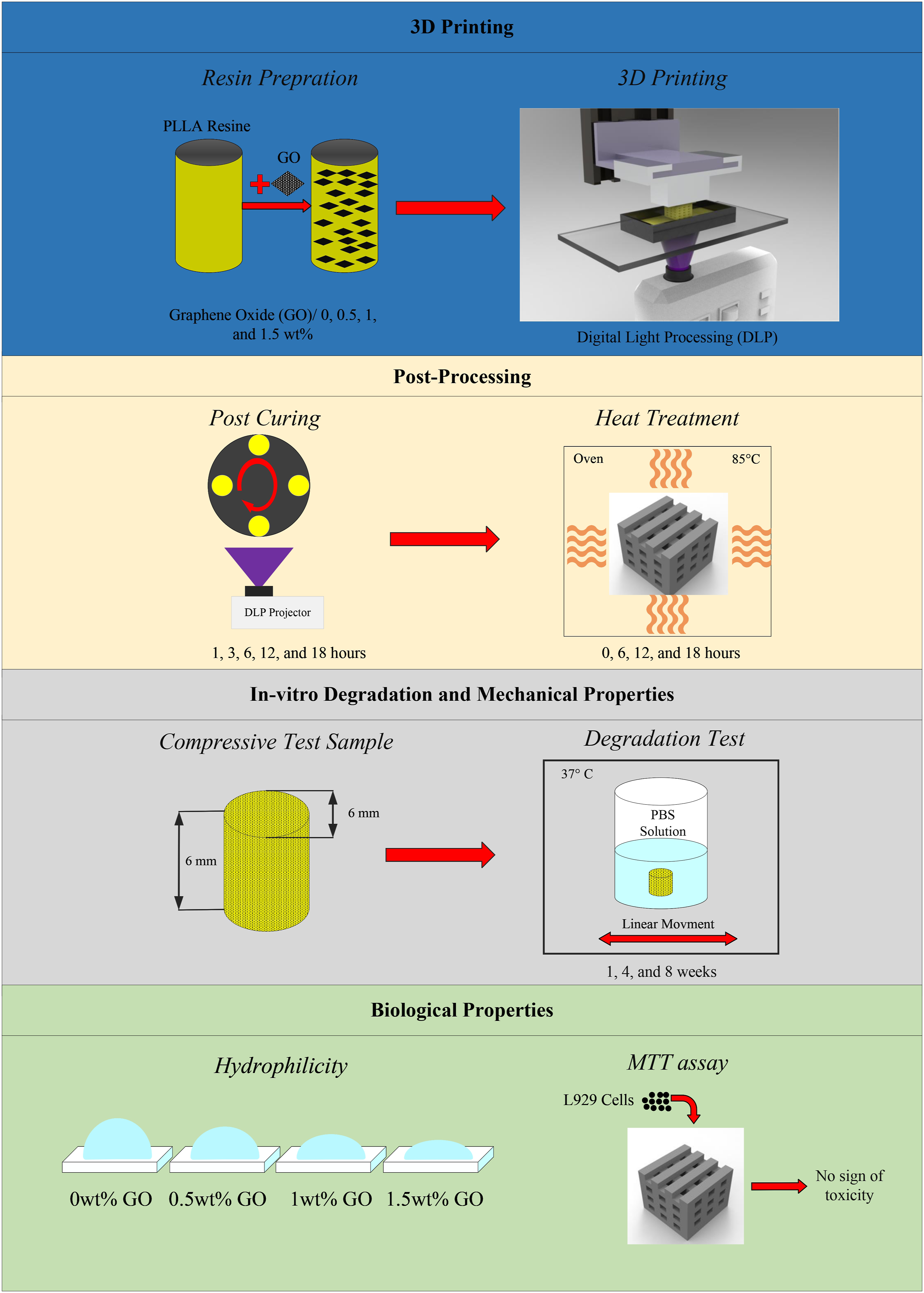
In this study, 3D printing of poly-l-lactic acid (PLLA) scaffolds reinforced with graphene oxide (GO) nanoparticles via Digital Light Processing (DLP) was investigated to mimic bone tissue. Stereolithography is one of the most accurate additive manufacturing methods, but the dominant available materials used in this method are toxic. In this research, a biocompatible resin (PLLA) was synthetized and functionalized to serve the purpose. Due to the low mechanical properties of the printed product with the neat resin, graphene oxide nanoparticles in three levels (0.5, 1, and 1.5 wt%) were added with the aim of enhancing the mechanical properties. At first, the optimum post cure time of the neat resin was investigated. Consequently, all the parts were post-cured for 3 h after printing. Due to the temperature-dependent structure of GO, all samples were placed in an oven at 85°C for different time periods of 0, 6, 12, and 18 h to increase mechanical properties. The compression test of heat-treated samples reveals that the compressive strength of the printed parts containing 0.5,1, and 1.5% of GO increased by 151,162 ad 235%, respectively. Scaffolds with the designed pore sizes of 750 microns and a porosity of 40% were printed. Surface hydrophilicity test was performed for all samples showing that the hydrophilicity of the samples increased with increasing GO percentage. The degradation behavior of the samples was evaluated in a PBS environment, and it revealed that by increasing GO, the rate of component degradation increased, but the heat treatment had the opposite effect and decreased the degradation rate. Finally, besides improving biological properties, a significant increase in mechanical properties under compression can introduce the printed scaffolds as a suitable option for bone implants.
BoseSRoyMBandyopadhyayA. Recent advances in bone tissue engineering scaffolds. Trends Biotechnol2012; 30(10): 546–554.
2.
PintoP. Therapeutic strategies for bone regeneration: the importance of biomaterials testing in adequate animal models. Adv Compos Mater2016: 275–319.
3.
AgarwalRGarcíaAJ. Biomaterial strategies for engineering implants for enhanced osseointegration and bone repair. Adv Drug Deliv Rev2015; 94: 53–62.
4.
FillinghamYJacobsJ. Bone grafts and their substitutes. The Bone and Joint Journal2016; 98(1_Supple_A): 6–9.
5.
Florencio-SilvaRSassoGRSSasso-CerriE, et al. Biology of bone tissue: structure, function, and factors that influence bone cells. BioMed Res Int. 2015; 2015: 421746.
6.
MarenzanaMArnettTR. The key role of the blood supply to bone. Bone research2013; 1(1): 203–215.
7.
GruskinEDollBAFutrellFW, et al. Demineralized bone matrix in bone repair: history and use. Adv Drug Deliv Rev. 2012; 64(12): 1063–1077.
8.
ShegarfiHReikerasO. Bone transplantation and immune response. J Orthop Surg2009; 17(2): 206–211.
9.
BouëtGMarchatDCruelM, et al. In vitro three-dimensional bone tissue models: from cells to controlled and dynamic environment. Tissue Eng B Rev. 2015; 21(1): 133–156.
10.
HenkelJWoodruffMAEpariDR, et al. Bone regeneration based on tissue engineering conceptions—a 21st century perspective. Bone research. 2013; 1(1): 216–248.
11.
Abou NeelEAChrzanowskiWSalihVM, et al. Tissue engineering in dentistry. J Dent. 2014; 42(8): 915–928.
12.
AravamudhanARamosDMNadaAA, et al. Natural and synthetic biomedical polymers. The Netherlands: Elsevier Amsterdam, 2014.
13.
ChanBLeongK. Scaffolding in tissue engineering: general approaches and tissue-specific considerations. Eur Spine J2008; 17(4): 467–479.
14.
O'brienFJ. Biomaterials & scaffolds for tissue engineering. Mater Today2011; 14(3): 88–95.
15.
Gil-CastellOBadiaJDOntoria-OviedoI, et al. In vitro validation of biomedical polyester-based scaffolds: Poly (lactide-co-glycolide) as model-case. Polym Test. 2018; 66: 256–267.
16.
ThavornyutikarnBChantarapanichNSitthiseripratipK, et al. Bone tissue engineering scaffolding: computer-aided scaffolding techniques. Progress in biomaterials. 2014; 3(2–4): 61–102.
17.
CaoHKuboyamaN. A biodegradable porous composite scaffold of PGA/β-TCP for bone tissue engineering. Bone2010; 46(2): 386–395.
18.
BajajPSchwellerRMKhademhosseiniA, et al. 3D biofabrication strategies for tissue engineering and regenerative medicine. Annu Rev Biomed Eng. 2014; 16: 247–276.
19.
TorabiKFarjoodEHamedaniS. Rapid prototyping technologies and their applications in prosthodontics, a review of literature. J Dent2015; 16(1): 1.
20.
HedayatiSKBehraveshAHHasanniaS, et al. 3D printed PCL scaffold reinforced with continuous biodegradable fiber yarn: A study on mechanical and cell viability properties. Polym Test. 2020; 83: 106347.
21.
RosetiLParisiVPetrettaM, et al. Scaffolds for bone tissue engineering: state of the art and new perspectives. Mater Sci Eng C. 2017; 78: 1246–1262.
22.
BhattacharjeeNUrriosAKangS, et al. The upcoming 3D-printing revolution in microfluidics. Lab Chip. 2016; 16(10): 1720–1742.
23.
FiedlerBGojnyFHWichmannMHG, et al. Fundamental aspects of nano-reinforced composites. Compos Sci Technol. 2006; 66(16): 3115–3125.
24.
LeeCWeiXKysarJW, et al. Measurement of the elastic properties and intrinsic strength of monolayer graphene. Science. 2008; 321(5887): 385–388.
25.
TerronesMMartínOGonzálezM, et al. Interphases in graphene polymer‐based nanocomposites: achievements and challenges. Wiley Online Library, 2011.
26.
LinD. A laser sintered layer of metal matrix consisting of 0D, 1D and 2D nanomaterials and its mechanical behaviors. Purdue University, 2013.
27.
DasariBLMorshedMNouriJM, et al. Mechanical properties of graphene oxide reinforced aluminium matrix composites. Compos B Eng. 2018; 145: 136–144.
28.
SaedABBehraveshAHHasanniaS, et al. Functionalized poly l-lactic acid synthesis and optimization of process parameters for 3D printing of porous scaffolds via digital light processing (DLP) method. J Manuf Process. 2020; 56: 550–561.
29.
SaedABBehraveshAHHasanniaS, et al. An in vitro study on the key features of Poly L-lactic acid/biphasic calcium phosphate scaffolds fabricated via DLP 3D printing for bone grafting. Eur Polym J. 2020; 141: 110057.
30.
KarageorgiouVKaplanD. Porosity of 3D biomaterial scaffolds and osteogenesis. Biomaterials2005; 26(27): 5474–5491.
31.
HedayatiSKBehraveshAHHasanniaS, et al. Additive manufacture of PCL/nHA scaffolds reinforced with biodegradable continuous Fibers: Mechanical Properties, in-vitro degradation Profile, and cell study. Eur Polym J. 2022; 162: 110876.
32.
ManapatJZMangadlaoJDTiuBDB, et al. High-strength stereolithographic 3D printed nanocomposites: graphene oxide metastability. ACS Appl Mater Interfaces. 2017; 9(11): 10085–10093.
KaniyoorABabyTTRamaprabhuS. Graphene synthesis via hydrogen induced low temperature exfoliation of graphite oxide. J Mater Chem2010; 20(39): 8467–8469.
35.
GengL-HPengXFJingX, et al. Investigation of poly (L-lactic acid)/graphene oxide composites crystallization and nanopore foaming behaviors via supercritical carbon dioxide low temperature foaming. J Mater Res. 2016; 31(3): 348–359.
36.
ShuaiCLiYYangW, et al. Graphene oxide induces ester bonds hydrolysis of poly-l-lactic acid scaffold to accelerate degradation. International Journal of Bioprinting. 2020; 6(1).
37.
EckhartKEHoltBDLaurencinMG, et al. Covalent conjugation of bioactive peptides to graphene oxide for biomedical applications. Biomater Sci. 2019; 7(9): 3876–3885.
38.
SalvoPMelaiBCalisiN, et al. Graphene-based devices for measuring pH. Sensor Actuator B Chem. 2018; 256: 976–991.