
Abstract

Introduction
Dual-energy subtraction relies on the differential attenuation of low- and high-energy photons by body tissues, enabling enhanced imaging to selectively highlight bones and soft tissues. Historically, the application of dual-energy X-ray imaging has been constrained by heightened radiation exposure and motion artefacts stemming from the necessity for multiple exposures during image acquisition. 1 However, the emergence of portable, single-exposure dual-energy X-ray imaging has mitigated these concerns. The visibility of lines and tubes on dual-energy radiography is improved due to their naturally higher density, which becomes more apparent when the surrounding soft tissues are subtracted in the bone image. This study aims to evaluate the augmented diagnostic confidence in a non-radiological review setting of portable dual-energy chest X-ray compared to conventional radiography, particularly focusing on the visibility of the tip of lines/tubes, and overall confidence among diverse medical professionals.
Methods
A portable, single-exposure, dual-energy X-ray triple-layer detector (SpectralDR™, KA Imaging, Waterloo, Ontario, Canada) was employed, enabling simultaneous acquisition of a conventional radiograph, a soft tissue image, and a bone image. Twenty-eight consecutive chest X-rays were obtained in an intensive care unit using the portable dual-energy detector (exposure: 120 kVp and 2.0-3.6 mAs), following institutional research ethics board approval and participant consent. PICC lines were present in 10 patients (Navilyst, AngioDynamics), a port system (BARD) in 2, and an additional tunnelled jugular catheter (Cardinal Health) in 2 patients. Images underwent pre-processing to remove annotations and anonymize cases before distribution. A web-based survey-style review involving 9 medical professionals and trainees with varying expertise levels was conducted, encompassing one medical student, 4 residents, one fellow, and 3 chest radiologists. Reviewers initially assessed cases using only the conventional radiograph, grading the likelihood of pneumonia, pneumothorax, bone fractures, pulmonary nodules, and the visibility of the tip of lines/tubes on a 5-point scale. After a 1-week washout period, reviewers re-evaluated cases using the same scale after reviewing additional soft tissue and bone images, with no clinical history provided. Reviewers reported the presence of motion artefacts and indicated whether their diagnostic confidence increased with the additional dual-energy X-ray images, with review time recorded.
Results
While all reviewers identified the tip of lines/tubes on the composite image in all 14 cases, 8 out of 9 reviewers reported improved visibility (P < .001). Figure 1 demonstrates an example of increased catheter visibility. Notably, in 16 out of 28 cases (57%), the reviewers reported an increased overall diagnostic confidence when dual-energy X-ray images were available for interpretation. No motion artefacts were noted in the acquired images. The median total reading time for the composite and dual-energy images showed no significant difference (median, 29:07 vs 29:52 minutes, P = .32).
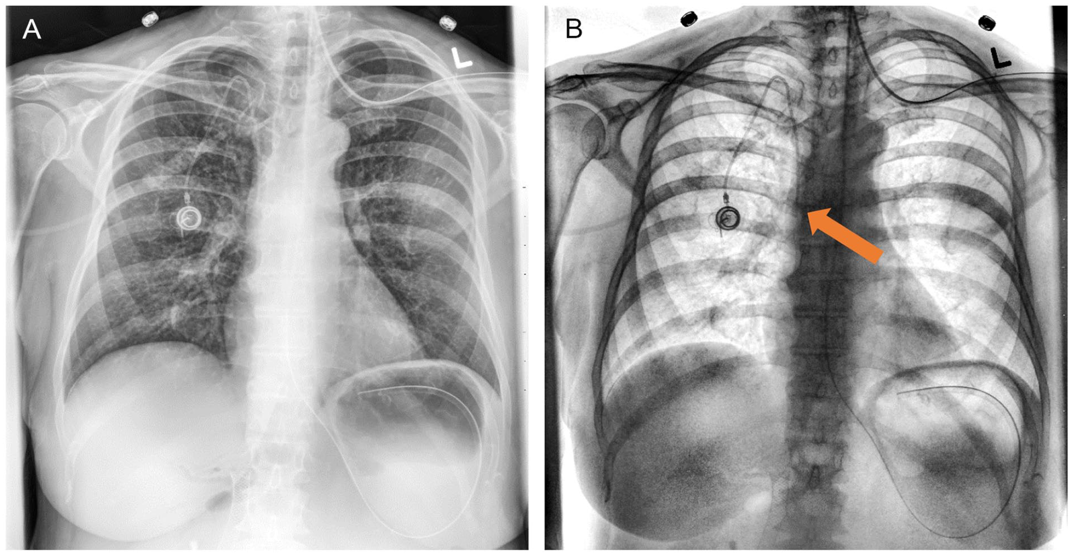
Case demonstrating a conventional radiograph (composite image) with the catheter tip of the port system not clearly visualized (A). The bone image obtained from the single-exposure dual-energy X-ray detector increases the conspicuity of the catheter tip (B, arrow).
Discussion
The enhanced visibility of the tip of lines/tubes and improved self-reported diagnostic confidence with dual-energy images underscore the clinical promise of portable dual-energy chest X-rays. Previous studies have also noted increased accuracy and interpretation speed for lung nodule identification with dual-energy subtraction. 2 Additionally, dual-energy X-ray imaging has been demonstrated to have a variety of clinical use cases beyond identification of lines, tubes, and pulmonary nodules, including identification of calcifications and vascular diseases, and bone, pleural, and chest wall abnormalities.3-5 With single-exposure dual-energy X-ray detectors eliminating motion artefacts and reducing radiation doses, the potential for widespread adoption of dual-energy X-ray imaging has been re-opened. Consistent with previous findings, this study demonstrates improved clinical sensitivity and reader confidence with dual-energy X-ray images. 6 Our study acknowledges limitations, including a small sample size and inclusion of only chest X-rays. The results may not be generalizable to imaging of other body parts, and the diagnostic confidence outcome in this study used a subjective self-reported measure.
In conclusion, portable, single-exposure, dual-energy X-ray technology offers opportunities to enhance chest X-ray capabilities across diverse clinical settings. Future research should explore its broader applications and potential impact on patient outcomes.
Footnotes
Declaration of Conflicting Interests
The author(s) declared the following potential conflicts of interest with respect to the research, authorship, and/or publication of this article: Dr. Patrik Rogalla holds a research grant from Ontario Bioscience Innovation Organization, Dual-Energy X-ray. Drs. Jay Potipcoe, Nikhil S. Patil, and Karim S. Karim are employees at KA Imaging Inc.
Funding
The author(s) received no financial support for the research, authorship, and/or publication of this article.