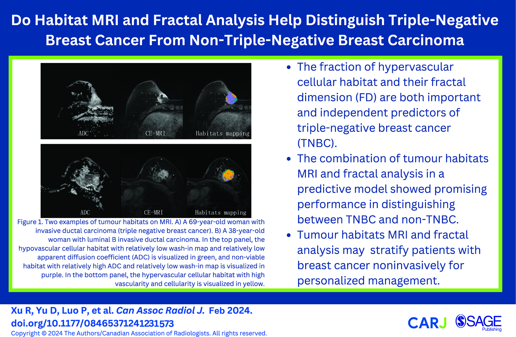
Purpose: To determine whether multiparametric MRI-based spatial habitats and fractal analysis can help distinguish triple-negative breast cancer (TNBC) from non-TNBC. Method: Multiparametric DWI and DCE-MRI at 3T were obtained from 142 biopsy- and surgery-proven breast cancer with 148 breast lesions (TNBC = 26 and non-TNBC = 122). The contrast-enhancing lesions were divided into 3 spatial habitats based on perfusion and diffusion patterns using K-means clustering. The fractal dimension (FD) of the tumour subregions was calculated. The accuracy of the habitat segmentation was measured using the Dice index. Inter- and intra-reader reliability were evaluated with the intraclass correlation coefficient (ICC). The ability to predict TNBC status was assessed using the receiver operating characteristic curve. Results: The Dice index for the whole tumour was 0.81 for inter-reader and 0.88 for intra-reader reliability. The inter- and intra-reader reliability were excellent for all 3 tumour habitats and fractal features (ICC > 0.9). TNBC had a lower hypervascular cellular habitat and higher FD 1 compared to non-TNBC (all P < .001). Multivariate analysis confirmed that hypervascular cellular habitat (OR = 0.88) and FD 1 (OR = 1.35) were independently associated with TNBC (all P < .001) after adjusting for rim enhancement, axillary lymph nodes status, and histological grade. The diagnostic model combining hypervascular cellular habitat and FD 1 showed excellent discriminatory ability for TNBC, with an AUC of 0.951 and an accuracy of 91.9%. Conclusions: The fraction of hypervascular cellular habitat and its FD may serve as useful imaging biomarkers for predicting TNBC status.
WuJCaoGSunX, et al. Intratumoral spatial heterogeneity at perfusion MR imaging predicts recurrence-free survival in locally advanced breast cancer treated with neoadjuvant chemotherapy. Radiology. 2018;288(1):26-35. doi:10.1148/radiol.2018172462
2.
GotoMLe BihanDSakaiKYamadaK. The diffusion MRI signature index is highly correlated with immunohistochemical status and molecular subtype of invasive breast carcinoma. Eur Radiol. 2022;32(7):4879-4888. doi:10.1007/s00330-022-08562-4
3.
AbramsonVGLehmannBDBallingerTJPietenpolJA. Subtyping of triple-negative breast cancer: implications for therapy. Cancer. 2015;121(1):8-16. doi:10.1002/cncr.28914
4.
SuttonEJOhJHDashevskyBZ, et al. Breast cancer subtype intertumor heterogeneity: MRI-based features predict results of a genomic assay. J Magn Reson Imaging. 2015;42(5):1398-1406. doi:10.1002/jmri.24890
5.
GilliesRJBalagurunathanY. Perfusion MR imaging of breast cancer: insights using “habitat imaging”. Radiology. 2018;288(1):36-37. doi:10.1148/radiol.2018180271
6.
LeeDHParkJEKimN, et al. Tumor habitat analysis by magnetic resonance imaging distinguishes tumor progression from radiation necrosis in brain metastases after stereotactic radiosurgery. Eur Radiol. 2022;32(1):497-507. doi:10.1007/s00330-021-08204-1
7.
Juan-AlbarracinJFuster-GarciaEPerez-GirbesA, et al. Glioblastoma: vascular habitats detected at preoperative dynamic susceptibility-weighted contrast-enhanced perfusion MR imaging predict survival. Radiology. 2018;287(3):944-954. doi:10.1148/radiol.2017170845
8.
WuHTongHDuX, et al. Vascular habitat analysis based on dynamic susceptibility contrast perfusion MRI predicts IDH mutation status and prognosis in high-grade gliomas. Eur Radiol. 2020;30(6):3254-3265. doi:10.1007/s00330-020-06702-2
9.
KazerouniASHormuthDA2ndDavisT, et al. Quantifying tumor heterogeneity via MRI habitats to characterize microenvironmental alterations in HER2+ breast cancer. Cancers (Basel). 2022;14(7):1837. doi:10.3390/cancers14071837
10.
ParkJEKimHSKimN, et al. Low conductivity on electrical properties tomography demonstrates unique tumor habitats indicating progression in glioblastoma. Eur Radiol. 2021;31(9):6655-6665. doi:10.1007/s00330-021-07976-w
11.
SyedAKWhisenantJGBarnesSLSoraceAGYankeelovTE. Multiparametric analysis of longitudinal quantitative MRI data to identify distinct tumor habitats in preclinical models of breast cancer. Cancers (Basel). 2020;12(6):1682. doi:10.3390/cancers12061682
12.
ParkYWKimSAhnSS, et al. Magnetic resonance imaging-based 3-dimensional fractal dimension and lacunarity analyses may predict the meningioma grade. Eur Radiol. 2020;30(8):4615-4622. doi:10.1007/s00330-020-06788-8
13.
LiuSFanXZhangC, et al. MR imaging based fractal analysis for differentiating primary CNS lymphoma and glioblastoma. Eur Radiol. 2019;29(3):1348-1354. doi:10.1007/s00330-018-5658-x
14.
SmithaKAGuptaAKJayasreeRS. Fractal analysis: fractal dimension and lacunarity from MR images for differentiating the grades of glioma. Phys Med Biol. 2015;60(17):6937-6947. doi:10.1088/0031-9155/60/17/6937
15.
MatsudaMTsudaTEbiharaR, et al. Triple-negative breast cancer on contrast-enhanced MRI and synthetic MRI: a comparison with non-triple-negative breast carcinoma. Eur J Radiol. 2021;142:109838. doi:10.1016/j.ejrad.2021.109838
16.
DuSGaoSZhangLYangXQiXLiS. Improved discrimination of molecular subtypes in invasive breast cancer: comparison of multiple quantitative parameters from breast MRI. Magn Reson Imaging. 2021;77:148-158. doi:10.1016/j.mri.2020.12.001
17.
ChoHHKimHNamSY, et al. Measurement of perfusion heterogeneity within tumor habitats on magnetic resonance imaging and its association with prognosis in breast cancer patients. Cancers (Basel). 2022;14(8):1858. doi:10.3390/cancers14081858
18.
Sales BarrosRTolhuisenMLBoersAM, et al. Automatic segmentation of cerebral infarcts in follow-up computed tomography images with convolutional neural networks. J Neurointerv Surg. 2020;12(9):848-852. doi:10.1136/neurintsurg-2019-015471
19.
PedregosaFVaroquauxGGramfortA, et al. Scikit-learn: machine learning in Python. J Mach Learn Res. 2011;12:2825-2830.
20.
KanungoTMountDMNetanyahuNSPiatkoCDSilvermanRWuAY. An efficient k-means clustering algorithm: analysis and implementation. IEEE Trans Pattern Anal Mach Intell. 2002;24(7):881-892. doi:10.1109/TPAMI.2002.1017616
21.
MaJWangRYuYXuXDuanHYuN. Is fractal dimension a reliable imaging biomarker for the quantitative classification of an intervertebral disk?Eur Spine J. 2020;29(5):1175-1180. doi:10.1007/s00586-020-06370-2
22.
OnaygilCKayaHUgurluMUAribalE. Diagnostic performance of diffusion tensor imaging parameters in breast cancer and correlation with the prognostic factors. J Magn Reson Imaging. 2017;45(3):660-672. doi:10.1002/jmri.25481
23.
PratAPinedaEAdamoB, et al. Clinical implications of the intrinsic molecular subtypes of breast cancer. Breast. 2015;24 Suppl 2:S26-S35. doi:10.1016/j.breast.2015.07.008
24.
ParkJEKimHSKimNParkSYKimYHKimJH. Spatiotemporal heterogeneity in multiparametric physiologic MRI is associated with patient outcomes in IDH-wildtype glioblastoma. Clin Cancer Res. 2021;27(1):237-245. doi:10.1158/1078-0432.CCR-20-2156
25.
NagasakaKSatakeHIshigakiSKawaiHNaganawaS. Histogram analysis of quantitative pharmacokinetic parameters on DCE-MRI: correlations with prognostic factors and molecular subtypes in breast cancer. Breast Cancer. 2019;26(1):113-124. doi:10.1007/s12282-018-0899-8
26.
KimJJKimJYKangHJ, et al. Computer-aided diagnosis-generated kinetic features of breast cancer at preoperative MR imaging: association with disease-free survival of patients with primary operable invasive breast cancer. Radiology. 2017;284(1):45-54. doi:10.1148/radiol.2017162079
27.
Jardim-PerassiBVHuangSDominguez-ViqueiraW, et al. Multiparametric MRI and coregistered histology identify tumor habitats in breast cancer mouse models. Cancer Res. 2019;79(15):3952-3964. doi:10.1158/0008-5472.CAN-19-0213
28.
KimSYShinJKimDH, et al. Correlation between electrical conductivity and apparent diffusion coefficient in breast cancer: effect of necrosis on magnetic resonance imaging. Eur Radiol. 2018;28(8):3204-3214. doi:10.1007/s00330-017-5291-0
29.
MoffaGGalatiFCollalungaE, et al. Can MRI biomarkers predict triple-negative breast cancer?Diagnostics (Basel). 2020;10(12):1090. doi:10.3390/diagnostics10121090
30.
NamSYKoESLimY, et al. Preoperative dynamic breast magnetic resonance imaging kinetic features using computer-aided diagnosis: association with survival outcome and tumor aggressiveness in patients with invasive breast cancer. PLoS One. 2018;13(4):e0195756. doi:10.1371/journal.pone.0195756
31.
KalaCAtharMKalaSKhanLJauhariRKSatsangiA. Clinical and cyto-morphological characterization of triple negative breast cancer. J Cytol. 2019;36(2):84-88. doi:10.4103/joc.Joc_47_18
32.
SungHWieseDJatoiIJemalA. State variation in racial and ethnic disparities in incidence of triple-negative breast cancer among US women. JAMA Oncol. 2023;9(5):700-704. doi:10.1001/jamaoncol.2022.7835
Supplementary Material
Please find the following supplemental material available below.
For Open Access articles published under a Creative Commons License, all supplemental material carries the same license as the article it is associated with.
For non-Open Access articles published, all supplemental material carries a non-exclusive license, and permission requests for re-use of supplemental material or any part of supplemental material shall be sent directly to the copyright owner as specified in the copyright notice associated with the article.