
Abstract
The Canadian Association of Radiologists (CAR) Head and Neck Expert Panel consists of radiologists, a laryngologist and laryngeal surgeon, a patient advisor, and an epidemiologist/guideline methodologist. After developing a list of 11 clinical/diagnostic scenarios, a systematic rapid scoping review was undertaken to identify systematically produced referral guidelines that provide recommendations for one or more of these clinical/diagnostic scenarios. Recommendations from 17 guidelines and contextualization criteria in the Grading of Recommendations, Assessment, Development, and Evaluations (GRADE) for guidelines framework were used to develop 26 recommendation statements across the 11 scenarios. This guideline presents the methods of development and the referral recommendations for sinus disease, tinnitus, thyroid and parathyroid disease, neck mass of unknown origin, acute sialadenitis, chronic salivary conditions, and temporomandibular joint dysfunction.
Introduction
Beginning in May 2022, an Expert Panel (EP) comprised of radiologists, a laryngologist and laryngeal surgeon, a patient advisor, and an epidemiologist/guideline methodologist met to develop a new set of recommendations specific to referral pathways for clinical scenarios related to the head and neck. Through discussion (via a virtual meeting) followed by offline communication, the EP developed a list of 11 clinical/diagnostic scenarios to be covered by this guideline. These recommendations are intended primarily for referring clinicians (eg, family physicians, specialty physicians, nurse practitioners); however, they may also be used by radiologists, individuals/patients, and patient representatives.
Our methods describing the guideline development process, including the rapid scoping review to identify the evidence base, has been published in CMAJ Open 1 and an editorial to this series of guideline publications is available in CARJ. 2 The application of well-established scoping review and rapid review guidance (JBI, 3 Cochrane Handbook, 4 Cochrane Rapid Review Methods Group 5 ) and guideline methodology (ie, Grading of Recommendations Assessment, Development, and Evaluation or GRADE6,7) were used to identify the evidence-base and to guide the Expert Panel in determining the strength and direction of the recommendations for each clinical scenario (Table 1). The quality of conduct and reporting of the included guidelines identified in the scoping review were evaluated with the AGREE-II checklist, 8 using a modified scoring system. In instances where guidelines were lacking, expert consensus was used to develop the recommendation. Contextualization to the Canadian health care system was considered for each recommendation, with discussion around the factors found in the Evidence to Decision framework in GRADE for guidelines (eg, balance of desirable and undesirable outcomes, values and preferences, resources implications). 7
Recommendation Text, Symbol, and Interpretation.
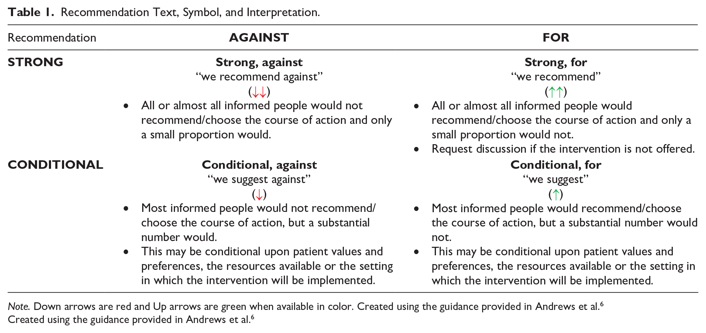
Note. Down arrows are red and Up arrows are green when available in color. Created using the guidance provided in Andrews et al. 6
Created using the guidance provided in Andrews et al. 6
A systematic search for guidelines (with an a priori defined inclusion criteria) was run in Medline and Embase on July 4, 2022. The search was limited to publications from 2016 onward (Supplemental Appendix 1). Supplemental searching included the following national radiology and/or guideline groups: the American College of Radiology, the National Institute for Health and Care Excellence, and the Royal College of Radiologists 8th Edition (2017). Recommendations for each clinical scenario were formulated during one in-person/virtual hybrid meeting in December 2022. External review and feedback were obtained from radiologists, a nuclear medicine radiologist, an emergency physician, a family physician, and a nurse practitioner. The full guideline can be found on the CAR website (www.car.ca).
Results
Systematic Scoping Review
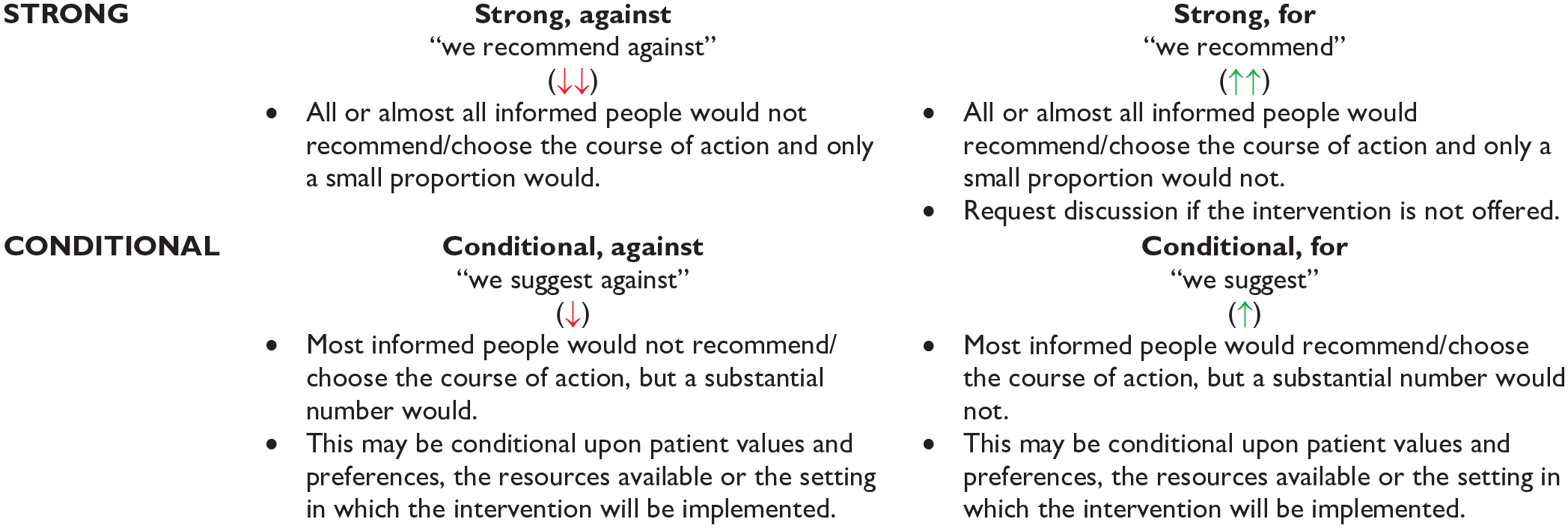
A total of 5214 records were identified through the electronic database and 6 additional records were added from the supplemental search. Seventeen guidelines, plus 6 companion papers, were included (Figure 1). Potentially relevant guidelines published in languages other than English can be found in Supplemental Appendix 2. A list of excluded records including justifications for exclusion is available upon request. Most guidelines were rated as moderate or high quality, using the modified AGREE-II checklist 8 (Supplemental Appendix 3). The number of guidelines included per clinical/diagnostic scenario ranged from 1 to 7, with a median of 2 guidelines per clinical scenario.
PRISMA flow diagram.
Recommendations
Additional details of the included guidelines, including which imaging modalities (eg, computed tomography [CT], magnetic resonance angiography [MRA], magnetic resonance imaging [MRI], nuclear medicine [NM], radiograph [XR], ultrasound [US]) that were discussed can be found in Supplemental Appendix 4.
A guideline is intended to guide and not be an absolute rule. Medical care is complex and should be based on evidence, a clinician’s expert judgment, the patient’s circumstances, values, preferences, and resource availability. Not all imaging modalities are available in all clinical environments, particularly in rural or remote areas of Canada. Decisions about patient transfer, use of alternative imaging or serial clinical examination and observation can be difficult. Therefore, the expected benefits of recommended imaging, risks of travel, patient preference, and other factors must be considered. The guideline recommendations are to assist the choice of imaging modality in situations where it is deemed clinically necessary to obtain imaging.
Recommendations do not specify when contrast should or should not be used, as this may vary based on clinical presentation, regional practice preferences, preference of the referring clinician, radiologist and the patient, and resource availability.
We reviewed relevant recommendations related to the 11 clinical/diagnostic scenarios previously published by radiology and specialty societies, including: the Canadian Association of Radiologists, 9 the American College of Radiology,10-15 the American Thyroid Association, 16 the European Thyroid Association, 17 the German Association of Endocrine Surgeons, 18 the International consensus statement on Allergy and Rhinology, 19 the Korean Society of Radiology/National Evidence-Based Healthcare Collaborating Agency, 20 the Korean Society of Thyroid Radiology, 21 the National Institute for Health and Care Excellence,22-27 the Neck Mass Guideline Development Group,28-30 and the Royal College of Radiologists. 31
Recommendations are presented in 2 tables: Sinus disease and tinnitus (Table 2), Thyroid and parathyroid disease, neck mass of unknown origin, acute sialadenitis, chronic salivary conditions, and temporomandibular joint dysfunction (Table 3).
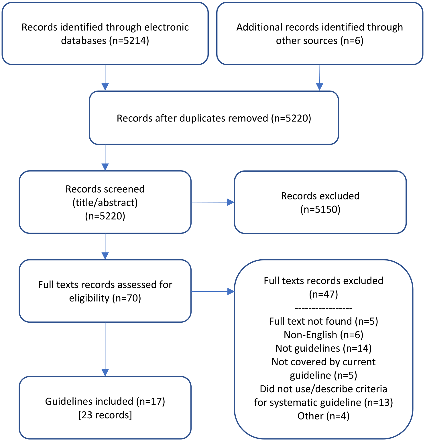
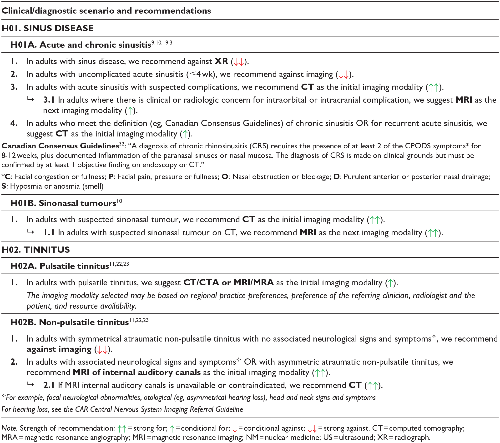
Sinus Disease and Tinnitus Recommendations.
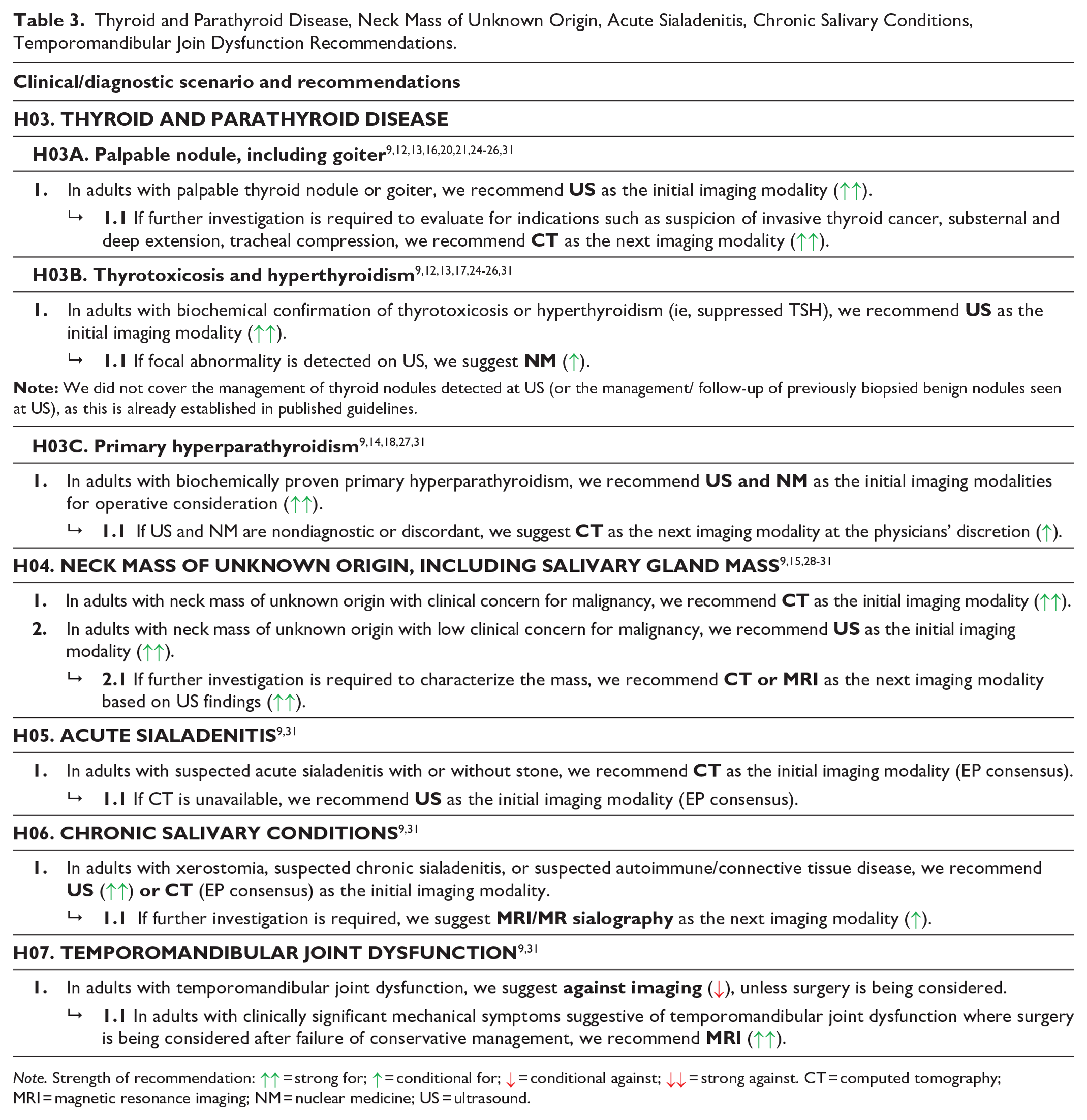
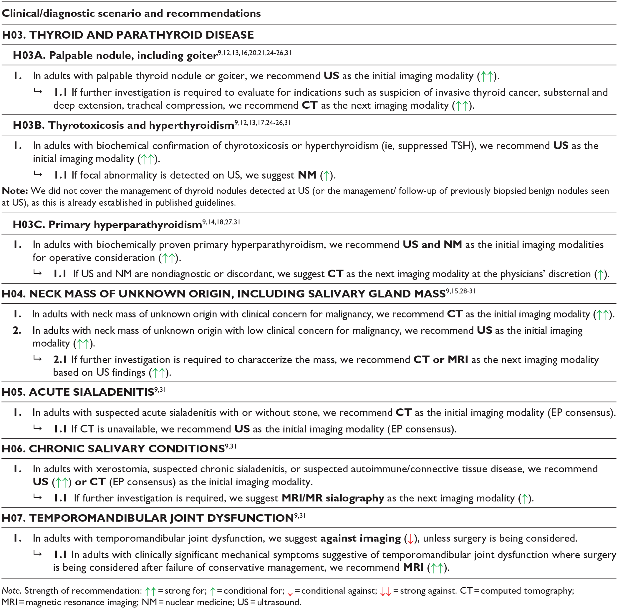
Thyroid and Parathyroid Disease, Neck Mass of Unknown Origin, Acute Sialadenitis, Chronic Salivary Conditions, Temporomandibular Join Dysfunction Recommendations.
Supplemental Material
sj-pdf-1-caj-10.1177_08465371231217212 – Supplemental material for Canadian Association of Radiologists Head and Neck Imaging Referral Guideline
Supplemental material, sj-pdf-1-caj-10.1177_08465371231217212 for Canadian Association of Radiologists Head and Neck Imaging Referral Guideline by Candyce Hamel, Barb Avard, Ross Campbell, Mario Kontolemos and Amanda Murphy in Canadian Association of Radiologists Journal
Footnotes
Acknowledgements
We would like to thank: Becky Skidmore for creating the search strategies for the systematic scoping review, Christopher Buckle who was a member of the Expert Panel, and the following individuals on the Diagnostic Imaging Referral Guidelines Working Group and external stakeholders for providing feedback on the guideline (listed alphabetically): Steve Burrell, Ryan Margau (WG co-chair), Cathy MacLean, Paul Pageau (WG co-chair), Charlotte Yong-Hing, and Kaitlin Zaki-Metias.
Declaration of Conflicting Interests
The author(s) declared no potential conflicts of interest with respect to the research, authorship, and/or publication of this article.
Funding
The author(s) disclosed receipt of the following financial support for the research, authorship, and/or publication of this article: This work was supported by the Canadian Medical Association.
Supplemental Material
Supplemental material for this article is available online.
References
Supplementary Material
Please find the following supplemental material available below.
For Open Access articles published under a Creative Commons License, all supplemental material carries the same license as the article it is associated with.
For non-Open Access articles published, all supplemental material carries a non-exclusive license, and permission requests for re-use of supplemental material or any part of supplemental material shall be sent directly to the copyright owner as specified in the copyright notice associated with the article.