
Abstract
The Canadian Association of Radiologists (CAR) Thoracic Expert Panel consists of radiologists, respirologists, emergency and family physicians, a patient advisor, and an epidemiologist/guideline methodologist. After developing a list of 24 clinical/diagnostic scenarios, a rapid scoping review was undertaken to identify systematically produced referral guidelines that provide recommendations for one or more of these clinical/diagnostic scenarios. Recommendations from 30 guidelines and contextualization criteria in the Grading of Recommendations, Assessment, Development, and Evaluations (GRADE) for guidelines framework were used to develop 48 recommendation statements across the 24 scenarios. This guideline presents the methods of development and the referral recommendations for screening/asymptomatic individuals, non-specific chest pain, hospital admission for non-thoracic conditions, long-term care admission, routine pre-operative imaging, post-interventional chest procedure, upper respiratory tract infection, acute exacerbation of asthma, acute exacerbation of chronic obstructive pulmonary disease, suspect pneumonia, pneumonia follow-up, immunosuppressed patient with respiratory symptoms/febrile neutropenia, chronic cough, suspected pneumothorax (non-traumatic), clinically suspected pleural effusion, hemoptysis, chronic dyspnea of non-cardiovascular origin, suspected interstitial lung disease, incidental lung nodule, suspected mediastinal lesion, suspected mediastinal lymphadenopathy, and elevated diaphragm on chest radiograph.
Introduction
Beginning in November 2022, an Expert Panel (EP) comprised of radiologists, respirologists, emergency and family physicians, a patient advisor, and an epidemiologist/guideline methodologist met to develop a new set of recommendations specific to referral pathways for conditions related to the thorax, including asymptomatic individuals, symptomatic patients, and other scenarios requiring imaging of the thorax. Through discussion (via a virtual meeting) followed by offline communication, the EP developed a list of 24 clinical/diagnostic scenarios to be covered by this guideline. These recommendations are intended primarily for referring clinicians (eg, family physicians, specialty physicians, nurse practitioners); however, they may also be used by radiologists, individuals/patients, and patient representatives.
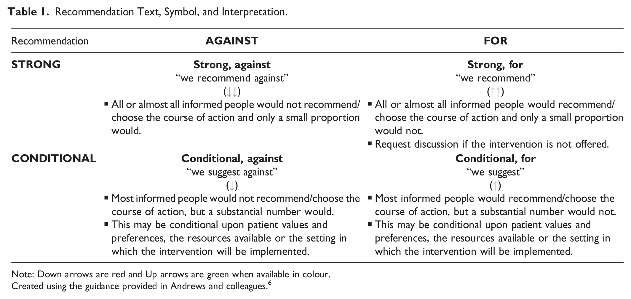
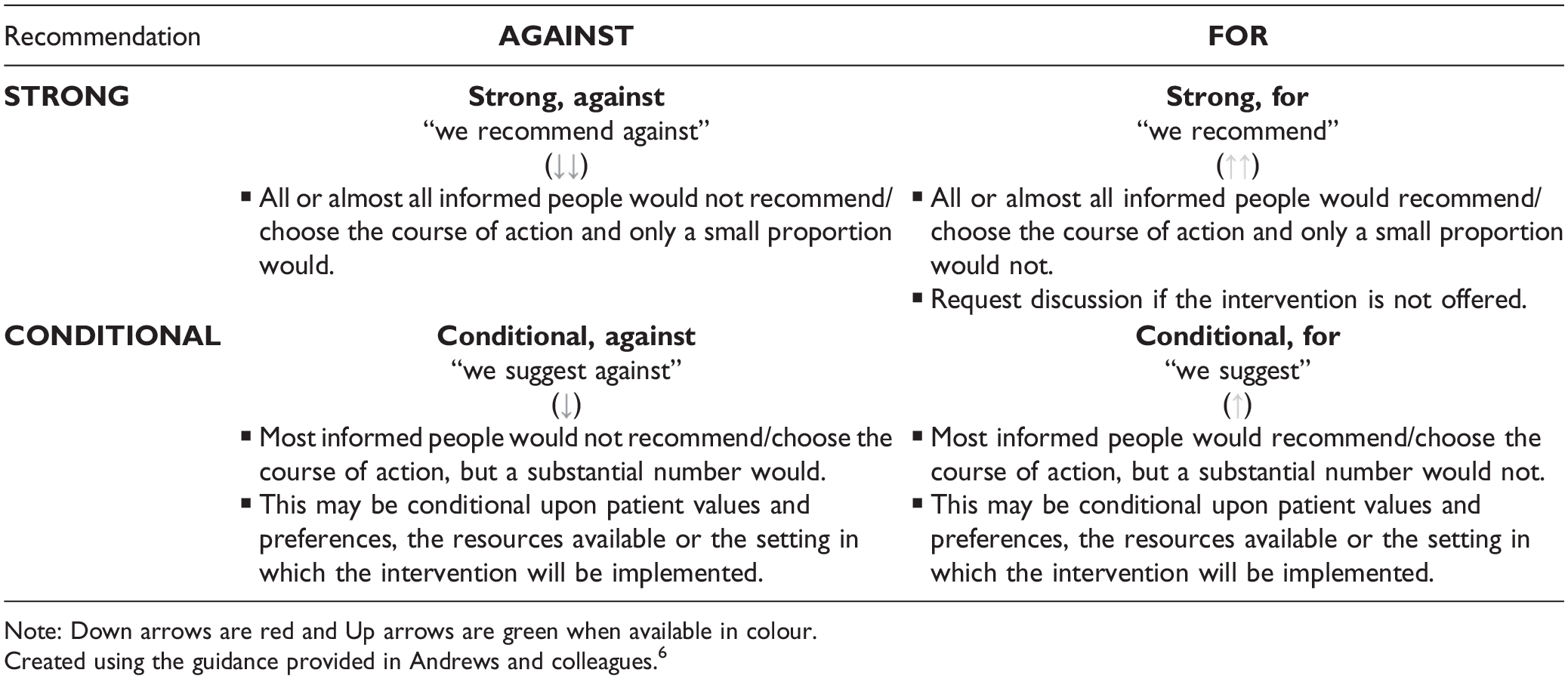
Our methods describing the guideline development process, including the rapid scoping review to identify the evidence base, has been published in CMAJ Open 1 and an editorial to this series of guideline publications is available in CARJ. 2 The application of well-established scoping review and rapid review guidance (JBI, 3 Cochrane Handbook, 4 Cochrane Rapid Review Methods Group 5 ) and guideline methodology (ie, Grading of Recommendations Assessment, Development, and Evaluation or GRADE6,7) were used to identify the evidence-base and to guide the Expert Panel in determining the strength and direction of the recommendations for each clinical scenario (Table 1). The quality of conduct and reporting of the included guidelines identified in the scoping review were evaluated with the AGREE-II checklist, 8 using a modified scoring system. In instances where guidelines were lacking, expert consensus was used to develop the recommendation. Contextualization to the Canadian health care system was considered for each recommendation, with discussion around the factors found in the Evidence to Decision framework in GRADE for guidelines (eg, balance of desirable and undesirable outcomes, values and preferences, resources implications). 7
Recommendation Text, Symbol, and Interpretation.
A systematic search for guidelines (with an a priori defined inclusion criteria) was run in Medline and Embase on January 5, 2023. The search was limited to publications from 2017 onward (Supplemental Appendix 1). Supplemental searching included the following national radiology and/or guideline groups: the American College of Radiology, the National Institute for Health and Care Excellence, and the Royal College of Radiologists 8th Edition (2017). Recommendations for each clinical scenario were formulated over 2 virtual meetings in April 2023. External review and feedback were obtained from radiologists, emergency and family physicians, and a nurse practitioner. The full guideline can be found on the CAR website (www.car.ca).
Results
Systematic Scoping Review
A total of 8479 records were identified through the electronic database and 7 additional records were added from the supplemental search. Thirty guidelines, plus 2 companion papers, were included (Figure 1). Potentially relevant guidelines published in languages other than English can be found in Supplemental Appendix 2. A list of excluded records with justifications for exclusion is available upon request. Most guidelines were rated as moderate or high quality, using the modified AGREE-II checklist 8 (Supplemental Appendix 3). The number of guidelines included per clinical/diagnostic scenario ranged from 0 to 10, with a median of 3 guidelines per clinical scenario.
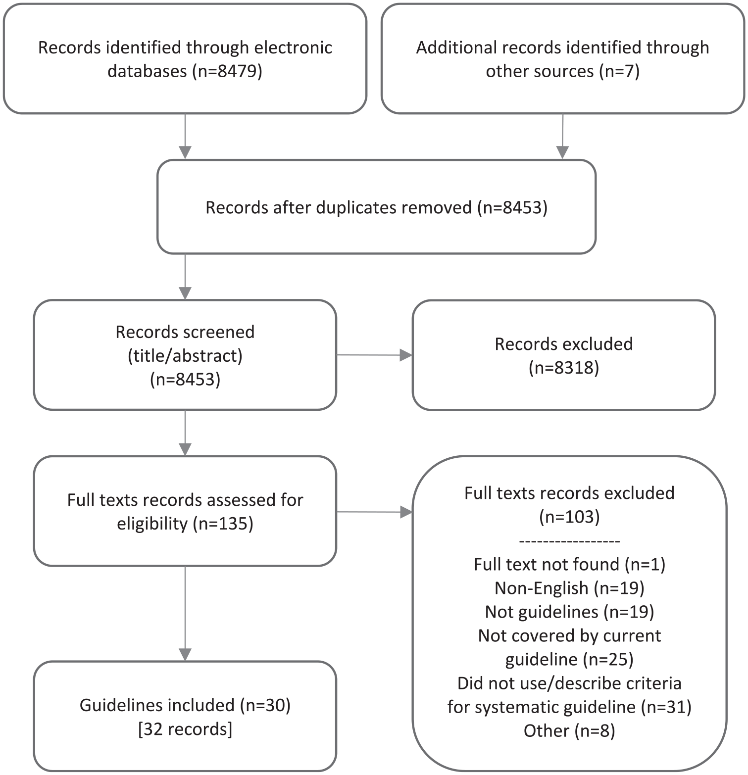
PRISMA flow diagram.
Recommendations
Additional details of the included guidelines, including which imaging modalities (eg, computed tomography [CT], magnetic resonance imaging [MRI], radiograph [XR], ultrasound [US]) that were discussed can be found in Supplemental Appendix 4.
A guideline is intended to guide and not be an absolute rule. Medical care is complex and should be based on evidence, a clinician’s expert judgment, the patient’s circumstances, values, preferences, and resource availability. Not all imaging modalities are available in all clinical environments, particularly in rural or remote areas of Canada. Decisions about patient transfer, use of alternative imaging or serial clinical examination and observation can be complex and difficult. Therefore, the expected benefits of recommended imaging, risks of travel, patient preference, and other factors must be considered. The guideline recommendations are designed to assist the choice of imaging modality in situations where it is deemed clinically necessary to obtain imaging.
Recommendations do not specify when contrast should or should not be used, as this may vary based on clinical presentation, regional practice preferences, preference of the referring clinician, radiologist and/or the patient, and resource availability.
We reviewed relevant recommendations related to the 24 clinical/diagnostic scenarios previously published by radiology and specialty societies, including: the Canadian Association of Radiologists, 9 the American College of Radiology,10-19 the American Thoracic Society and Infectious Diseases Society of America, 20 the CHEST Expert Panel,21-23 the Emergency Medicine Association of Turkey/Turkish Thoracic Society, 24 the European Respiratory Society, 25 the Fleischner Society,26,27 the French Language Pulmonology Society, 28 the German S3 guideline, 29 the Indian Chest Society National College of Chest Physicians, 30 the Indian Society of Anesthaesiologists, 31 the Italian intersociety consensus,32,33 the Korean guideline, 34 the National Institute for Health and Clinical Excellence, 35 the Polish recommendations for lung ultrasound in internal medicine, 36 the S2K guideline, 37 the combined guideline by the Société Française de Médecine d’Urgence, the Société de Réanimation de Langue Française and the French Group for Pediatric Intensive Care and Emergencies, 38 the Spanish Society of Medical Oncology, 39 and the Royal College of Radiologists. 40
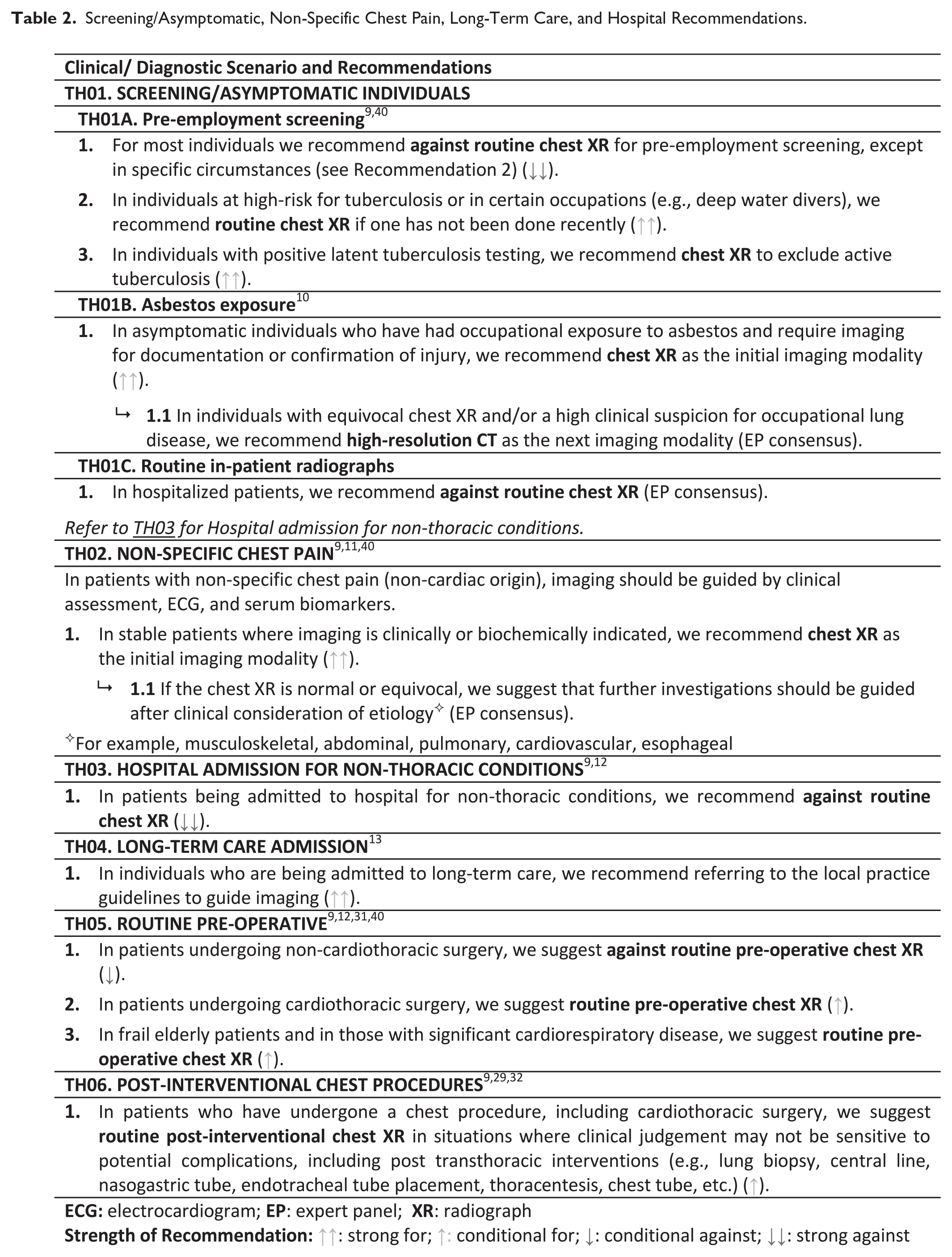
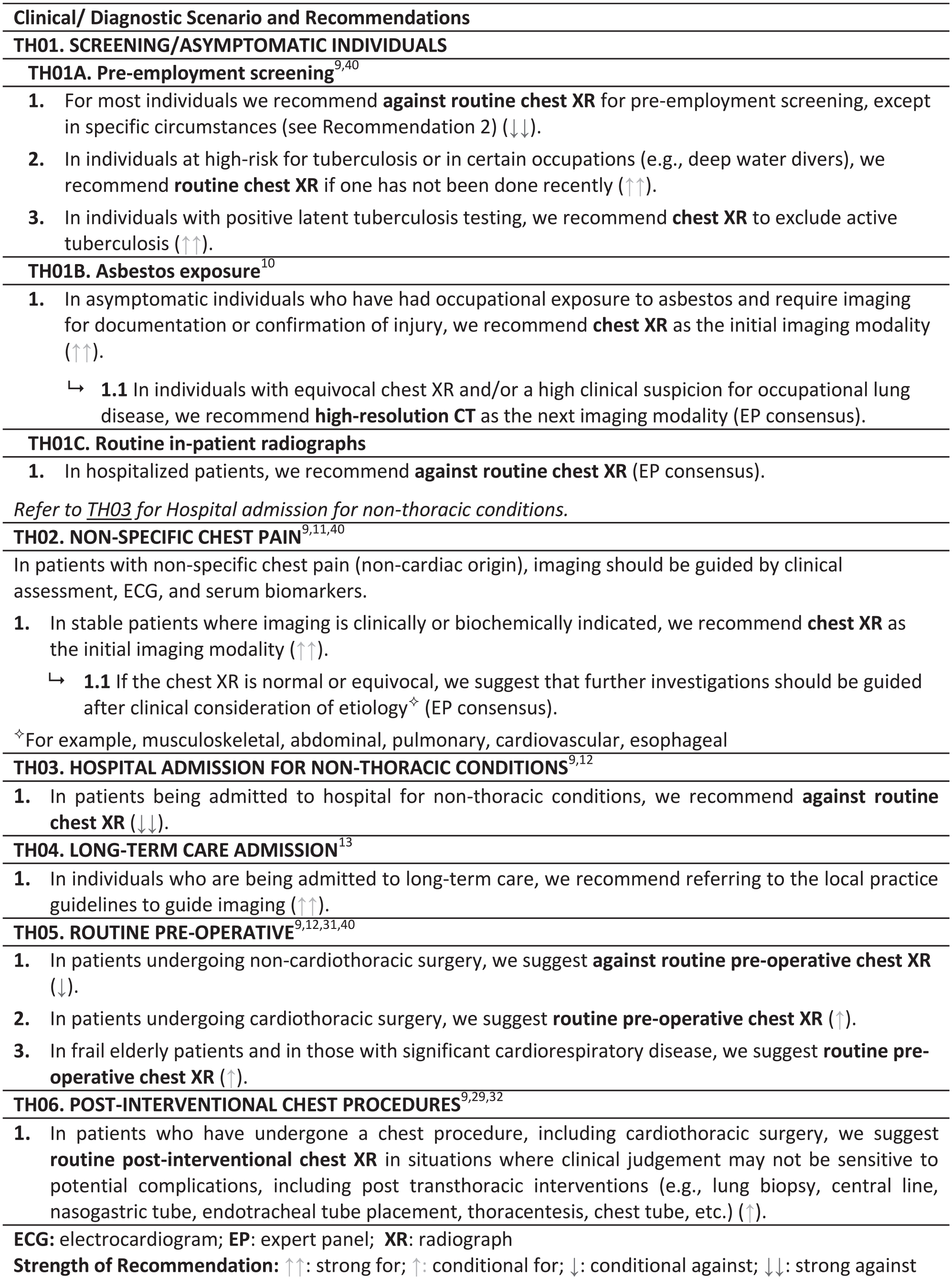
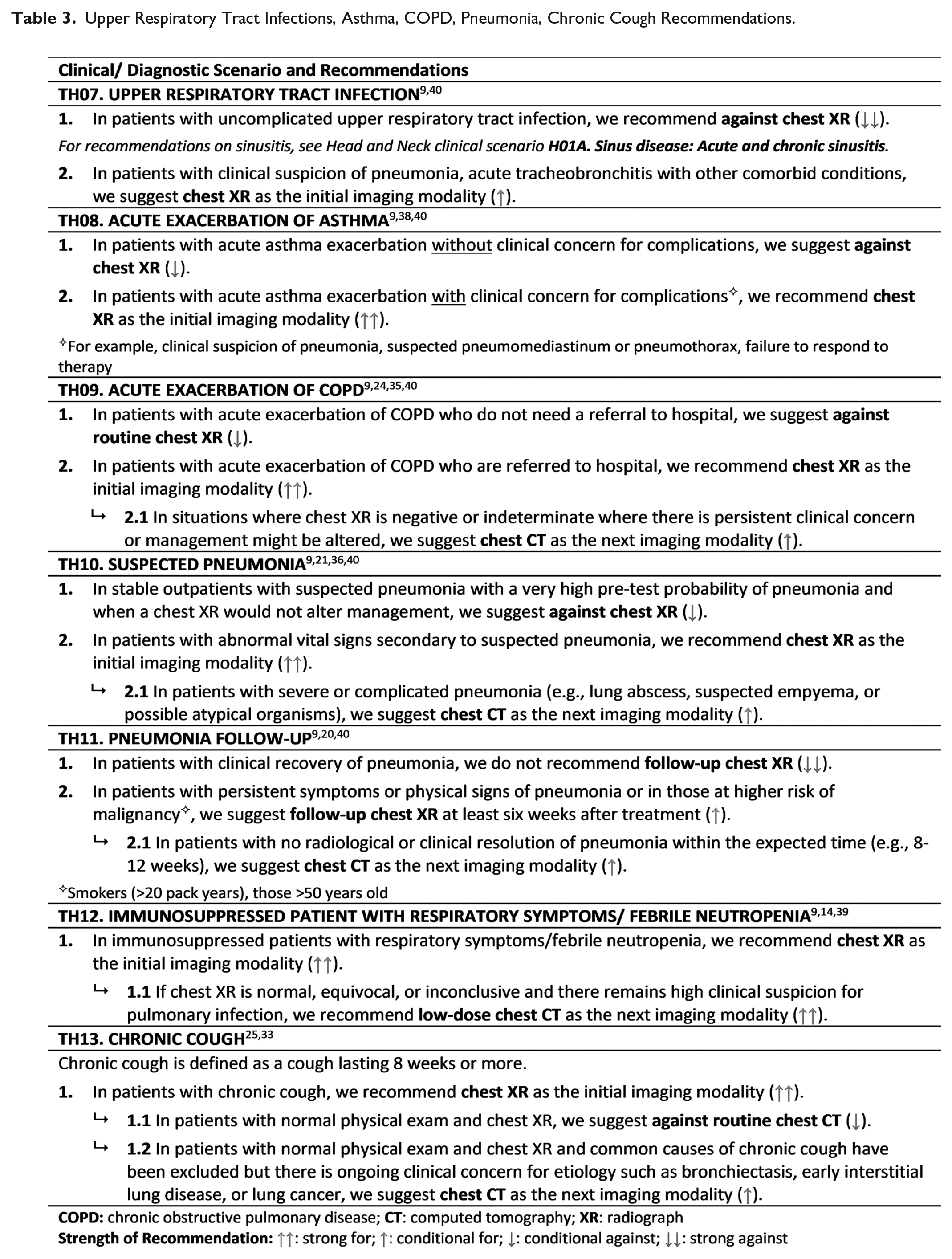
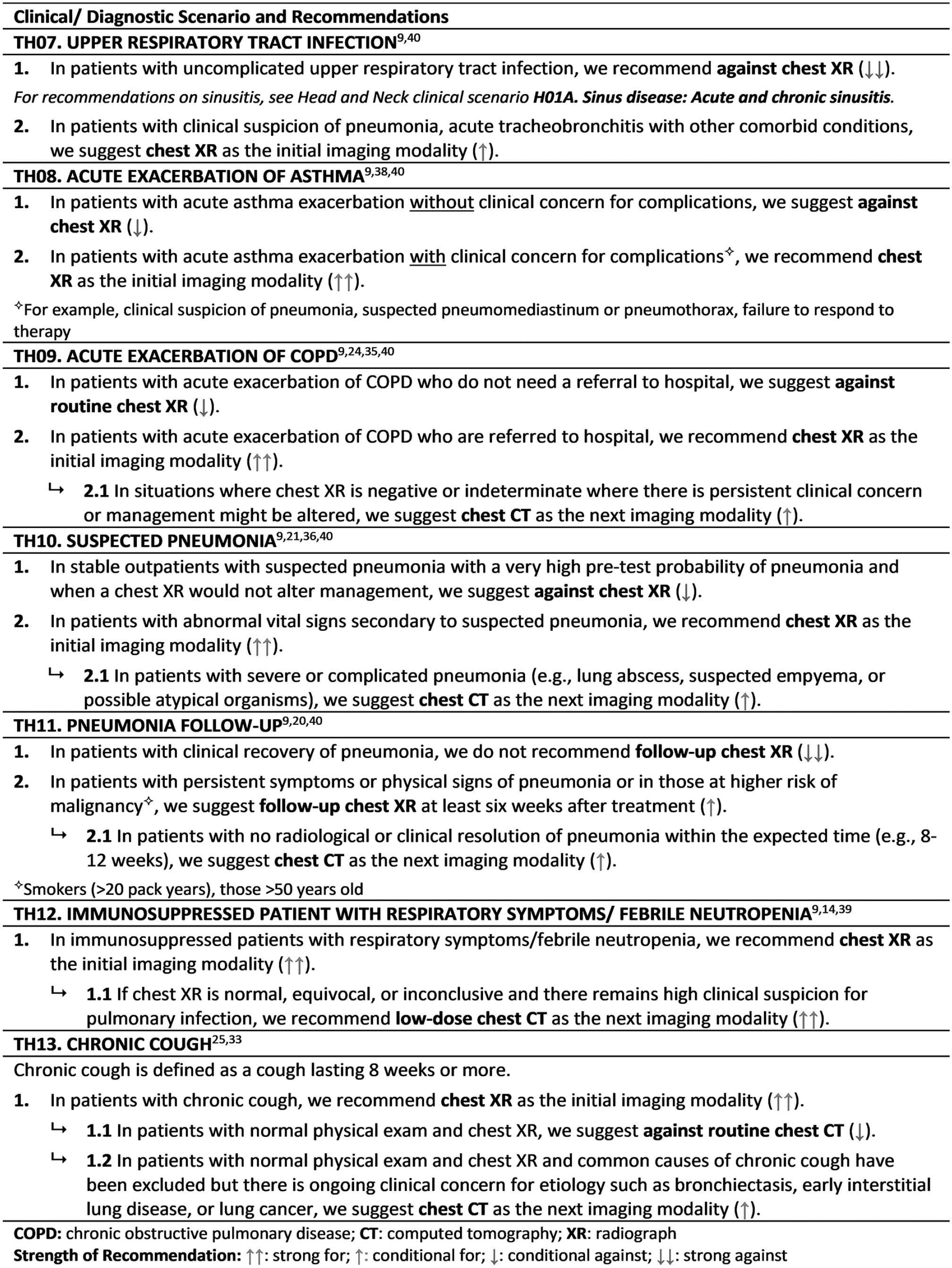
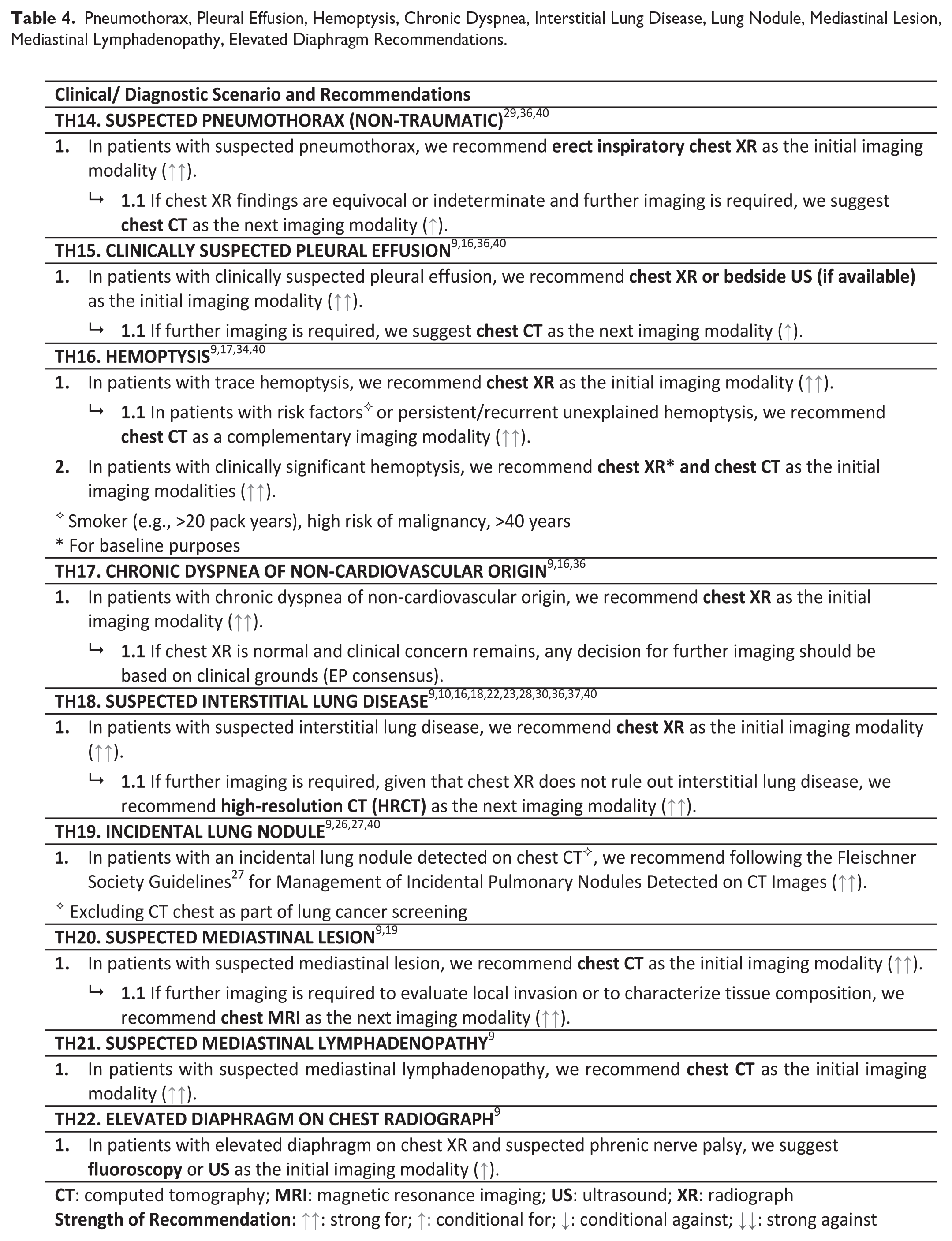
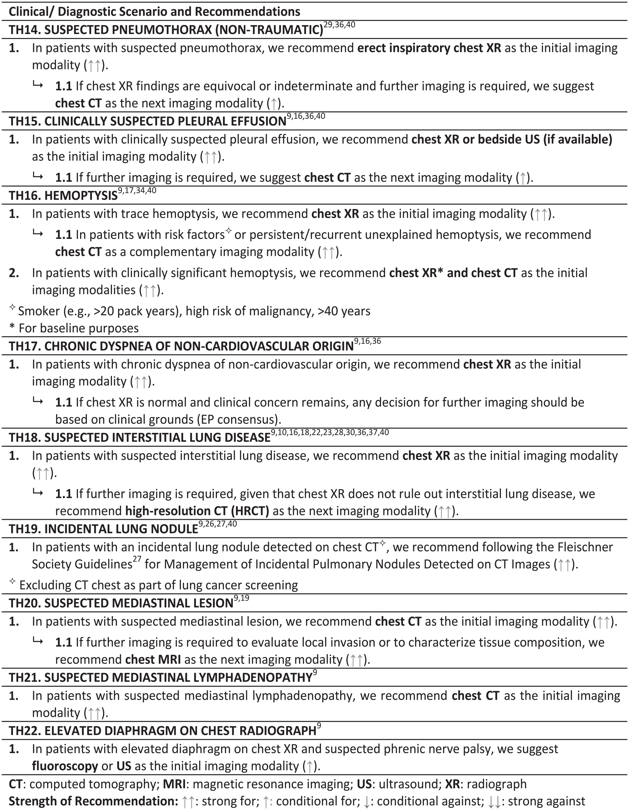
Recommendations are presented in 3 tables: Non-specific chest pain, long-term care, and hospital-based scenarios (Table 2), Upper respiratory tract infections, asthma, chronic obstructive pulmonary disease (COPD), pneumonia, and chronic cough scenarios (Table 3), Pneumothorax, pleural effusion, hemoptysis, chronic dyspnea, interstitial lung disease, lung nodule, mediastinal lesion, mediastinal lymphadenopathy, and elevated diaphragm scenarios (Table 4).
Screening/Asymptomatic, Non-Specific Chest Pain, Long-Term Care, and Hospital Recommendations.
Upper Respiratory Tract Infections, Asthma, COPD, Pneumonia, Chronic Cough Recommendations.
Pneumothorax, Pleural Effusion, Hemoptysis, Chronic Dyspnea, Interstitial Lung Disease, Lung Nodule, Mediastinal Lesion, Mediastinal Lymphadenopathy, Elevated Diaphragm Recommendations.
Supplemental Material
sj-pdf-1-caj-10.1177_08465371231214699 – Supplemental material for Canadian Association of Radiologists Thoracic Imaging Referral Guideline
Supplemental material, sj-pdf-1-caj-10.1177_08465371231214699 for Canadian Association of Radiologists Thoracic Imaging Referral Guideline by Candyce Hamel, Barb Avard, Catherine Belanger, Patrick Bourgouin, Stephen Lam, Daria Manos, Alan Michaud, Brian H. Rowe, Kevin Sanders and Ana-Maria Bilawich in Canadian Association of Radiologists Journal
Footnotes
Acknowledgements
We would like to thank: Becky Skidmore for creating the search strategies for the systematic scoping review, and the following individuals on the Diagnostic Imaging Referral Guidelines Working Group and external stakeholders for providing feedback on the guideline (listed alphabetically): Carole Dennie, Cameron Hague, Ryan Margau (WG co-chair), Cathy MacLean, Elsie Nguyen, Paul Pageau (WG co-chair), Erin Sarrazin, Carolina Souza, Jana Taylor, Charlotte Yong-Hing, and Kaitlin Zaki-Metias.
Declaration of Conflicting Interests
The author(s) declared no potential conflicts of interest with respect to the research, authorship, and/or publication of this article.
Funding
The author(s) disclosed receipt of the following financial support for the research, authorship, and/or publication of this article: This work was supported by the Canadian Medical Association.
Supplemental Material
Supplemental material for this article is available online.
References
Supplementary Material
Please find the following supplemental material available below.
For Open Access articles published under a Creative Commons License, all supplemental material carries the same license as the article it is associated with.
For non-Open Access articles published, all supplemental material carries a non-exclusive license, and permission requests for re-use of supplemental material or any part of supplemental material shall be sent directly to the copyright owner as specified in the copyright notice associated with the article.