Preoperative Dynamic Contrast-Enhanced and Diffusion-Weighted Breast Magnetic Resonance Imaging Findings for Prediction of Lymphovascular Invasion of the Lesions in Node-Negative Invasive Breast Cancer
Restricted accessResearch articleFirst published online May, 2024
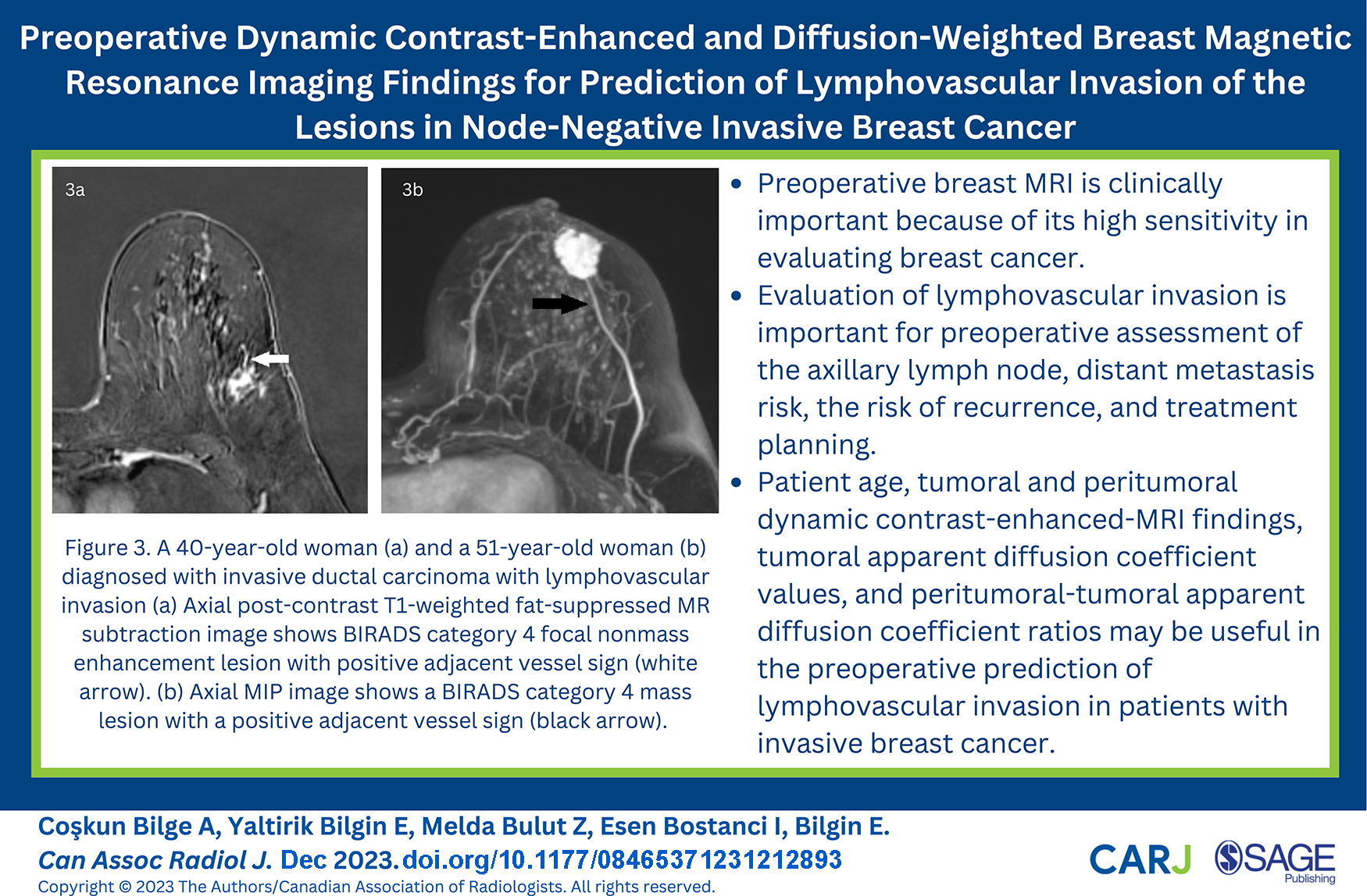
Preoperative Dynamic Contrast-Enhanced and Diffusion-Weighted Breast Magnetic Resonance Imaging Findings for Prediction of Lymphovascular Invasion of the Lesions in Node-Negative Invasive Breast Cancer
Purpose: Our single-centre retrospective study aimed to investigate the relationship between preoperative dynamic contrast-enhanced magnetic resonance imaging (DCE-MRI) findings and apparent diffusion coefficient (ADC) values and lymphovascular invasion (LVI) status of the lesions in patients with clinically-radiologically lymph node-negative invasive breast cancer. Methods: A total of 250 breast lesions diagnosed in preoperative magnetic resonance imaging were identified. All patients were divided into 2 subgroups: LVI-negative and LVI-positive according to the pathological findings of surgical specimens. The 2 groups’ DCE-MRI findings, ADC values, and histopathological results of lesions were compared. Results: LVI was detected in 100 of 250 lesions. Younger age than 45 years and larger lesion size than 20 mm were found to be associated with the presence of LVI (P < .001). High histological and nuclear grade (P = .001), HER2-enriched molecular subtype (P = .001), and Ki-67 positivity (P = .016) were significantly associated with LVI. The LVI positivity rate was significantly higher in the lesions with medium-rapid initial phase kinetic curve and washout delayed phase kinetic curve (P = .001). The presence of LVI was significantly associated with the presence of peritumoural edema, sentinel lymph node metastasis, adjacent vessel sign, and increased whole breast vascularity (P < .001). When diffusion-weighted imaging findings were evaluated, it was determined that tumoural ADC values lower than 1068 × 10−6 mm2/second (P = .002) and peritumoural-tumoural ADC ratios higher than 1.5 (P = .001) statistically increased the probability of LVI. Conclusion: The patient’s age, various histopathological and DCE-MRI findings, tumoural ADC value, and peritumoural-tumoural ADC ratio may be useful in the preoperative prediction of LVI status in breast cancer lesions.
HouvenaeghelGCohenMClasseJM, et al. Lymphovascular invasion has a significant prognostic impact in patients with early breast cancer, results from a large, national, multicenter, retrospective cohort study. ESMO Open. 2021;6(6):100316.
2.
TiradaNAujeroMKhorjekarG, et al. Breast cancer tissue markers, genomic profiling, and other prognostic factors: a primer for radiologists. Radiographics. 2018;38(7):1902-1920.
3.
OshiroCYamasakiMNodaYNishimaeATakahashiHInajiH.Comparative evaluation of nuclear and histological grades as prognostic factors for invasive breast cancer. Breast Cancer. 2020;27(5):947-953.
4.
KuhnEGambiniDDespiniLAsnaghiDRunzaLFerreroS.Updates on lymphovascular invasion in breast cancer. Biomedicines. 2023;11(3):968.
5.
MorkavukŞBGünerMÇulcuSEroğluABayarSÜnalAE. Relationship between lymphovascular invasion and molecular subtypes in invasive breast cancer. Int J Clin Pract. 2021;75(4): e13897.
6.
RyuYJKangSJChoJSYoonJHParkMH.Lymphovascular invasion can be better than pathologic complete response to predict prognosis in breast cancer treated with neoadjuvant chemotherapy. Medicine (Baltimore). 2018;97(30):e11647.
7.
KombakFEŞahinHMollamemişoğluH, et al. Concordance of immunohistochemistry between core needle biopsy and surgical resection of breast cancer. Turk J Med Sci. 2017;47(6):1791-1796.
8.
MoriNMugikuraSTakasawaC, et al. Peritumoral apparent diffusion coefficients for prediction of lymphovascular invasion in clinically node-negative invasive breast cancer. Eur Radiol. 2016;26(2):331-339.
9.
QuRFGuoDRChangZX, et al. Differential diagnosis of benign and malignant breast tumors using apparent diffusion coefficient value measured through diffusion-weighted magnetic resonance imaging. J Comput Assist Tomogr. 2015;39(4):513-522.
10.
ChoiBB.Associations between apparent diffusion coefficient values and the prognostic factors of breast cancer. J Comput Assist Tomogr. 2019;43(6):931-936.
11.
LiuZLiRLiangK, et al. Value of digital mammography in predicting lymphovascular invasion of breast cancer. BMC Cancer. 2020;20(1):274.
12.
IgarashiTFurubeHAshidaHOjiriH.Breast MRI for prediction of lymphovascular invasion in breast cancer patients with clinically negative axillary lymph nodes. Eur J Radiol. 2018;107:111-118.
13.
CheonHKimHJLeeSM, et al. Preoperative MRI features associated with lymphovascular invasion in node-negative invasive breast cancer: a propensity-matched analysis. J Magn Reson Imaging. 2017;46(4):1037-1044.
14.
ChoiBB.Dynamic contrast enhanced-MRI and diffusion-weighted image as predictors of lymphovascular invasion in node-negative invasive breast cancer. World J Surg Oncol. 2021;19(1):76.
15.
MorrisEAComstockCELeeCH, et al. ACR BI-RADS® Magnetic resonance imaging. In: ACR BI-RADS® Atlas, Breast Imaging Reporting and Data System. Reston, VA: American College of Radiology; 2013:56-71.
16.
ChoiWJChaJHKimHHShinHJChaeEY.The accuracy of breast MR imaging for measuring the size of a breast cancer: analysis of the histopathologic factors. Clin Breast Cancer. 2016;16(6):e145-e152.
17.
HanMKimTHKangDKKimKSYimH.Prognostic role of MRI enhancement features in patients with breast cancer: value of adjacent vessel sign and increased ipsilateral whole-breast vascularity. AJR Am J Roentgenol. 2012;199(4):921-928.
18.
SardanelliFIozzelliAFaustoACarrieroAKirchinMA.Gadobenate dimeglumine-enhanced MR imaging breast vascular maps: association between invasive cancer and ipsilateral increased vascularity. Radiology. 2005;235(3):791-797.
19.
OkumaHSudahMKettunenT, et al. Peritumor to tumor apparent diffusion coefficient ratio is associated with biologically more aggressive breast cancer features and correlates with the prognostication tools. PLoS One. 2020;15(6):e0235278.
20.
RadeckaBLitwiniukM.Breast cancer in young women. Ginekol Pol. 2016;87(9):659-663.
21.
AndersCKFanCParkerJS, et al. Breast carcinomas arising at a young age: unique biology or a surrogate for aggressive intrinsic subtypes?J Clin Oncol. 2011;29(1):e18-e20.
22.
NishimuraROsakoTOkumuraY, et al. An evaluation of lymphovascular invasion in relation to biology and prognosis according to subtypes in invasive breast cancer. Oncol Lett. 2022;24(2):245.
23.
ShenSWuGXiaoG, et al. Prediction model of lymphovascular invasion based on clinicopathological factors in Chinese patients with invasive breast cancer. Medicine (Baltimore). 2018;97(43):e12973.
24.
YuCCCheungYCHsuehCChenSC.Predictors of sentinel lymph node metastasis in postoperatively upgraded invasive breast carcinoma patients. Cancers (Basel). 2021;13(16):4099.
25.
AbdullaHASalmanAZAlaraibiSJ, et al. Risk factors associated with sentinel lymph node metastasis in clinically node-negative breast cancer. Eur J Breast Health. 2023;19(3):229-234.
26.
VialeGZurridaSMaioranoE, et al. Predicting the status of axillary sentinel lymph nodes in 4351 patients with invasive breast carcinoma treated in a single institution. Cancer. 2005; 103(3):492-500.
27.
CapdetJMartelPCharitanskyH, et al. Factors predicting the sentinel node metastases in T1 breast cancer tumor: an analysis of 1416 cases. Eur J Surg Oncol. 2009;35(12):1245-1249.
28.
MannRMChoNMoyL.Breast MRI: state of the art. Radiology. 2019;292(3):520-536. doi:10.1148/radiol.2019182947
29.
SungJSLiJDa CostaG, et al. Preoperative breast MRI for early-stage breast cancer: effect on surgical and long-term outcomes. AJR Am J Roentgenol. 2014;202(6):1376-1382.
30.
ChoiEJChoiHChoiSAYoukJH.Dynamic contrast-enhanced breast magnetic resonance imaging for the prediction of early and late recurrences in breast cancer. Medicine (Baltimore). 2016;95(48):e5330.
31.
KooJSJungWHKimH.Epithelial displacement into the lymphovascular space can be seen in breast core needle biopsy specimens. Am J Clin Pathol. 2010;133(5):781-787.
32.
Thomassin-NaggaraISilesPTropI, et al. How to measure breast cancer tumoral size at MR imaging?Eur J Radiol. 2013;82(12):e790-e800.
33.
ChangYWKwonKHChoiDL, et al. Magnetic resonance imaging of breast cancer and correlation with prognostic factors. Acta Radiol. 2009;50(9):990-998.
34.
CheonHKimHJKimTH, et al. Invasive breast cancer: prognostic value of peritumoral edema identified at preoperative MR imaging. Radiology. 2018;287(1):68-75.