
Abstract
The Canadian Association of Radiologists (CAR) Obstetrics and Gynecology Expert Panel consists of radiologists specializing in obstetrics and gynecology, obstetrics and gynecology physicians, a patient advisor, and an epidemiologist/guideline methodologist. After developing a list of 12 clinical/diagnostic scenarios, a systematic rapid scoping review was undertaken to identify systematically produced referral guidelines that provide recommendations for one or more of these clinical/diagnostic scenarios. Recommendations from 46 guidelines and contextualization criteria in the Grading of Recommendations, Assessment, Development and Evaluations (GRADE) for guidelines framework were used to develop 68 recommendation statements across the 12 scenarios related to the evaluation of obstetrics and gynecology clinical and diagnostic scenarios. This guideline presents the methods of development and the imaging recommendations for a variety of obstetrical and gynecological conditions including pregnancy assessment, recurrent first trimester pregnancy loss, post-partum indications, disorders of menstruation, localization of intra-uterine contraceptive device, infertility assessment, assessment of adnexal mass, pelvic pain of presumed gynecological origin, and pelvic floor evaluation.
Introduction
Beginning in October 2021, an Expert Panel (EP) comprised of radiologists specializing in obstetrics and gynecology, obstetrics and gynecology physicians, a patient advisor, and an epidemiologist/guideline methodologist met to develop a new set of recommendations specific to referral pathways for obstetrics and gynecology. Through discussion (via virtual meetings), the EP developed a list of 12 clinical/diagnostic scenarios to be covered by this guideline. These recommendations are intended primarily for referring clinicians (e.g., family physicians, nurse practitioners); however, they may also be used by radiologists, other clinicians in allied specialties, patients, and patient representatives.
Recommendation text, symbol, and interpretation.
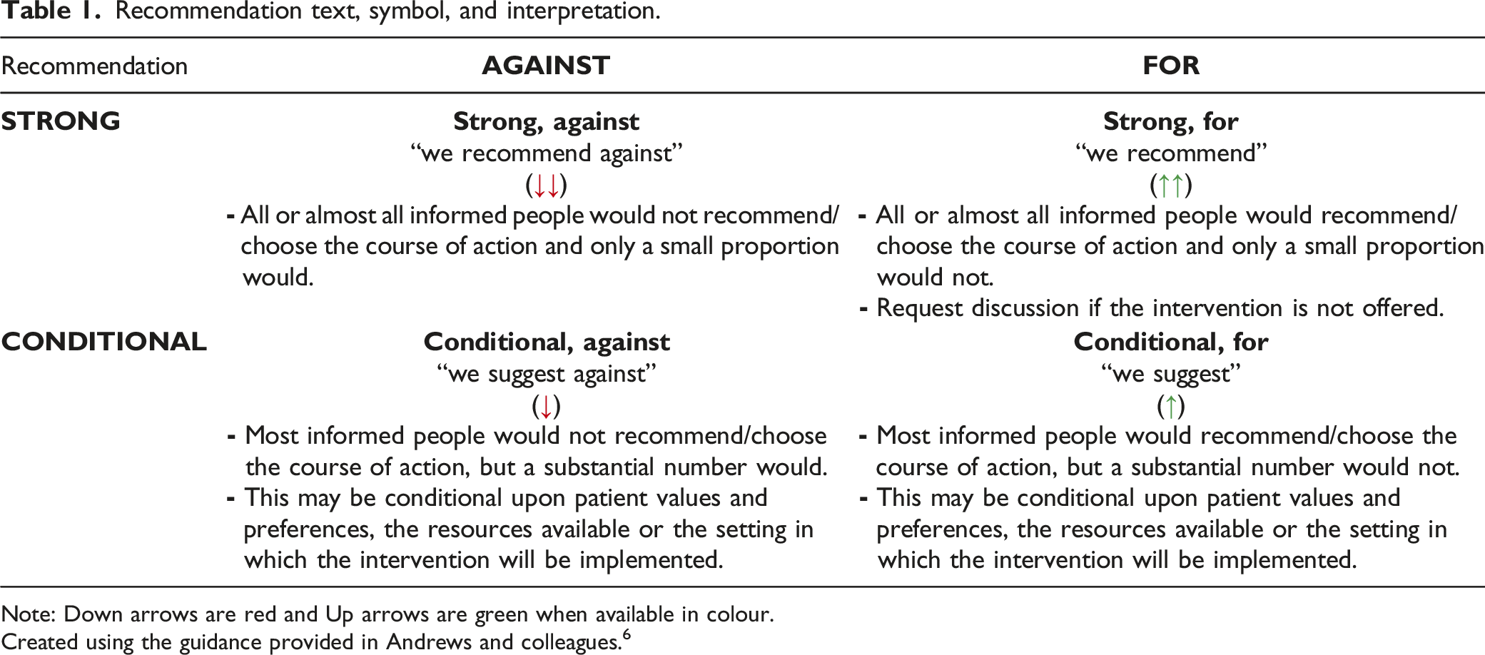
Note: Down arrows are red and Up arrows are green when available in colour.
Created using the guidance provided in Andrews and colleagues. 6
A systematic search for guidelines (with an a priori defined inclusion criteria) was run in Medline and Embase on November 24, 2021. The search was limited to publications from 2016 onward (Supplementary file Appendix 1). Supplemental searching included the following national radiology and/or guideline groups: the American College of Radiology, the National Institute for Health and Care Excellence, the Society of Obstetricians and Gynaecologists of Canada, and the Royal College of Radiologists 8th Edition (2017). Recommendations for each clinical scenario were formulated over six virtual meetings from February to April 2022. External review and feedback were obtained from radiologists, emergency medicine physicians, family physicians, obstetrics and gynaecology physicians, and a nurse practitioner. The full guideline can be found on the CAR website (www.car.ca).
Results
Systematic Scoping Review
A total of 4279 records were identified through the electronic database and nine additional records were added from the supplemental search. A total of 46 guidelines, plus seven companion papers, were included (Figure 1 PRISMA flow diagram). Potentially relevant guidelines published in languages other than English can be found in Supplementary file Appendix 2. A list of excluded records including justifications for exclusion is available upon request. Most guidelines were rated as moderate or high quality, using the modified AGREE-II checklist (Supplementary file Appendix 3). The number of guidelines included per clinical/diagnostic scenario ranged from zero to 16, with a median of five guidelines per clinical scenario. PRISMA flow diagram.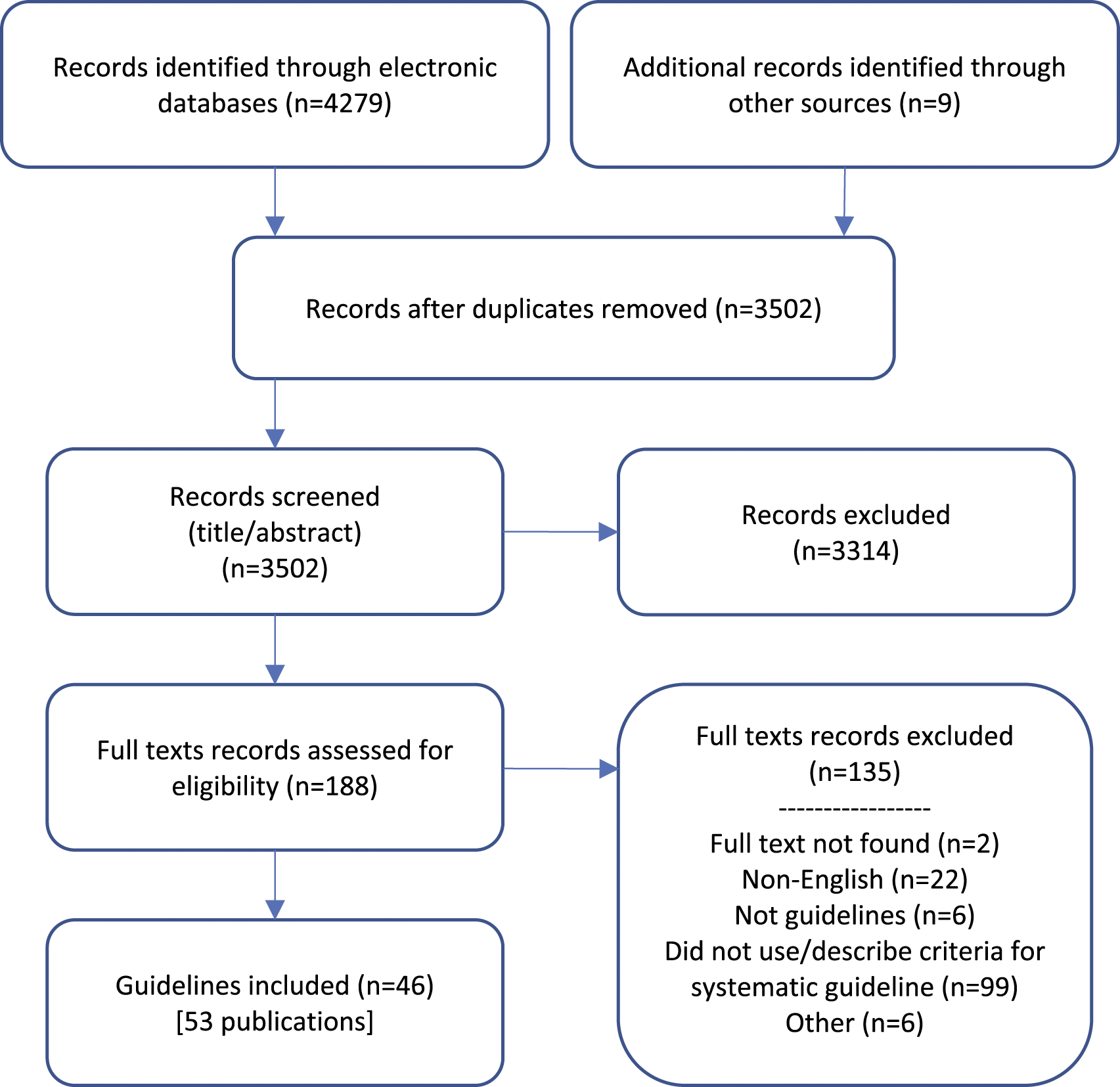
Recommendations
Additional details of the included guidelines, including which imaging modalities (e.g., computed tomography [CT], magnetic resonance imaging [MRI], radiograph [XR], saline infusion sonohysterogram [SIS], ultrasound [US]) that were discussed can be found in Supplementary file Appendix 4. Recommendations do not specify when contrast should or should not be used, as this may vary based on clinical presentation, regional practice preferences, preference of the referring clinician, radiologist and the patient, and resource availability.
A guideline is intended to guide and not be an absolute rule. Medical care is complex and should be based on evidence, a clinician’s expert judgment, the patient’s circumstances, values, preferences, and resource availability. Not all imaging modalities are available in all clinical environments, particularly in rural or remote areas of Canada. Decisions about patient transfer, use of alternative imaging or serial clinical examination and observation can be difficult. Therefore, the expected benefits of recommended imaging, risks of travel, patient preference, and other factors must be considered. The guideline recommendations are to assist the choice of imaging modality in situations where it is deemed clinically necessary to obtain imaging.
We reviewed relevant recommendations related to the 12 clinical/diagnostic scenarios previously published by radiology and specialty societies, including: the Canadian Association of Radiologists, 9 the American College of Obstetricians and Gynecologists, 10,11 the American College of Radiology, 12 -28 the American Society of Clinical Oncology, 29 the European Society of Urogenital Radiology Scrotal and Penile Imaging Working Group, 30,31 the European Society of Urogenital Radiology, 32 the French College of Gynecologists and Obstetricians, 33 the International Society of Ultrasound in Obstetrics and Gynecology, 34 the International guideline on polycystic ovarian syndrome, 35 -38 the National Institute for Health and Care Excellence, 39 -47 the Society for Maternal-Fetal Medicine, 48 the Society of Abdominal Radiology, 49 the Society of Obstetricians and Gynaecologists of Canada, 50 -57 the Royal College of Obstetricians and Gynaecologists, 58 -60 and the 2017 Royal College of Radiologists. 61
Pregnancy assessment recommendations.
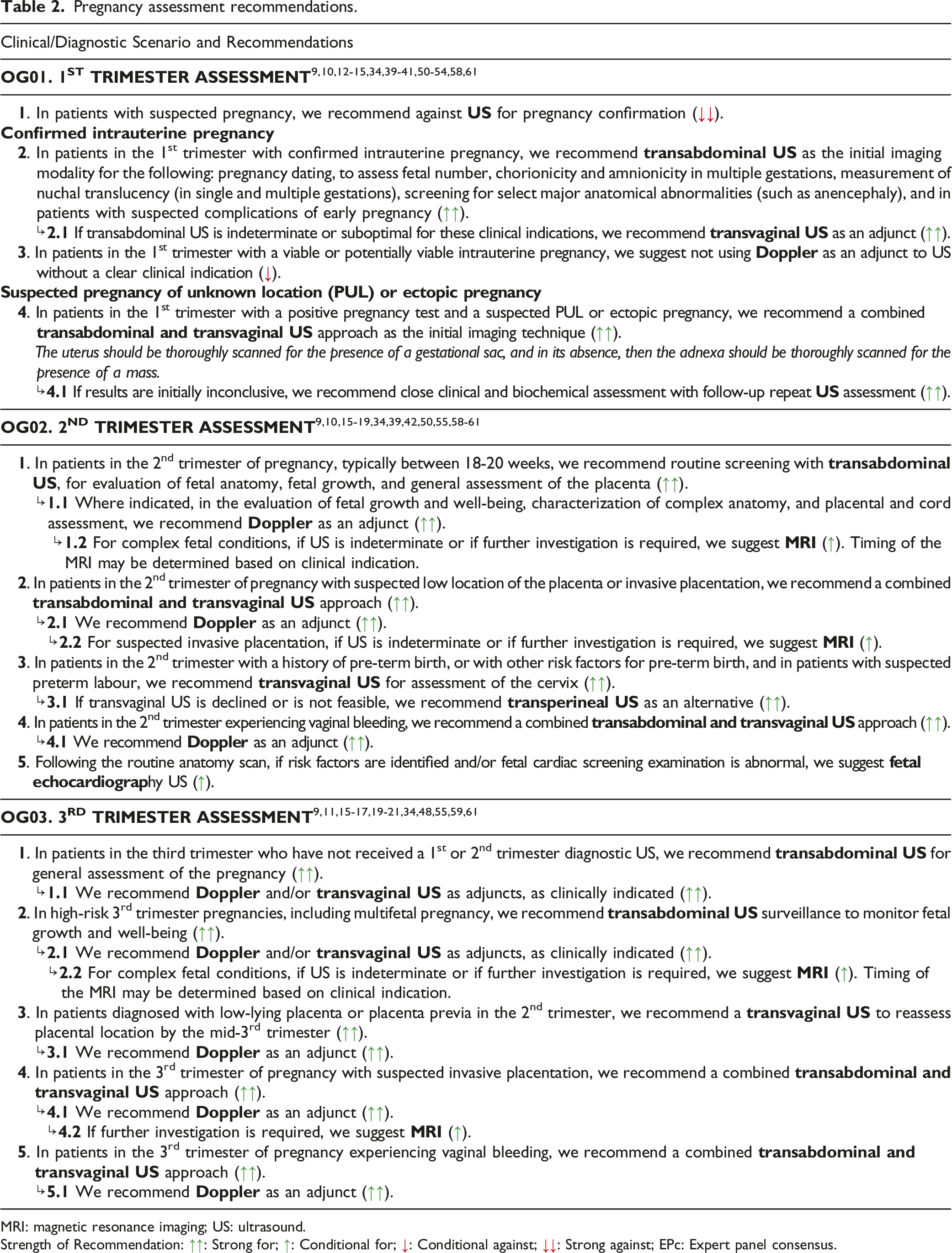
MRI: magnetic resonance imaging; US: ultrasound.
Strength of Recommendation: ↑↑: Strong for; ↑: Conditional for; ↓: Conditional against; ↓↓: Strong against; EPc: Expert panel consensus.
Post-partum, recurrent pregnancy loss, and infertility clinical/diagnostic scenario recommendations.
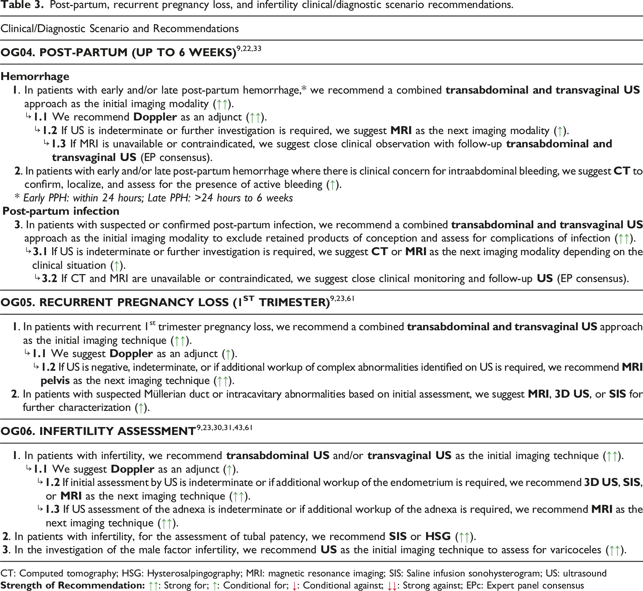
CT: Computed tomography; HSG: Hysterosalpingography; MRI: magnetic resonance imaging; SIS: Saline infusion sonohysterogram; US: ultrasound
Gynecology clinical/diagnostic scenario recommendations.
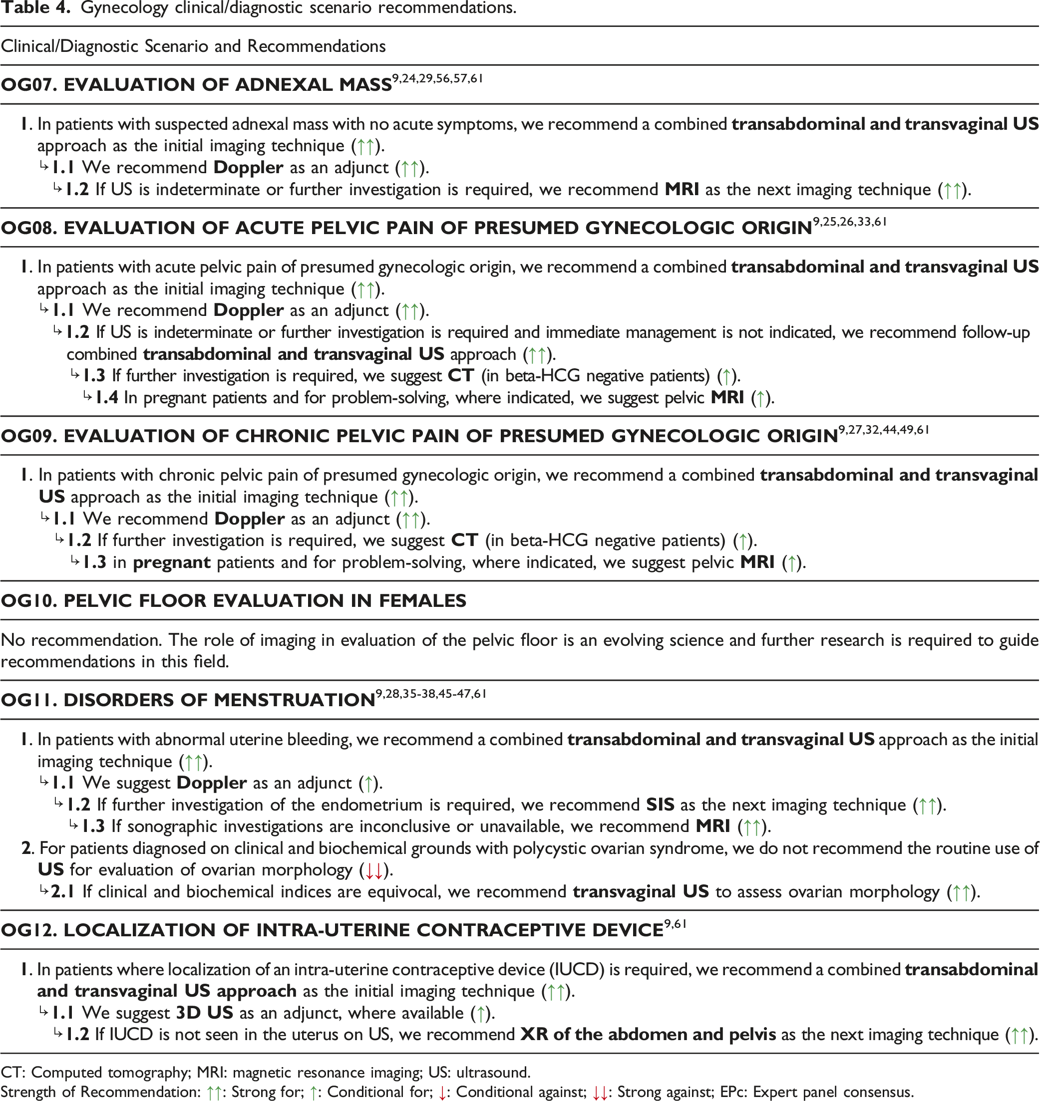
CT: Computed tomography; MRI: magnetic resonance imaging; US: ultrasound.
Strength of Recommendation: ↑↑: Strong for; ↑: Conditional for; ↓: Conditional against; ↓↓: Strong against; EPc: Expert panel consensus.
There are clinical/diagnostic scenarios that may pertain to more than one CAR guideline section. For example, pelvic floor evaluation can be found in both the Obstetrics & Gynecology guideline and will be in the Urology, Adrenal, and Genitourinary guideline. As not all guidelines are available concurrently, it is possible these scenarios may be updated in subsequent guideline sections to include a newer evidence-base.
Supplemental Material
Supplemental Material - Canadian Association of Radiologists Obstetrics and Gynecology Diagnostic Imaging Referral Guideline
Supplemental Material for Canadian Association of Radiologists Obstetrics and Gynecology Diagnostic Imaging Referral Guideline by Candyce Hamel, Baharak Amir, Barb Avard, Karen Fung Kee Fung, Beth Furey, Juliette Garel, and Hournaz Ghandehar in Canadian Association of Radiologists Journal
Footnotes
Acknowledgments
We would like to thank: Becky Skidmore for creating the search strategies for the systematic scoping review, Leila Esmaeilisaraji for her role as a reviewer on the scoping review, and the following individuals on the Diagnostic Imaging Referral Guidelines Working Group and external stakeholders for providing feedback on the guideline (listed alphabetically): Samuel Campbell, Elisa Cohen, Noel Corser, Cathy MacLean, Ryan Margau (WG co-chair), Lucie Morin, Paul Pageau (WG co-chair), Erin Sarrazin, Charlotte Yong-Hing, Kaitlin Zaki-Metias.
Declaration of Conflicting Interests
The author(s) declared no potential conflicts of interest with respect to the research, authorship, and/or publication of this article.
Funding
The author(s) disclosed receipt of the following financial support for the research, authorship, and/or publication of this article: This work was supported by the Canadian Medical Association
Supplemental Material
Supplemental material for this article is available online.
References
Supplementary Material
Please find the following supplemental material available below.
For Open Access articles published under a Creative Commons License, all supplemental material carries the same license as the article it is associated with.
For non-Open Access articles published, all supplemental material carries a non-exclusive license, and permission requests for re-use of supplemental material or any part of supplemental material shall be sent directly to the copyright owner as specified in the copyright notice associated with the article.