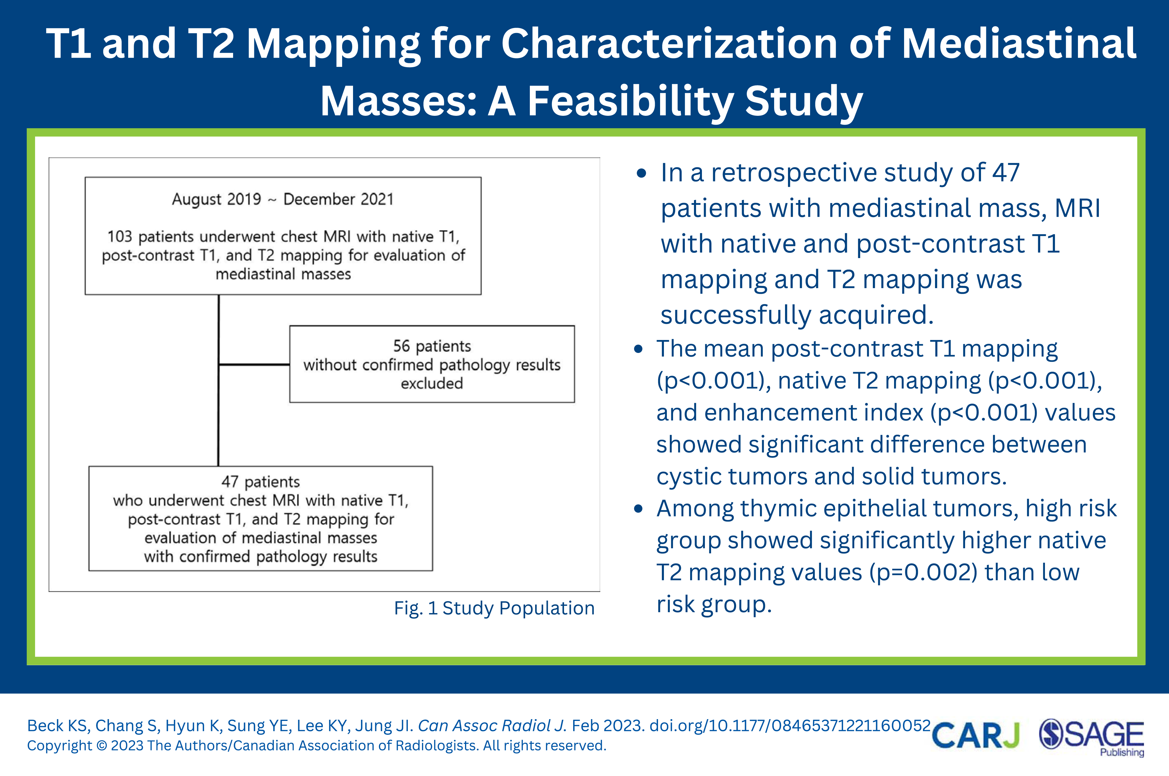
Purpose: To evaluate the feasibility and usefulness of T1 and T2 mapping in characterization of mediastinal masses. Methods: From August 2019 through December 2021, 47 patients underwent 3.0-T chest MRI with T1 and post-contrast T1 mapping using modified look-locker inversion recovery sequences and T2 mapping using a T2-prepared single-shot shot steady-state free precession technique. Mean native T1, native T2, and post-contrast T1 values were measured by drawing the region of interest in the mediastinal masses, and enhancement index (EI) was calculated using these values. Results: All mapping images were acquired successfully, without significant artifact. There were 25 thymic epithelial tumors (TETs), 3 schwannomas, 6 lymphomas, and 9 thymic cysts, and 4 other cystic tumors. TET, schwannoma, and lymphoma were grouped together as “solid tumor,” to be compared with thymic cysts and other tumors (“cystic tumors”). The mean post-contrast T1 mapping (P < .001), native T2 mapping (P < .001), and EI (P < .001) values showed significant difference between these two groups. Among TETs, high risk TETs (thymoma types B2, B3, and thymic carcinoma) showed significantly higher native T2 mapping values (P = .002) than low risk TETs (thymoma types A, B1, and AB). For all measured variables, interrater reliability was good to excellent (intraclass coefficient [ICC]: .869∼.990) and intrarater reliability was excellent (ICC: .911∼.995). Conclusion: The use of T1 and T2 mapping in MRI of mediastinal masses is feasible and may provide additional information in the evaluation of mediastinal masses.
KimPKHongYJImDJ, et al.Myocardial T1 and T2 mapping: Techniques and clinical applications. Korean J Radiol. 2017;18(1):113-131. doi:10.3348/kjr.2017.18.1.113
2.
SaeedMLiuHLiangCHWilsonMW. Magnetic resonance imaging for characterizing myocardial diseases. Int J Cardiovasc Imag. 2017;33(9):1395-1414. doi:10.1007/s10554-017-1127-x
PiechnikSKFerreiraVMLewandowskiAJ, et al.Normal variation of magnetic resonance T1 relaxation times in the human population at 1.5 T using ShMOLLI. J Cardiovasc Magn Reson. 2013;15:13. doi:10.1186/1532-429X-15-13
5.
CremaMDRoemerFWMarraMD, et al.Articular cartilage in the knee: current MR imaging techniques and applications in clinical practice and research. Radiographics. 2011;31(1):37-61. doi:10.1148/rg.311105084
6.
GuermaziARoemerFWAlizaiH, et al.State of the Art: MR imaging after knee cartilage repair surgery. Radiology. 2015;277(1):23-43. doi:10.1148/radiol.2015141146
7.
LiXMajumdarS. Quantitative MRI of articular cartilage and its clinical applications. J Magn Reson Imag. 2013;38(5):991-1008. doi:10.1002/jmri.24313
8.
AndreisekGWeigerM. T2* mapping of articular cartilage: current status of research and first clinical applications. Invest Radiol. 2014;49(1):57-62. doi:10.1097/RLI.0b013e3182a574e1.
9.
Thomaides-BrearsHBLepeRBanerjeeRDunckerC. Multiparametric MR mapping in clinical decision-making for diffuse liver disease. Abdom Radiol. 2020;45(11):3507-3522. doi:10.1007/s00261-020-02684-3
10.
SchaapmanJJTushuizenMECoenraadMJLambHJ. Multiparametric MRI in patients with nonalcoholic fatty liver disease. J Magn Reson Imag. 2021;53:1623-1631. doi:10.1002/jmri.27292
11.
CarterBWBenvenisteMFMadanR, et al.ITMIG classification of mediastinal compartments and multidisciplinary approach to mediastinal masses. Radiographics. 2017;37(2):413-436. doi:10.1148/rg.2017160095
12.
MadanRRatanaprasatpornLRatanaprasatpornLCarterBWAckmanJB. Cystic mediastinal masses and the role of MRI. Clin Imag. 2018;50:68-77. doi:10.1016/j.clinimag.2017.12.011
13.
TomiyamaNHondaOTsubamotoM, et al.Anterior mediastinal tumors: diagnostic accuracy of CT and MRI. Eur J Radiol. 2009;69(2):280-288. doi:10.1016/j.ejrad.2007.10.002
14.
AckmanJB. MR imaging of mediastinal masses. Magn Reson Imag Clin N Am. 2015;23(2):141-164. doi:10.1016/j.mric.2015.01.002
15.
AckmanJBWuCC. MRI of the thymus. AJR Am J Roentgenol. 2011;197(1):W15-W20. doi:10.2214/AJR.10.4703
16.
KishidaYKoyamaHSekiS, et al.Comparison of fat suppression capability for chest MR imaging with Dixon, SPAIR and STIR techniques at 3 Tesla MR system. Magn Reson Imaging. 2018;47:89-96. doi:10.1016/j.mri.2017.11.012
17.
ShinKEYiCAKimTS, et al.Diffusion-weighted MRI for distinguishing non-neoplastic cysts from solid masses in the mediastinum: problem-solving in mediastinal masses of indeterminate internal characteristics on CT. Eur Radiol. 2014;24(3):677-684. doi:10.1007/s00330-013-3054-0
18.
CasparTEl GhannudiSOhanaM, et al.Magnetic resonance evaluation of cardiac thrombi and masses by T1 and T2 mapping: an observational study. Int J Cardiovasc Imag. 2017;33(4):551-559. doi:10.1007/s10554-016-1034-6
19.
WongTCPiehlerKMKangIA, et al.Myocardial extracellular volume fraction quantified by cardiovascular magnetic resonance is increased in diabetes and associated with mortality and incident heart failure admission. Eur Heart J. 2014;35(10):657-664. doi:10.1093/eurheartj/eht193.
20.
OkumuraMOhtaMTateyamaH, et al.The world health organization histologic classification system reflects the oncologic behavior of thymoma: A clinical study of 273 patients. Cancer. 2002;94(3):624-632. doi:10.1002/cncr.10226
21.
OkumuraMMiyoshiSFujiiY, et al.Clinical and functional significance of WHO classification on human thymic epithelial neoplasms: a study of 146 consecutive tumors. Am J Surg Pathol. 2001;25(1):103-110. doi:10.1097/00000478-200101000-00012
22.
NakagawaKAsamuraHMatsunoY, et al.Thymoma: a clinicopathologic study based on the new World Health Organization classification. J Thorac Cardiovasc Surg. 2003;126(4):1134-1140. doi:10.1016/s0022-5223(03)00798-0.
23.
SadoharaJFujimotoKMullerNL, et al.Thymic epithelial tumors: comparison of CT and MR imaging findings of low-risk thymomas, high-risk thymomas, and thymic carcinomas. Eur J Radiol. 2006;60(1):70-79. doi:10.1016/j.ejrad.2006.05.003
24.
CarterBWBetancourtSLBenvenisteMF. MR imaging of mediastinal masses. Top Magn Reson Imag. 2017;26(4):153-165. doi:10.1097/RMR.0000000000000134
25.
BardoDMEBiyyamDRPatelMCWongKvan TasselDRobisonRK. Magnetic resonance imaging of the pediatric mediastinum. Pediatr Radiol. 2018;48(9):1209-1222. doi:10.1007/s00247-018-4112-1
26.
HuangSYSeethamrajuRTPatelPHahnPFKirschJEGuimaraesAR. Body MR Imaging: Artifacts, k-space, and solutions. Radiographics. 2015;35(5):1439-1460. doi:10.1148/rg.2015140289
27.
WuWMillerKL. Image formation in diffusion MRI: A review of recent technical developments. J Magn Reson Imaging. J Magn Reson Imag. 2017;46(3):646-662. doi:10.1002/jmri.25664
28.
YanagawaMTomiyamaN. Prediction of thymoma histology and stage by radiographic criteria. Thorac Surg Clin. 2011;21(1):1-12. doi:10.1016/j.thorsurg.2010.08.008
29.
JeongYJLeeKSKimJShimYMHanJKwonOJ. Does CT of thymic epithelial tumors enable us to differentiate histologic subtypes and predict prognosis?AJR Am J Roentgenol. 2004;183(2):283-289. doi:10.2214/ajr.183.2.1830283
30.
YakushijiSTateishiUNagaiS, et al.Computed tomographic findings and prognosis in thymic epithelial tumor patients. J Comput Assist Tomogr. 2008;32(5):799-805. doi:10.1097/RCT.0b013e31815896df
31.
MoonJCMessroghliDRKellmanP, et al.Myocardial T1 mapping and extracellular volume quantification: a society for cardiovascular magnetic resonance (SCMR) and CMR working group of the European society of cardiology consensus statement. J Cardiovasc Magn Reson. 2013;15:92. doi:10.1186/1532-429X-15-92
32.
YabuuchiHMatsuoYAbeK, et al.Anterior mediastinal solid tumours in adults: Characterisation using dynamic contrast-enhanced MRI, diffusion-weighted MRI, and FDG-PET/CT. Clin Radiol. 2015;70(11):1289-1298. doi:10.1016/j.crad.2015.07.004