
Abstract
Our study intended to explore Hesp antioxidant and anti-inflammatory effects against TAA hepatic fibrosis in rats. Hesperidin (Hesp), is a pharmacologically active flavonoid, found abundantly in citrus species. Our present research attempts to inspect the potential hepatoprotective role of Hesp against thioacetamide (TAA)-induced hepatic fibrosis. Thirty-two male albino rats were split up into four equal groups, each with eight rats: Cont group was treated with ip saline. Every other day, the TAA group was injected 100 mg/kg BW ip TAA, Hesp group received every day oral Hesp 200 mg/kg BW as well as TAA + Hesp group received both therapies (TAA, Hesp) for eight successive weeks. Hesp in TAA treated group reduces ALT, AST, and ALP activities, total, direct bilirubin, total cholesterol, and triglycerides, meanwhile TP, Alb, globulin, A/G ratio levels were insignificantly differed. The antioxidant capacity of Hesp was pronounced by a marked reduction in MDA level. While the antioxidant markers (SOD, CAT, GSH) were insignificantly changed after Hesp treatment. A strong significant correlation in treated rats between fibrosis score and CD34 and FGF23 gene expression. Liver sections from dual-treated rats showed a moderately decreased hepatic lesion and the dense, bluish-stained fibrous tissue by Masson’s trichrome. Elevated gene expressions of CD34 and FGF23 after TAA hepatotoxicity were diminished by the antifibrotic effect of Hesp. Also, immunohistochemical expression showed reduction of TGF-β and α-SMA in hepatocytes in the dual therapy group. Hesp possesses a potent antioxidant, and antifibrotic activities against TAA induced hepatic fibrosis by modulating TGF-β/α-SMA pathways.
Introduction
Many chemical compounds have been emitted into the environment over the past century. Certain chemicals serve a useful purpose despite being toxic, and their negative effects on the environment exceed their advantages for humanity. 1 Hepatotoxins predominance damage the liver cells to harmful intermediates via cytochrome P450 oxidases which is followed by an increase in reactive Oxygen Species (ROS), malondialdehyde (MDA), and pro-inflammatory cytokines. 2 The DNA of nuclei, adipocytes, and proteins are all harmed by an increase in free radicals. 3 The organosulfur compound, thioacetamide (TAA) is a fungicide that is categorized as a human carcinogen (class 2B) and is frequently used to stop fruit from decaying. 1 Hepatic cells were damaged through a severe pathway that includes CYP4502E1-facilitated biotransformation once it breaks down to TAA sulphone. 4 Liver fibrosis was caused by the stimulation of hepatic stellate cells (HSCs) via lipid peroxide production, cytotoxicity, and mitochondrial damage. Thus, apoptosis, necrosis, cholangiocarcinoma, and an excessive build-up of extracellular matrix proteins (ECM) may result from these processes. 5 According to some research, TAA has a role in the beginning of liver fibrosis.6–10
Different studies clarified that the experimental induction of liver fibrosis by chronic exposure to TAA results in biochemical and histological changes similar to the mechanisms involved in human liver fibrosis. 11
TGF-β has cytostatic and apoptotic effects in hepatocytes, promoting liver differentiation during embryogenesis and physiological liver regeneration. However, high levels of TGF-β, as a consequence of chronic liver damage, result in activation of stellate cells to myofibroblasts and massive hepatocyte cell death, which contributes to the promotion of liver fibrosis and later cirrhosis. 12
Hesperidin (Hesp) is a pharmacologically active flavonoid prevalent in citrus species. 13 Nowadays, citrus flavonoids’ positive impacts on health have just come to light. These substances have demonstrated biological activity in studies using animals, cell lines, and in vitro tests linked to cancer, cardiovascular, inflammatory, allergy, and bleeding disorders. 14 Recently, Hesp showed hepatoprotective impacts that were attributed to the restoration of oxidative stress, pro-inflammatory mediators, and antioxidant biomarker levels besides reducing ECM buildup in the liver.15–17
Despite the hepatoprotective action of Hesp has been documented. The antifibrotic effects of Hesp against TAA were not previsously explored. Hence, this study intended to investigate the antioxidant and anti-inflammatory effects of Hesp beside the TGF-β/α-SAM pathways against pre-existing hepatic fibrosis in experimental rats, which was confirmed by novel fibrotic markers, cluster differentiation 34 (CD34) and fibroblast growth factor 23 (FGF23).
Materials and methods
Chemicals
TAA was acquired from Germany’s Sigma-Aldrich Munich, and Hesp was acquired from Sigma—Aldrich Co, UK.
Design experimentation
Thirty-two male albino rats, weighing between 100 and 120 g. were procured from Zagazig University’s research animal unit. In accordance with the institutional rules of Mansoura Egyptian University’s Faculty of Veterinary Medicine’s Animal Research Ethics Committee (Ph. D/46), During the acclimatization phase, the housing used for the rats had a 12-hour cycle of light and dark, as well as consistent humidity and temperature. Their access to water was unrestricted. and a conventional mouse pellet meal was supplied to them without restriction.
Following a 2-week period of conditioning, the rats were kept for 8 weeks after being handled with care and split into four equal groups of eight rats each:
Control (
Dual treatment of TAA and Hesp (
Animal care and experimental procedures were approved by the Mansoura University under the “Guide for the Care and Use of Laboratory Animals.”
Handling of biological samples
At the conclusion of the eighth week of our study, an ip injection containing a 50 mg/kg dosage of a ketamine and xylazine mixture was given for animal anesthesia. For biochemical examination, serum was drawn and stored at −20°C in Eppendorf tubes. After blood samples from the retro-orbital plexus were drawn and centrifuged at 1500 ×g for 10 min. Three sections of liver tissue were divided: the initial sample used for tissue homogenate preparation in order to assess hepatic oxidative and antioxidant biomarkers. For RNA extraction for gene expression studies, the second portion of the sample was stored in RNA later then kept at −80°C. The final portion was embedded in formalin for examination of the hepatic building construction by H&E and special stain (Masson’s trichrome) for staining collagen and analysis using immunohistochemistry.
Evaluation of serum biochemical parameters
Spectrophotometric measurements were made of alkaline phosphatase (ALP) (EliTech Company, Paris, France Ref. No AlPI-0230), aspartate aminotransferase (AST) (Ref. No. 12031) and alanine aminotransferase (ALT) (Ref. No. 12032) (Human Company, Wiesbaden, Germany). In order to determine indirect bilirubin, the calorimetric measurements of bilirubin (Diamond Company, Cairo, Egypt, Cat. No. 202141) were made and then deducted. Evaluations of total protein (Ref. No. 1001290), albumin (Cat. No. 1001023), total triglycerides (TG) (Cat. No. LIQ370), and total cholesterol (TC) (Cat. No. LIQ428) were also conducted using Spinreact Co., Girona, Spain.
Assessment of hepatic lipid peroxidation indicators and antioxidants
Using readily accessible Biodiagnostics kits (Cairo, Egypt), Oxidative stress and antioxidant indicators were evaluated using hepatic homogenate as reduced glutathione (GSH) (Cat. No. GR.2511), catalase (CAT) (Cat. No. CA 2517), MDA (Cat. No. MD 2529), as well as superoxide dismutase (SOD) (Cat. No. SD 2521). The measurements were carried out in accordance with the manufacturer’s instructions.
Analysis of hepatic expression of genes
Hepatocyte RNA extraction was performed using a RNeasy Mini Kit (Cat. No. 74104; Qiagen, Hilden, Germany), depending on the manufacturer guidelines. The RNA samples were verified utilizing a NanoDrop spectrophotometer, two milliliters of RNase-free water, one milliliter of 2× QuantiTect SYBR Green PCR Master Mix, the QuantiTect SYBR Green PCR kit (Cat. No. 204141), and Revert Aid Reverse Transcriptase. SYBR Green real-time PCR was used to identify CD34 and FGF23 using oligonucleotide primers and probes (Metabion, Steinkirchen, Germany). Table 1 contains a list of primer sequences. Amplification curves and CT values were computed using Stratagene Mx3005P software.
Primer sequences for RT-PCR.

Using the Ct approach, the CT of each sample was contrasting with the control group in order to assess the change in the RNA samples gene expression described in Yuan et al. 24 As a housekeeping reference gene, β-actin was used. 21
Liver histopathology assessment
Sections of a liver fixed in formalin were cut, then embedded in paraffin, for H&E then stained by Masson’s trichrome in accordance with a previously published method in Bancroft and Gamble. 25 This allowed for the assessment of the extent of liver damage. The degree of hepatic lobular and portal inflammation, the fibrosis score, and the steatosis grade were assessed according to Ishak et al. 26
Expression of TGF-β and α-SMA by immunohistochemistry
The liver tissue was sliced into 5µ thick sections then deparaffinized, the slides were then autoclaved in a pH-6 citrate buffer for 10 min at 120°C in order to remove the antigen. A 3% H2O2 solution was used to block the portions. Primary antibodies were applied to the sidewalls and treated for 1 h at room temperature with (ready to use) TGF-β rabbit polyclonal (1:150) and α-SMA rabbit polyclonal (Wuhan Servicebio Technology Co. Ltd., Hubei, China). The slides underwent a 30-min incubation at room temperature with peroxidase-conjugated anti-rabbit secondary antibodies, followed by a 5-min incubation with the chromogen 3,3′ diaminobenzidine tetrahydrochloride to “visualize” the labeling. 27 According to Alkreathy and Esmat, 28 In every group, the level of both TGF-β and α-SMA positive immunoexpression was ranked from 0 to 4 respectively.
Statistical analysis
The data in all groups were indicated as mean ± standard error (SEM) using SPSS software, (USA, version 26). The results were then determined statistically significant after ANOVA and Duncan multiple comparison tests were run at P 0.05. 29
Results
Effects of Hesp and TAA on serum biochemical markers
When liver damage was induced by TAA, ALT, AST, ALP, total bilirubin, and direct bilirubin blood values were significantly increased (P < 0.05), whereas TP and Alb significantly decreased when compared to the control group (Figures 1 and 2). Treatment with Hesp significantly reduced both ALT, AST, and ALP activities as well total and direct bilirubin values in the TAA + Hesp group in contrast to the TAA-impaired group, however they didn’t get back to their typical state. Meanwhile TP and Alb levels were insignificantly differed in TAA + Hesp group in contrast to the TAA group (Figures 1 and 2). Our findings demonstrate that the group of rats treated with TAA alone had considerably higher levels of TC and TG, then triglyceride improved after treatment with Hesp but TC was insignificantly changed (Figure 2). In all investigated groups, the serum indirect bilirubin, globulin level and A/G ratio were insignificantly changed. Moreover, the safety of Hesp has been approved in the present study, where all estimated parameters were insignificantly changed in Hesp treated group with respect to the control group (Figures 1 and 2).

Serum liver function indicators after 8 weeks of Hesp therapy in contrast to TAA-intoxicated rats (mean ± SE) containing dissimilar superscripts) that distinct from one another statistically (P < 0.05): (a) ALT, (b) AST, (c) ALP, and (d) total bilirubin, (e) direct bilirubin, and (f) indirect bilirubin.

Biochemical indicators measured in blood at the conclusion of the eighth week after Hesp therapy in contrast to TAA-intoxicated rats (mean ± SE): (a) total protein, (b) albumin, (c) globulin, and (d) A/G ratio, (e) cholesterol, and (f) triglyceride, with distinct superscripts denoting statistically significant differences (P < 0.05). A/G, albumin/globulin ratio.
Impact of TAA and Hesp on both hepatic lipid peroxidation and antioxidant indicators
As seen in Figure 3, TAA-treated group had significantly higher hepatic MDA levels than the control group. Also, the lowest antioxidant biomarkers SOD, CAT, and GSH were observed in the TAA group. Moreover, the dual therapy with Hesp and TAA reduced the elevated MDA levels, while the antioxidant markers were insignificantly changed in comparison to the TAA intoxicated group. Furthermore, we found no variations in oxidative and antioxidant indices between the Hesp treated and control groups.

Liver oxidative/antioxidative indicators showing significant variations (P < 0.05) (mean ± SE) with distinct superscripts: (a) MDA, (b) SOD, (c) GSH, and (d) catalase.
Effects of TAA and Hesp on the expression of the CD34 and FGF23 genes in the liver
FGF23 and CD34, both fibrotic markers, were raised in the TAA-treated group in contrast to the control group. While they were markedly lowered in the dual therapy group in comparison with the TAA-treated group alone but remain comparable to the control rats. In rats treated with Hesp alone, FGF23 was insignificantly affected but CD34 was significantly decreased when compared with the control group (Figure 4).

(a) CD34 and (b) FGF23 gene expression (mean ± SE) with distinct different superscripts (P < 0.05).
Correlation between hepatic fibrosis score and hepatic CD34 and FGF23 gene expression
The correlation between fibrosis score and hepatic CD34 and FGF23 gene expression displayed a strong significant correlation in the TAA-treated group against the control rats (Figure 5a, b).
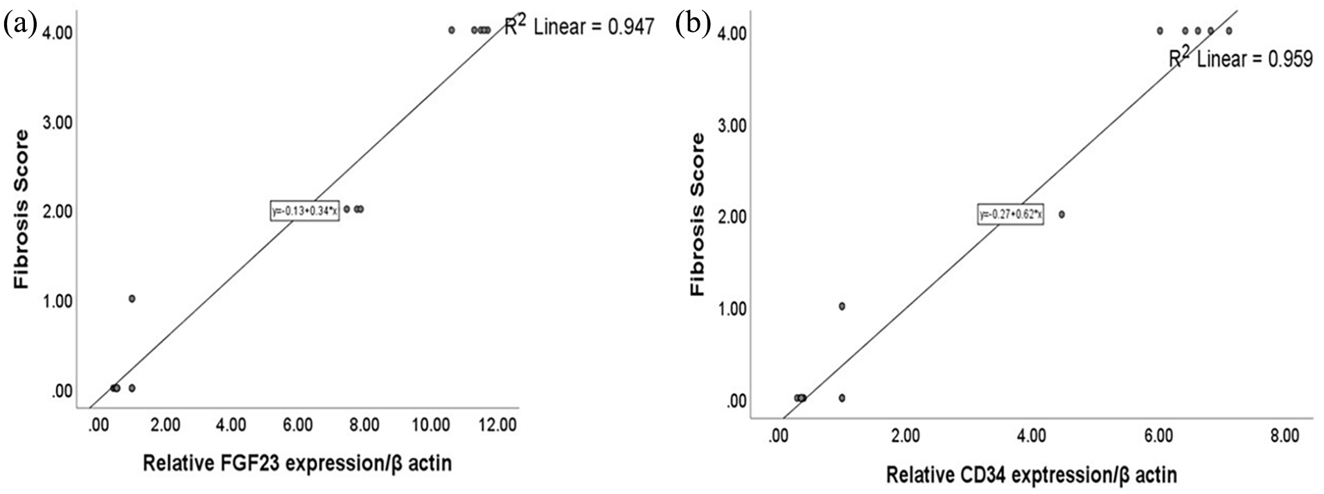
(a) Correlation between fibrosis score and FGF23 and (b) Correlation between fibrosis score and CD 34.
H&E staining for histopathological analysis of liver sections
The liver sections from both the control and Hesp groups showed normal arrangements of the hepatic cords, central veins, portal regions, and sinusoids (Figure 6a, b, e, f). Hepatic sections from the TAA rats demonstrated significantly altered portal regions, central veins, and hepatic cord arrangements. Thick anastomosing fibrous tissue deposition, hemosiderin-loaded macrophages, leukocytes, and many apoptotic hepatocytes producing multiple different entire hepatic nodules were the characteristics of the hepatic cords. The TAA + Hesp group’s liver sections had moderately reduced lesions in the liver, which were characterized by fibrous strands that anastomosed from the portal regions and contained a small number of dilated blood vessels as well as a small number of apoptotic hepatocytes that formed a few whole nodules in the liver (Figure 6c, d, g, h). The TAA group showed a substantial increase in lobular inflammation, portal inflammation, hepatic steatosis grade, and fibrosis score when compared with the control group. Meanwhile, Hesp therapy (TAA + Hesp) decreased these scores in the interim, but it did not bring them back to baseline (Figure 6i–l).

Histopathological analysis of H&E-stained liver sections demonstrates (a, b) normal control group, (c, d) markedly disrupted arrangement of hepatic cords, central veins, and portal areas, with thick anastomosing fibrous tissue deposition infiltrated with leukocytes and hemosiderin-laden macrophages (thick black arrows) filled with congested blood vessels (red arrows) and (thin arrows) refers to many apoptotic hepatocytes forming separate complete hepatic nodules (*) in TAA group. (e, f) Hesp group. (g, h) moderately diminished hepatic lesions with anastomosing fibrous strands extending from the portal areas (thick black arrows) filled with few dilated blood vessels and very few apoptotic hepatocytes (thin arrows) creating few complete hepatic nodules (*). Few dispersed fat vacuoles can be seen in these groups (yellow arrows). Magnification (µm): 100 bar = 100×; 400 bar = 50×. (i) Hepatic steatosis, (j) hepatic lobular inflammation, (k) hepatic portal inflammation, and (l) hepatic fibrosis grades in different experimental groups.
Masson’s trichrome to examine liver sections histopathologically
The control and Hesp groups’ liver sections did not exhibit any fibrosis (Figure 7a, b, e, f), On the other hand, the TAA group exhibited hepatic nodules that were surrounded by a thick layer of fibrous tissue that was stained blue. There was a considerable reduction in fibrous tissue deposition with blue staining in the TAA + Hesp group (Figure 7c, d, g, h). In comparison to the control group, the TAA group had a substantial rise in fibrosis score as determined by Masson’s trichrome. Although Hesp treatment (TAA + Hesp) decreased this score, it did not go back to its baseline (Figure 7i).

Hepatic histopathological examination by Masson’s trichrome stain. (a, b) the normal control group shows no fibrosis. (c, d) TAA group that demonstrates a strong layer of fibrous tissue deposits tinged blue encircling hepatic nodules (black arrows). (e, f) The Hesp group does not have fibrosis. (g, h) In the TAA + Hesp group, the deposited bluish-stained fibrous tissue showed a moderate reduction in thickness. Magnifications (µm): 400 bar equals 50×, and 100 bar equals 100×. scoring of hepatic fibrosis in the experimental groups by Masson’s trichrome with distinct superscript characters (P < 0.05).
The immunohistochemistry expression of both TGF-β and α-SMA
As shown in Figures 8 and 9 the control and Hesp rats were stained negatively against both TGF-β and α-SMA. In the meanwhile, some hepatocytes in the TAA-treated rats exhibited increased brown cytoplasmic expression in opposition to TGF-β, whereas numerous neighboring hepatocytes and fibrous tissue also displayed markedly positive brown expression in opposition to α-SMA. In fibrous tissue, the TAA + Hesp group exhibited α-SMA expression and very rare nearby hepatic cells, as well as somewhat reduced positive brown cytoplasmic expression against TGF-β in a rare number of liver cells.

Hepatic sections immunohistochemical stain. (a, b) represent the control group. (c, d) several hepatocytes (black arrows) in the TAA-intoxicated group exhibit elevated brown cytoplasmic expression in opposition to TGF-β. (e, f) are negative staining in the Hesp rats against TGF-β. A small number of hepatocytes (black arrows) in the TAA + Hesp group. (g, h) are showing a somewhat reduced positive brown cytoplasmic expression against TGF-β. IHC used Mayer’s hematoxylin as a counterstain. Magnification (µm): 400 bar equals 50×, and 100 bar equals 100×. TGF-β positive immune-expression in the various experimental groups; each bar shows the mean ± standard error (n = 8). Significant differences are shown by bars with distinct superscript characters (P < 0.05).

Microscopic pictures of immunohistochemical stains. (a, b) the control group. (c, d) markedly positive brown expression in opposition to α-SMA in TAA intoxicated group. (e, f) Hesp group. (g, h) Significantly reduced positive brown cytoplasmic expression in fibrous tissue in comparison to α-SMA expression, and very few neighboring hepatocytes (black arrows) in the TAA + Hesp group. A black arrow indicates a positive expression using Mayer’s hematoxylin as a counterstain. Magnification (µm): 400 bar equals 50× and 100 bar equals 100×. positive immune-expression in the various experimental groups (P < 0.05).
Discussion
Many etiologists emphasize that persistent liver damage leads to hepatic fibrosis. Although liver fibrosis often goes away after an injury is stopped, if the underlying condition is not well addressed, it can worsen and eventually turn into cirrhosis. Liver insufficiency is brought on by progressive fibrosis, which also increases the chance of hepatic cell carcinoma (HCC) Norušis. 30
TAA serves as a sulfur donor in both pharmacological and industrial applications. Henceforth, TAA was first used as a fungicide to avoid the rot of citrus fruits, particularly oranges. Recently, TAA metabolites like sulfines and sulfene have been discovered to have a powerful hepatotoxic and carcinogenic effect. 31 The hepatic enzymes ALT, AST, and ALP are sensitive markers used to assess the status of liver injury. 32 In the current study, Serum levels of total protein and albumin were significantly lower in TAA-mediated liver damage than in control rats, and there was a substantial rise in ALT, AST, ALP and bilirubin (total and direct) activity. TAA diminished hepatic function capacity attributed to enhanced free radical production and inflammatory mediators in the hepatocytes, while decreased antioxidant protection mechanisms with severe damage to the hepatic cells.33–35,6 Additionally, TAA causes damage that hinders RNA’s drive from the nucleus to the cytoplasm, raising serum liver indications. In addition to centrilobular necrosis, hepatocyte destruction, and a significant decline in albumen and total protein synthesis from hepatocytes.36–38 Our results confirmed histopathological findings and Masson’s trichrome stain that revealed many complete apoptotic nodules.
Our results disclose that the administration of Hesp significantly ameliorated the damaging effects of TAA on hepatocytes and liver function through downregulated AST, ALT, and ALP activities and both total and direct bilirubin compared to the group that received TAA treatment, but did not reach levels considered normal. Hesp may prevent the harmful effects of TAA and restore damaged hepatic cellular membranes, both of which could lower the amount of enzyme leakage into the bloodstream, 39 As well as, this hepatoprotective effect of Hesp might be attributed to its anti-inflammatory and antioxidant activities as Because of its ability to scavenge free radicals and enhance endogenous antioxidant enzymes like GSH or SOD, it offers effective cellular antioxidant defense against the harmful effects of hydrogen peroxide.40–43 The histological findings, which showed mild liver lesions and a small number of apoptotic hepatocytes, supported these conclusions.
Liver fibrosis is thought to have a close relationship with lipid profile. TG and TC levels. The TAA treated rats exhibited markedly elevated values than the control one, this might be linked to abnormalities in the metabolism of fat. 44 TAA metabolites reduce the activity of mitochondria and change the permeability of cells, which hinders lipoprotein production and secretion. 45 Hesp has the ability to lower serum TC and TG levels in treated TAA rats, as Hesp has inhibitory effects through suppressing free radicals as lipids are the first target of free radicals’ peroxidation. 46 Additionally, inhibition of hepatic hydroxy methyl glutaryl-coenzyme A reductase and cholesterol acyltransferase activities. 47
An imbalance between the production of ROS locally and antioxidant systems is known as oxidative stress. 48 In this work, TAA-induced cytotoxic injury exhibited by a notable increase in MDA levels in liver cells and a marked reduction in antioxidant biomarkers levels as CAT, SOD, and GSH in hepatic tissue compared to control rats. Reactive oxidative agents are produced during the oxidative metabolism of TAA by the hepatocyte cytochrome P-450 monooxygenase systems. TAA-S-dioxide is a particularly reactive molecule that targets tissue lipids, protein, and DNA, causing tissue oxidative damage and necrosis.44,49 Our results are in agreement with Özdemir-Kumral et al.; Alkreathy and Esmat.37,28 In our investigation, the suppression of lipid peroxidation and lower MDA levels in hepatic homogenates served as the main indicator for assessing the antioxidant ability of Hesp against the injured liver in TAA -exposed group. Hesp is a natural polyphenolic compound, has antioxidant activity and protective effects through its ability to neutralize ROS, and scavenge free radicals. It protects against both inflammation and oxidative damage by interrupting the vicious cycle contributing to the pathophysiology of hepatic injury at later stages. 41
In current work, fibrotic indicators markers, CD34 and FGF23 expression displayed higher elevation in TAA-intoxicated group than the control rats. CD34 is a cell surface glycoprotein expressed on hematopoietic progenitor cells, normal vascular endothelium, and fibroblasts. 50 Endothelial antigen CD34 is utilized to separate well-differentiated HCC from non-neoplastic liver and to emphasize the density of vessels as a direct indicator of the degree of neoangiogenesis in malignancies. The first argument in support of the hypothesis that angiogenesis may play a major role in fibrogenesis and the advancement of the illness is the observation that vascular remodeling is a common feature in human livers with advanced fibrosis, regardless of the underlying cause. 51 Additionally, the liver sinusoids do not express CD34 while pathological conditions can alter their phenotype and express this marker. 52
It has been shown that several distinct FGF subfamilies affect fibrogenesis. Even yet, in a healthy liver, it is only very weakly expressed, damaged hepatic tissue also expresses FGF23. 53 It’s been demonstrated that FGF23 encourages renal and heart fibrosis.54,55 During liver injury, inflammatory factors induce Kupffer cell production of FGF23 that may have a paracrine pro-inflammatory effect on hepatocytes. Liver produced FGF23 may have systemic hormonal effects as well that influence diseases in other organs. 56 In addition, the promotion of inflammation in non-hepatic organs by hepatic FGF23 may result in the release of other pro-inflammatory factors from these organs which in turn triggers the progression of liver disease.57,58 Acute and chronic liver injury induces hepatic production of FGF23 through the stimulation of the resident liver macrophages or Kupffer cells by injury-associated inflammatory factors. 56 Additionally, in rats treated with TAA, the histology revealed a notable number of apoptotic hepatocytes producing many separate entire hepatic nodules. Hepatic nodules were surrounded by thick, fibrous tissue deposition, as shown by Masson’s trichrome staining. Furthermore, because they are inflammatory molecules and indicators of HSC activity, TGF-β and α-SMA play a crucial role in the development of liver fibrosis. They also further promote collagen transcription as well as other ECM constituents.59,60 TAA produced ROS, which in turn stimulated HSC, a key component of the extracellular matrix (ECM) in chronic liver fibrosis by the upregulation of TGF-beta and alpha-SMA. The development of ECM synthesis and cell division, along with the presence of alpha-SMA in myofibroblasts, signal the start of HSC. 38
Hesp therapy decreased CD34 and FGF23 in comparison with TAA intoxicated rats. The strong positive effects of Hesp administration on diabetic toxicity in the liver and kidneys suggest that it may be included in the alternative treatment methods in the future.56,61 Our results are in line with Kumar et al. and Dokumacioglu et al.56,61 who discovered that Hesp administration reversed the increase in FGF23 levels in diabetic rats, by enhancing the antioxidant system, and removing disorders in the axis of FGF23, production in liver, and kidney. The FGF-23 pathway is a promising bioindicator in various cases of systemic toxicity and pathology.56,61 Also, the histopathology and immunohistochemistry studies in our work, approved the protective effect of Hesp against TAA induced liver fibrosis. Hesp could have hepatic antifibrotic effect by the inhibition of HSC proliferation and activation by modulating TGF-β1/ α-SMA signaling pathway.62,63 Also, the decreased circulating levels of TGF-β1 has been correlated with inhibiting fibrogenesis in a rat model of ethanol-induced liver fibrosis. 64 Outstanding of the pathways of FGF23 and CD34 in hepatocytes damage, in the present study, the fibrotic marker of CD34 and FGF23 has been investigated. There was a significant positive correlation between fibrotic score with FGF23 and CD34 hepatic expression in TAA exposed group.
The limitations of our study are recommended more features studies to investigate the pharmacokinetics of Hesp in liver fibrosis conditions and its bioavailability to precise dosage regimens for treating experimental liver fibrosis models. Also, investigate the Heps treatment of liver fibrosis with variable doses and methods of administration in different animal species to explore the physiological interpretation of species differences in drug treatment. Sample size calculation was not done in our study as its experimental one.
Conclusions
Hesp stabilizes the antioxidant system with reduced oxidative damage and inflammatory conditions, by modulating fibrotic molecular pathways as the TGF-β and α-SMA pathways, in TAA-induced hepatotoxicity and fibrosis in experimental animal study. Also, in the future study, the FGF23 and CD34 are dependable fibrotic markers.
Footnotes
Acknowledgements
Authors like to express my deep thanks to Prof. Dr. Walaa awadin, professor of Pathology, Faculty of Vet. Medicine, Mansoura University for her faithful help to finish the histopathological findings in this study.
Author contributions
Aya Megahed: methodology, formal analysis, data curation, writing original draft, review, and editing.
Hatem Sembawa,Talat A. Albukhari, Waheed A Filimban, Rehab M Bagadood and Ghadir Sindi: conceptualization, validation, visualization, data curation and analysis.
Mohamed E. El-Boshy: data analysis, reviewing and revising the completed manuscript.
Fatma M. Abdelhamid, Hossam Gadalla, and Engy F. Risha: conceptualization, validation, visualization, and supervision. E.R.. Preparation of the manuscript for publication. All authors read and accepted the final manuscript.
Declaration of conflicting interests
The author(s) declared no potential conflicts of interest with respect to the research, authorship, and/or publication of this article.
Funding
The author(s) received no financial support for the research, authorship, and/or publication of this article.
Institutional review board statement
The Faculty of Veterinary Medicine at Mansoura Egyptian University’s Animal Research Ethics Committee (Ph. D/46) institutional guidelines provided ethical approval.
Ethics approval
In accordance with the institutional rules of Mansoura Egyptian University’s Faculty of Veterinary Medicine’s Animal Research Ethics Committee (Ph. D/46)
Animal welfare
Animal welfare The study was conducted in accordance with the “ guidelines for the care and use of laboratory animals” all animal procedures were approved by the Faculty of Veterinary medicine Ethics Committee at Mansoura University, Egypt.