
Abstract
IL-9, which may be an inflammatory or regulatory cytokine, can be experimentally produced in a Th17 or modified Th2 context in the presence of T cell receptor (TCR) stimulation. The primary aim of this study was to measure serum IL-9 levels in patients with inflammatory bowel disease (IBD), and evaluate their relationships with the patients’ clinical characteristics. The secondary aim was to determine the levels of interferon-γ (IFN (interferon)-γ), Th2 cytokines (IL-4, IL-5 and IL-13), and IL-6 in order to clarify the context of detectable peripheral cytokines in which IL-9 is produced.
Venous blood samples of 43 IBD patients (20 with Crohn’s disease [CD] and 23 with ulcerative colitis [UC]) were analysed by means of quantitative enzyme-linked immunosorbent assays using purified anti-human IL-4, IL-5, IL-13, IFN-γ, IL-9 and IL-6 antibodies, and the laboratory findings were statistically correlated with their clinical expression.
None of the patients showed the peripheral presence of IL-4, IL-5 and IL-13. Forty (93%) were positive for IFN-γ, thus confirming the presence of Th1 in both UC and CD, and IFN-γ levels correlated with disease activity (P = 0.045). Eighteen patients (41%) were positive for IL-9, which was associated with a severe prognosis (P <0.001), and 72.2% of the IL-9-positive patients were also IL-6 positive. There was a significant correlation between disease severity and IL-9 in the CD patients (P <0.001), but not in the UC patients (P = 0.1).
Our findings confirm the presence of common Th1 cytokines in UC and CD. However the IL-9 positivity indicates the presence of an alternative population of T cells that respond to antigen stimulation and condition the prognosis of IBD. The fact that the same serum IL-9 levels were differentially associated with clinical measures of CD and UC activity suggest that the same cytokine can be produced in different contexts.
Keywords
Introduction
The differentiation of CD4 T helper (Th) cells plays a critical role in the immune response. Four major Th cell subsets have been described: Th1 produce IFN-γ; Th2 are characterised by the secretion of interleukin (IL)-4, IL-5 and IL-13; Th17 produce IL-17; and Treg cell subsets secrete transforming growth factor-β (TGF-β).
Th1 and Th2 have been the sole paradigm of T helper functional diversification for many years. 1 Both cell types arise after the sustained activation of uncommitted Th0 cell precursors and are characterised by the expression of mutually exclusive sets of cytokines. This cellular dichotomy has been mechanistically explained by the mutual inhibition of the master transcription factors T-bet and GATA-3 at individual cell level, and by cross-regulatory mechanisms at cell population level. 2 However, the identification of Th17 and TGF-secreting Treg cells has complicated the picture of the CD4+ effector T cell programme. Neither have yet been rigorously analysed, but recent acquisitions suggest that memory cells need to be flexible in order to be able to protect against a re-encountered pathogen. 3 Th17 cells express the transcription factor retinoic acid-related orphan receptor γt (RORγt) and IL-17A, which not only protect against fungi and various other extracellular bacteria, but also play a pathogenetic role in the development of autoimmune inflammatory disease.
Treg-controlled self-reactive T cells escape negative selection, and control excessive effector T cell responses against exogenous antigens when these become dangerous for the body. They suppress both Th1 and Th2 responses by means of contact-dependent mechanisms and/or the production of IL-10 and TGF-β. 4 It has been shown that the IL-l0 secreted by these cells effectively prevents chronic intestinal inflammation, and it has been postulated that TGF-β is involved in maintaining tolerance to dietary antigens. 5 TGF-β1 can promote the development of Th17 effector and adaptive Treg cells, whereas the simultaneous production of IL-6 contributes to development of Th17 cells but inhibits Treg cells.4,6 The central transcription factors for Th17 (RORγt) and Treg (Foxp3) both depend on TGF-β signalling, and their direct interactions establish a competitive antagonism that determines the specification of a Th17 or induced Treg lineage. 7 Tyrosine phosphatase SHP-1 is a critical regulator of Th17: its impairment promotes Th17 development, and the ex vivo disruption of its activity in T cells leads to a hyper-response to stimulation by IL6 and IL21. It has also been found that tyrosine phosphatase SHP-1 decreases the overall cytokine-induced phosphorylation of STAT3 in primary CD4+ T cells. 8
One important characteristic of Th17 and Treg cells is their ability to produce IL-9. The IL-9 produced by Th17 is pro-inflammatory in a standard myelin oligodendrocyte glycoprotein peptide-induced model of experimental autoimmune encephalomyelitis, 9 whereas the IL-9 produced by Treg cells mediates graft tolerance by means of a mast cell-dependent process. 10 IL-9 may therefore be inflammatory or regulatory depending on the context.
Nevertheless, current evidence indicates that late Th cell differentiation pathways are more complex and probably include other, non-canonical cell types. Early phenomenological models represented the development of Th1 vs. Th2 responses from naïve Th0 cells using ordinary differential equations. However, although the stable states corresponding to canonical Th1, Th2, Th17 and Treg subtypes are readily identified, they also co-exist with other hybrid cell types that co-express a combination of Th1, Th2 and Treg markers in an enviroment-dependent manner. The in vitro stimulation of Th2 cells in the presence of TGF-β generates a non-canonical cell type expressing IL-10 and IL-9 in the absence of Foxp3, 2 and this subset of differentiated T cells has led to a re-evaluation of the classification of IL-9 as a type 2 cytokine. In vitro IL-9-producing T cells seem to be a distinct subset of ‘Th9’ cells that depend on TGF-β signalling. 11 TGF-β can alter Th2 cells to such an extent that they lose their previous characteristics, including the expression of GATA-3 and the ‘signature’ cytokines IL-4, IL-5 and IL-13. 3
It has also been shown that IL-25 (IL-17-E), a member of the IL-17 cytokine family, plays a role in the regulation of ‘Th9’ cells. IL-25 can be found in the large intestine (particularly in intestinal epithelial cells) and is involved in the maintenance of intestinal homeostasis, but is not expressed in germ-free mice. 12 The requirement of TGF-β and IL-4 for IL-25-dependent IL-9 production can be by-passed by the retroviral expression of IL-17RB in T cells or in transgenic mice in which IL-17RB expression is controlled by the CD4 promoter, thus suggesting that TGF-β and IL-4 render T cells IL-25-responsive by inducing IL-17RB expression. 11 It has been reported that a number of innate immune cells responsive to IL-25 (including monocytes and NKT cells) express functional IL-17RB, and that their treatment with IL-25 induces Th2 cytokine production. Three groups have recently and independently identified a new population of innate effector cells that are innate IL-25 responders, but it is still unclear whether these are the same population and function in other diseases associated to Th2-cells. 13
The primary aim of this study was to evaluate serum IL-9 levels in inflammatory bowel disease (IBD) patients, and their relationships with the clinical features of the disease. We also studied the levels of interferon-γ (IFN-γ), Th2 cytokines (IL-4, IL-5 and IL-13), and IL-6 in order to clarify the context in which IL-9 is produced.
Materials and methods
Patients
Blood samples were randomly obtained from 43 IBD patients attending Fatebenefratelli Hospital in Milan, Italy: five untreated patients with inactive disease, 29 mesalazine-treated patients with mild and moderate disease, and nine mesalazine-treated patients with severe disease. The diagnosis of IBD was confirmed by means of standard endoscopic and histological criteria (the additional haematoxylin and eosin staining of each sample). Their clinical and immunological data were obtained from their medical records; 97% of the patients did not have any other relevant symptoms.
The serum levels of the studied cytokines were also measured in 10 controls randomly selected from blood donors with a negative history for IBD.
The study was approved by the hospital’s Ethics Committee, and the patients were enrolled after signing an informed consent form in accordance with the Declaration of Helsinki.
Sample collection and management
The 10 mL venous blood samples for cytokine analysis were collected in pyrogen-free tubes between 08:00 and 10:00, and then immediately centrifuged at 3000 rpm for 15 min. The serum samples were aliquoted and stored at −80°C. The samples were frozen and thawed only once.
Enzyme-linked immunosorbent assays (ELISA)
Serum IL-9, IFN-γ, IL-4, IL-5, IL-13 and IL-6 levels were assayed in duplicate using commercial ELISA kits (Human IL ELISA Ready-Set-Go, eBioscience, San Diego, CA, USA) in accordance with the instructions of the manufacturer. After specific anti-human IL antibody adhesion to microwells, the human IL present in the sample or standard binds to the adsorbed antibodies, and then the addition of a biotin-conjugated anti-human IL antibody binds to the human IL captured by the first antibody. The reaction was revealed by means of streptavidin/horseradish peroxidase (HRP) staining, the coloured product of which is proportionate to the amount of human IL in the sample or standard. The reaction was terminated by adding H2SO4 (sulphuric) acid (3 mol/L), and absorbance was measured at 450 nm. A standard curve was prepared using seven human IL standard dilutions. The limit of detection was 6 pg/mL: values of 6–10 pg/mL were considered borderline, and those of >10 pg/mL positive.
Clinical scores
All of the patients were diagnosed as having Crohn’s disease (CD) or ulcerative colitis (UC) on the basis of standard endoscopic, histological, barium contrast enema, or other recognised criteria.14,15
The activity of CD was defined by means of the Crohn’s Disease Activity Index (CDAI) and markers of inflammation. 16 The CDAI is based on eight factors, the scores of which are totalled after adjustment with a weighting factor: the number of liquid or soft stools each day for 7 days; abdominal pain each day for 7 days (severity graded 0–3); subjectively assessed general wellbeing each day for 7 days (from 0 = well to 4 = terrible); the presence of complications such as arthralgia or frank arthritis, inflammation of the iris or uveitis, erythema nodosum, pyoderma gangrenosum or aphthous ulcers, anal fissures, fistulae or abscesses, fever; the need to use loperamide or opiates for diarrhoea; the presence of an abdominal mass (0 = absent, 2 = questionable, 5 = definite); haematocrit levels of <0.47 in men or <0.42 in women; and percentage deviation from standard weight. A score of ⩽150 was defined as inactive disease (remission), and scores of 151–220, 221–450 and >450 points as mildly, moderately and highly active disease, respectively. Disease activity was evaluated at the time of serum collection.
The activity of UC was defined using the 30-point Clinical Colitis Activity Index (CCAI): 17 a score of 0–4 was defined as inactive disease (remission), and scores of 5–10, 11–17 and ⩾18 as mildly, moderately and highly active disease, respectively.
Statistical analysis
The continuous variables are expressed as mean values and standard deviation, and were compared using the unpaired Student’s t test; the categorical variables are expressed as absolute and relative frequencies, and were compared using the chi-squared test. The data were analysed using the Statistical Package for Social Sciences (SPSS 13.0, SPSS Inc., Chicago, IL, USA). All of the tests were two-sided, and P values of <0.05 were considered statistically significant.
Results
Study population
Table 1 shows the demographic and clinical characteristics of the study population.
Demographic and clinical characteristics of study patients.

Cytokine background in IBD patients
All of the samples were negative for Th2 cytokines (IL-4, IL-5 and IL-13) (Table 2). Ninety-three percent of the patients were positive or borderline for IFN-γ, and their IFN-γ levels correlated with disease activity (P = 0.045) (Table 3). The absolute IFN-γ levels in the positive patients varied widely from a minimum of 11 pg/mL to a maximum of 500 pg/mL (average 83 ± 29.8 pg/mL). Furthermore, the fact that diagnosis, therapy and disease duration had no effect on cytokine levels suggests that Th1 plays a role in both bowel diseases.
Cytokine levels in all samples.

CD, Crohn’s disease; UC, ulcerative colitis.
Correlations between IFN-γ and disease activity.
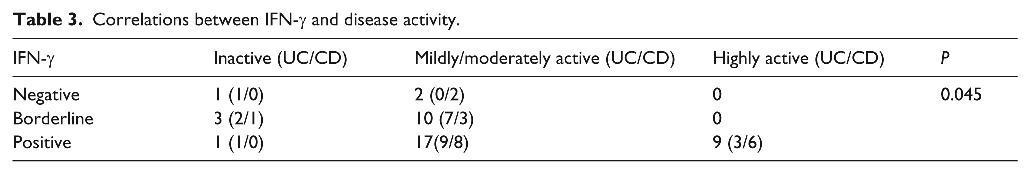
Twenty-three patients (53%) were positive for IL-6 (Table 2), the presence of which was not associated with disease activity (P = 0.5) (Table 4). The absolute levels of IL-6 in the positive patients were 20 ± 3.2 pg/mL, and were lower and more constant than IFN-γ levels. The role of IL-6 was independent of Th2 cytokines and did not correlate with IFN-γ levels. Furthermore, the levels of IL-6 did not distinguish the patients with UC from those with CD, although 72% of the IL-9 positive patients were also positive for IL-6. In this Th1 habitus, the simultaneous production of IL-6 and IL-9 suggests the development of Th17 cells and the inhibition of Treg cells, and the combination of these cytokines suggests the presence of a cell network that includes Th1 cells, monocytes and cells producing IL-9. Eighteen patients (41%) were positive for IL-9, which was associated with a severe prognosis (P <0.001). The levels of IL-9 were always >10 pg/mL (mean value, 40 ± 5 pg/mL), and never borderline, whereas IFN-γ and IL-6 levels were borderline in 13 and six cases, respectively (Table 2). There was a significant association between IL-9 levels and disease activity in the patients with CD (P <0.001), but not in those with UC (P = 0.1) (Table 5), thus confirming IL-9 may be inflammatory or regulatory.
Correlations between IL-6 and disease activity.

Correlations between IL-9 and disease activity by type of disease.

Using the same method, Th1, Th2 and IL-6 cytokines were all undetectable in the 10 control patients, whereas a borderline level of IL-9 was detected in two patients.
Discussion
There is considerable debate as to whether UC and CD express fully polarised immunophenotypes of IBD, but recent studies indicate markedly increased levels of Th1-related cytokines and Th17 in the inflamed mucosa of CD patients, whereas the inflamed mucosa of UC patients show an increased production of Th2 cytokines. 18
In this pilot study, IFN-γ was detected in 93% of the patients, only five of whom had inactive disease and were untreated. The remaining 38 were all being treated with mesalazine (patients treated with corticosteroids or immunosuppressors were excluded). Our findings seem to highlight a common Th1 cytokine pattern in UC and CD patients, thus supporting the hypothesis that the diseases have a common genetic background. It is known that protein tyrosine phosphatase non-receptor type 2 (PTPN2) gene variants are associated with susceptibility to both, 19 and PTPN2 regulates IFN-γ-induced signalling and barrier function in intestinal epithelial cells. The loss of PTPN2 promotes IFN-γ-induced STAT signalling and the secretion of IL-6 and macrophage chemoattractant protein 1 (MCP-1), 20 and PTPN2 protects epithelial barrier function in vitro by restricting the ability of IFN-γ to increase epithelial permeability and prevent the induction of the expression of the pore-forming protein claudin-2. 21
However, current evidence strongly suggests that IBD is a result of a more complex mechanism in genetically susceptible individuals that leads to alterations in the processing of enteric antigens, pathogenic T cell activation and chronic inflammation.22,23 In the absence of any input from the environment, resting Th0, Th1 and Th2 are the only three attractors but, in a strongly polarising environment, Tregs may differentiate into mixed cell types associated with the Th1 or Th2 cell constellations. Treg cells can be maintained only in the presence of TCR stimulation delivered by APC, and IL-2 produced by other T cells.2,24
The levels of Th2 cytokines (IL-4, IL-5 and IL-13) were undetectable in the blood of our IBD patients. However, high levels of a plethora of inflammatory cytokines have been found in inflamed mucosa, and the complex differentiation of Th17 cells may be due to differences in the local cytokine milieu. Recent studies have shown that lamina propria mononuclear cells isolated from CD patients produce significantly less TGF-β1 than those isolated from controls when stimulated via both the CD2 and CD28 pathways, whereas UC patients produce more than controls under the same conditions. 23 Other studies have shown that the levels of most of the transcripts for Th17-related cytokines are higher than normal in patients with UC or CD, but more abundant in those with UC than in those with CD; on the contrary, the upregulation of IFN-γ mRNA are more marked in the lamina propria CD4+ T cells of CD patients. 22
On the basis of our data, IBD patients have detectable blood levels of IL-9 and IL-6. Circulating IL-6 was detected in 23/43 serum samples (17 positive and six borderline), but there was no correlation between IL-6 positivity and disease activity (P = 0.5) (Table 4). Many authors have investigated IL-6 levels in serum and endoscopic biopsy samples taken from IBD patients,25,26 and an analysis of cytokine production by lamina propria mononuclear cells has revealed an increase in the spontaneous production of IL-6, TNF-α and IL-1. 27
IL-6 was detected in the serum of 13/18 IL-9-positive patients (72.2%). Unlike those of IFN-γ and IL-6, the serum levels of IL-9 were always >10 pg/mL without any borderline values, and correlated with highly active disease (P <0.001).
Only 41.8% of the patients in our study were positive for IL-9, which suggests that a variable that can sporadically affect the other responses associated with a rigid Th1 orientation may play a role in IBD. IL-9 can be produced by Treg or Th17 cells with different effects, but the IBD environment in which both active TGF-β and IL-6 are available promotes the development of Th17 cells. 7 However, despite the common genetic substratum and inflammatory role of IL-9, we found a correlation between disease activity and IL-9 positivity only in our CD patients, which suggests that the same cytokine can be produced in different contexts. This hypothesis is supported by the findings of an international study showing a close association between the IL2/IL21 locus on 4q27 and UC. 28 Interleukin-2 is the key cytokine supporting the survival and function of Treg cells, 29 and TGF-β and IL-2 can also be crucial in ‘reprogramming’ Th2 cells to lose their characteristic profile and switch to IL-9 secretion or, in combination with IL-4, drive the differentiation of Th9 cells directly. 3 In colonic mucosa, IL-25 (IL-17E), which has been experimentally found to be involved in this switch, can downregulate Th1/Th17 immune responses in an IL-10-dependent manner, 30 and it has also been found that the IL-10 31 and IL-17 families 22 are increased in the colonic mucosa of patients with active UC. It is therefore possible that, IL-9 can be produced by Th17 or modified Th2 cells in a Th1 habitus in the presence of TCR stimulation in vivo and is detectable in the peripheral blood of IBD patients. The in vitro and in vivo hierarchy of IL-9 production in mouse suggests that adaptive Treg cells consistently produce more IL-9 than Th2 cells, but produce less than cultured T cells derived from TGF-β and IL-4 (Th9) or IL-6 (Th17). 9
IL-9, an important regulatory cytokine in lung and the gastrointestinal tract that plays a role in inflammation and immunosuppression, is produced by Th17, Treg and modified Th2 cells.
The majority of our IBD patients were positive for IFN-γ, thus confirming the presence of Th1 in both UC and CD.
Contacts between the antigen and genetic substrate are crucial for the development, progression and different clinical expressions of IBD.
IL-9 positivity in some IBD patients suggests that factors other than the TPNP2 variant may be involved in worsening the pro-inflammatory response of the bowel T cell system. Furthermore, our findings confirm that IL-9 may be inflammatory or regulatory in human diseases.
Footnotes
Authors’ contributions
CD and FA participated in the design and coordination of the study and to draft the manuscript; PSP participated in the study in study design and critical review. CD carried out the enzyme-linked method. SS, SB, PD and SB participated in the design of the study and performed the statistical analysis. PLA conceived of the study, and participated in its design and coordination.
All of the authors have read and approved the final manuscript.
Declaration of conflicting interests
The author(s) declared no potential conflicts of interest with respect to the research, authorship, and/or publication of this article.
Funding
This project was supported by our Department.