
Abstract
Background:
In young patients with irreparable subscapularis deficiency (SSC-D) and absence of severe osteoarthritis, anterior latissimus dorsi transfer (aLDT) has been proposed as a treatment option to restore the anteroposterior muscular force couple to regain sufficient shoulder function. However, evidence regarding the biomechanical effect of an aLDT on glenohumeral kinematics remains sparse.
Purpose/Hypothesis:
The purpose of this study was to investigate the effects of an aLDT on range of glenohumeral abduction motion, superior migration of the humeral head (SM), and cumulative deltoid force (cDF) in a simulated SSC-D model using a dynamic shoulder model. It was hypothesized that an aLDT would restore native shoulder kinematics by reestablishing the insufficient anteroposterior force couple.
Study Design:
Controlled laboratory study.
Methods:
Eight fresh-frozen cadaveric shoulders were tested using a validated shoulder simulator. Glenohumeral abduction angle (gAA), SM, and cDF were compared across 3 conditions: (1) native, (2) SSC-D, and (3) aLDT. gAA and SM were measured using 3-dimensional motion tracking, while cDF was recorded in real time during dynamic abduction motion by load cells connected to actuators.
Results:
The SSC-D significantly decreased gAA (Δ–9.8°; 95% CI, –14.1° to −5.5°; P < .001) and showed a significant increase in SM (Δ2.0 mm; 95% CI, 0.9 to 3.1 mm; P = .003), while cDF was similar (Δ7.8 N; 95% CI, –9.2 to 24.7 N; P = .586) when compared with the native state. Performing an aLDT resulted in a significantly increased gAA (Δ3.8°; 95% CI, 1.8° to 5.7°; P < .001), while cDF (Δ–36.1 N; 95% CI, –48.7 to −23.7 N; P < .001) was significantly reduced compared with the SSC-D. For the aLDT, no anterior subluxation was observed. However, the aLDT was not able to restore native gAA (Δ–6.1°; 95% CI, –8.9° to −3.2°; P < .001).
Conclusion:
In this cadaveric study, performing an aLDT for an irreparable subscapularis insufficiency restored the anteroposterior force couple and prevented superior and anterior humeral head migration, thus improving glenohumeral kinematics. Furthermore, compensatory deltoid forces were reduced by performing an aLDT.
Clinical Relevance:
Given the favorable effect of the aLDT on shoulder kinematics in this dynamic shoulder model, performing an aLDT may be considered as a treatment option in patients with irreparable SSC-D.
Keywords
Isolated irreparable subscapularis (SSC) tendon tears still pose a significant challenge for surgeons and patients, especially in young patients with high functional demands. 7 In young patients with SSC deficiency (SSC-D) without the presence of severe osteoarthritis, tendon transfers enable shoulder surgeons to restore the anteroposterior force couple to regain sufficient shoulder function.14,15,18,20
Recent evidence propagates the use of a modification of the latissimus dorsi (LD) transfer with an anterior fixation at the lesser tuberosity (anterior LD transfer [aLDT]) to restore the anteroposterior force couple.12-14 The rationale for performing a tendon transfer in the setting of SSC insufficiency is to restore internal rotation strength and limit anterior humeral translation. 20 The aLDT has been hypothesized to hold the ideal anatomic location for restoring the physiological vector of the SSC muscle, 8 by mimicking the physiological line of pull of the SSC tendon. This notion is supported by recent clinical evidence, which demonstrates favorable clinical outcomes and overall shoulder function after the aLDT, although limited long-term data are available. 20 Acknowledging this evidence, the comprehensive effects of aLDT on glenohumeral kinematics in the sense of range of abduction motion, superior humeral head migration, and compensatory deltoid forces during dynamic testing have not yet been investigated in the setting of SSC-D.
Thus, the purpose of the present study was to investigate the effects of an aLDT on range of glenohumeral abduction motion, superior migration of the humeral head (SM), and cumulative deltoid force (cDF) in a simulated SSC insufficiency using a dynamic shoulder model. It was hypothesized that performing an aLDT would restore the insufficient anteroposterior force couple, enabling a restoration of native glenohumeral kinematics.
Methods
Eight fresh-frozen, cadaveric shoulders (mean age, 59.6 ± 12.7 years; 3 male, 5 female; 2 left, 6 right) were obtained from Medcure Inc and used for the study. All specimens underwent visual and radiographic inspection to detect and exclude those with tears of the rotator cuff tendons and capsule, joint contractures, moderate to severe osteoarthritis, or bony defects. The study was reviewed via Human Research Determination Form by the institutional review board (IRB) of the University of Connecticut, and it was concluded that no IRB approval was required.
Specimen Preparation
Specimen preparation was performed according to a previously described method.1,11,23 Dissection of the skin, subcutaneous tissue, and muscles was performed after they were thawed overnight at room temperature. The rotator cuff muscles, coracoacromial ligament, and humeral tendinous insertions of the LD muscle were carefully preserved. The anterior, middle, and posterior portions of the deltoid tendon were detached from the muscle belly at the deltoid tuberosity and preserved with anchor loops sutured to the tendinous insertions using a locking running stitch (No. 2 FiberWire; Arthrex Inc). Each of the 3 deltoid heads was then attached to an individual shoulder simulator actuator.1,11,26 Suture loops were placed at the humeral attachment of the LD covering the whole insertional footprint to ensure physiological load distribution. Subsequently, the attachments were then each attached to an individual actuator.
The rotator cuff muscles (supraspinatus, SSC, infraspinatus, and teres minor) were sharply detached from the scapula and separated from the underlying capsule. The infraspinatus and teres minor muscles were simulated as 1 unit as described. The supraspinatus, SSC, and the infraspinatus/teres minor muscles were sutured to pulley straps (No. 5 FiberWire; Arthrex Inc) to avoid pull-through during load application.1,11,17,26
A steel rod, loaded with 1.7 kg, was cemented into the distal humerus 30 cm distal from the center of the humeral head, representing a constant moment arm for each tested shoulder.17,29 The steel rod was positioned to place an anatomically correct moment arm on the shoulder. The glenohumeral joint capsule was vented by opening the rotator interval to prevent changes during testing as previously described.1,11,26 The scapular body was placed in a custom rectangular box with the medial border aligned perpendicular to the ground and the glenoid tilted 10° superiorly, while bone cement was poured in the box to ensure proper fixation.1,11,16,26,29
Testing Setup
For biomechanical testing, a validated dynamic cadaveric shoulder model was used (Figure 1). ¶ The shoulder simulator consisted of up to 6 linear screw-driven actuators (Bimba) connected to load cells (444 N; Futek). A universal strain gauge signal conditioner (Model CSG110; Futek) was linked to a panel mount display (Model IMP 650; Futek), while a test and measurement software (Sensit Version 2.5.1.0; Futek) was used for load cell data acquisition in real time.1,11,26
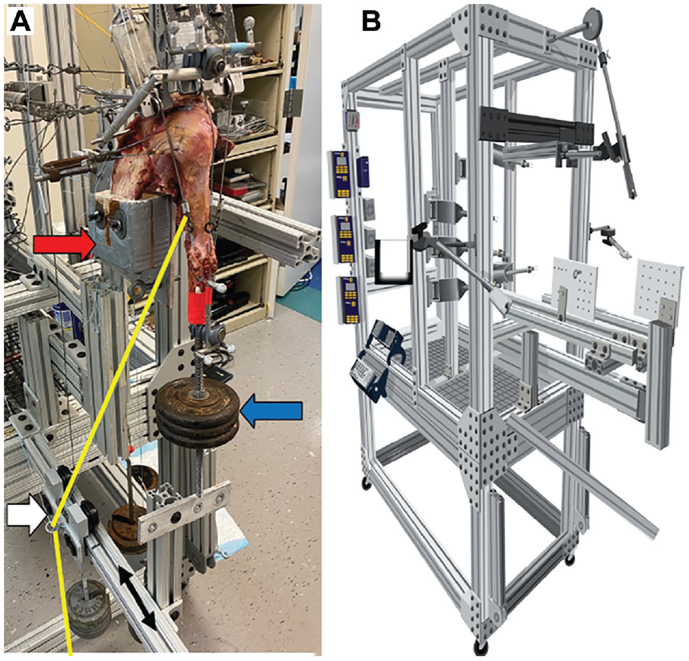
(A) The specimen was mounted to the simulator (upper arrow). A steel rod, loaded with 1.7 kg, was cemented into the distal humerus 30 cm distal from the center of the humeral head, representing a constant moment arm for each tested shoulder (lower arrow). The white arrow represents the pull of line of the latissimus dorsi tendon. Starting at the humeral insertion, the suture loop was routed over a guide pulley, which was placed on a leveled slide rail, allowing for medial and lateral (relative to the mounted shoulder specimen) motion of the guide pulley during dynamic abduction. (B) The dynamic biomechanical testing rig. 21
The potted scapular body was fixed to the shoulder simulator on a 6 degrees of freedom jig with the scapula in 10° of anteflexion and 10° superior tilt of the glenoid, resulting in a 110° angle between the scapular spine and vertical axis. 29 Then, the anatomic lines of action of the 3 portions of the deltoid muscle, SSC muscle, and infraspinatus/teres minor unit were routed using custom 7 mm–diameter frictionless pulleys. The cable attached to the supraspinatus tendon was aligned with a tilt of 10° to the horizontal plane. 29 Approximately 5 mm anteriorly to the anterolateral corner of the acromion, the pulley for the anterior deltoid was placed over the tip of the coracoid process. The middle deltoid pulley routed over a point 5 mm posterior to the anterolateral corner of the acromion. The posterior deltoid pulley was placed at the posterolateral edge of the acromion in line with the scapular spine to re-create the native force vectors.1,11,26,29
The lines of pull of the LD and pectoralis major (PM) muscles were placed according to their anatomic positions.21,24 Starting at the respective humeral insertions, the suture loops of each muscle were routed over a guide pulley, which was placed on a leveled slide rail, allowing for medial and lateral (relative to the mounted shoulder specimen) motion of the guide pulley during dynamic abduction.
Motion Analysis and Dynamic Biomechanical Testing
Four infrared cameras (Vero Version 1.3; Vicon Motion Capture Systems) were mounted around the shoulder simulator to cover a 180° field of view. 23 To establish 3-dimensional (3D) coordinates in the working space, a coordinate system and a calibration tool were used and assigned x, y, and z coordinates with precision. 11 The stationary triad, comprising 3 optical markers, was affixed to the acromion, aligning its center with the pulley of the middle deltoid. Additionally, a mobile triad was securely attached to the humeral shaft, with its longitudinal axis aligned to the center of the stationary triad placed on the acromion. The triads, as tracked within the working space, exhibited high accuracy, with an error of <1° in x and y coordinate movement. Additionally, a set of 3D coordinates was computed before testing, enabling the determination of the specimen's origin point. This approach ensured a consistent and reproducible starting position for each testing cycle, promoting objectivity in the experiments. 23
Computer software (SiNet Hub Programmer Version 1.29; Applied Motion Products Inc) was used to generate custom motion profiles (for each native specimen) for the individual actuator of the supraspinatus and anterior, middle, and posterior deltoid separately on a displacement-controlled setting.1,11,23,26 To ensure re-creation of physiological positional changes of the LD and PM vector during abduction motion, the distance from the starting position of the guide pulley on the slide rail (0° of abduction) to its respective position at 60° of abduction was measured. Subsequently, the guide pulley of the LD and PM muscles was attached to an individual actuator. For each specimen, the measured distance was used to calculate the velocity for the LD and PM guide pulley actuator. This allowed the guide pulley to move along the slide rail with the calculated velocity during dynamic abduction, while re-creating the physiological vectors of the LD and PM muscles at each abduction angle.
Three-dimensional motion tracking (Vicon Nexus 2.8; Vicon Motion Capture Systems) and 4 infrared cameras (Version 1.3; Vicon Vero; Vicon Motion Capture Systems) with a frame rate of 250 Hz and a position accuracy of 0.01 mm and 0.1° recorded each motion profile. 23 The shoulder was abducted in neutral rotation from 0° to 60° in the scapular plane with the scapula fixed, corresponding to approximately 90° of total shoulder abduction.1,11,23,26 The SSC muscle and infraspinatus/teres minor unit were loaded statically with a 1.36-kg hanging weight, allowing for a balanced abduction motion. 25 Based on previously determined cross-sectional area ratios, the LD and PM muscles were each statically loaded with a 24-N (2.45-kg) hanging weight.24,28
Testing Conditions
Each motion cycle was repeated 3 times, to generate reliable data of applied forces.1,11,26 To maintain centering of the glenohumeral joint at the resting position, 10 N was applied to the supraspinatus as well as to the anterior, middle, and posterior deltoid muscle, respectively.1,11,23,26 Every testing cycle started with the specimen in its resting position of 0° of abduction and neutral rotation. Individual tendon excursion was measured, and velocity (0.1 inch per second for the middle deltoid) was calculated to reach 60° of glenohumeral abduction as previously described.9,26 The force in each muscle was specified to increase linearly.23,26 For each specimen, an individual motion profile was generated in the native state and maintained throughout the following testing conditions. 23
Specimens remained in the shoulder simulator throughout all testing and surgical repairs. To avoid performance bias, all surgeries were performed by the same fellowship-trained surgeon (L.N.M.). In total, each specimen underwent the 3 following conditions, with each specimen being its own control: (1) native, (2) SSC-D, and (3) aLDT.
After being tested in the (1) native state, an (2) irreparable SSC tear was created by sharply dissecting the footprint of the SSC muscle at the lesser tuberosity (Figure 2). The SSC muscle belly was detached from the scapula to create an irreparably retracted tear. Subsequently, an aLDT was performed as previously described by Elhassan et al. 13 The (3) aLDT was simulated by unloading the LD at its humeral insertion and attaching an Achilles tendon allograft to the lesser tuberosity. Then, the loaded allograft (24 N) was aligned according to the anatomic line of pull of the LD to simulate an aLDT as previously described (Figure 3).24,28
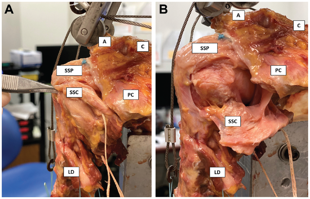
(A) Specimen mounted to the dynamic shoulder simulator in an intact condition. (B) Simulated irreparable subscapularis deficiency. A, acromion; C, clavicle; LD, latissimus dorsi; PC, processus coracoideus; SSC, subscapularis tendon; SSP, supraspinatus tendon.
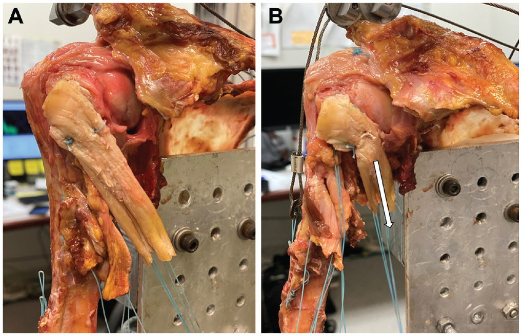
(A) To simulate the anterior latissimus dorsi transfer (aLDT), an Achilles tendon allograft was attached to the lesser tuberosity. (B) Then, the loaded allograft (24 N) was aligned to the initial pull of line of the latissimus dorsi (LD) to re-create an aLDT.
Outcome Measures
Outcome parameters included the (1) glenohumeral abduction angle (gAA; degrees), (2) SM (in millimeters) relative to the native state, and (3) cDF (in Newtons).1,11,26 Motion analysis software (ProCalc; Vicon Motion Capture Systems) was used to analyze the recorded 3D motion videos. 23 SM was calculated as the change in distance between the 2 tripods relative to the native state. The deltoid force was recorded in real time throughout range of motion by load cells (Futek) connected to the actuators.1,11,23,26 The cDF was calculated as the sum of the anterior, middle, and posterior deltoid forces.1,11,23,26 Specimens underwent 3 testing cycles for each condition.1,23,26
Statistical Analysis
An a priori power analysis was performed to determine detectable differences in the dependent variables given estimated standard deviations. 26 For the gAA, an error variance of 1° across all conditions with a correlation of 0.3 between measurements was assumed. A sample size of 6 specimens provided 80% power to detect a 1° difference in shoulder angle at an alpha level of .05.
Descriptive statistics including mean and standard deviation were calculated to characterize the kinematics. Repeated-measures analysis of variance was performed to examine differences in gAA, SM, and cDF among the various testing conditions. The distributions of the model residuals were examined to ensure that large deviations from normality were not present. When significant, post hoc paired t tests with a corrected alpha using the Holm-Bonferroni sequential correction method were performed to determine which pairwise comparisons were statistically significant. The alpha level for all analyses was set at .05. All statistical analyses were performed using commercial software (Stata Version 15.2; StataCorp).
Results
Glenohumeral Abduction Angle
The SSC-D significantly decreased the gAA compared with the native state (Δ–9.8°; P < .001). The aLDT showed a significantly increased gAA compared with the SSC-D condition (Δ3.8°; P < .001) (Tables 1 and 2). For the aLDT, no anterior subluxation or decentralized glenohumeral abduction was observed.
Glenohumeral Abduction Angle, Superior Humeral Head Migration Relative to the Native State, and Cumulative Deltoid Force Stratified by Testing Condition a
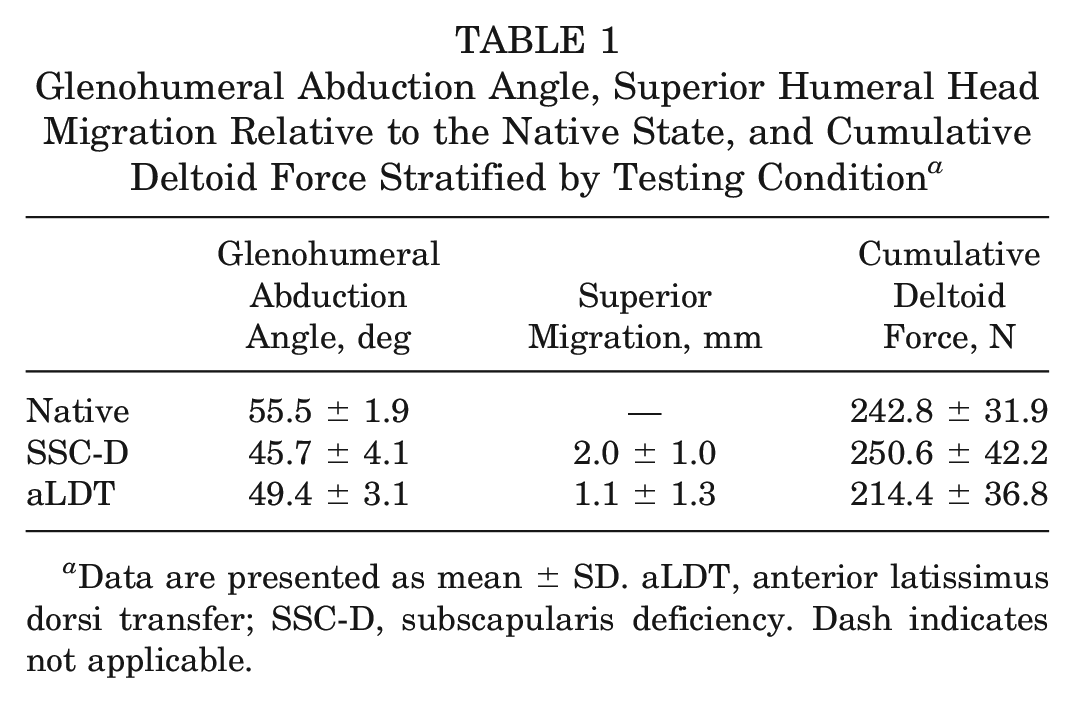
Data are presented as mean ± SD. aLDT, anterior latissimus dorsi transfer; SSC-D, subscapularis deficiency. Dash indicates not applicable.
Statistical Comparisons for Glenohumeral Abduction Angle Among Testing Conditions a
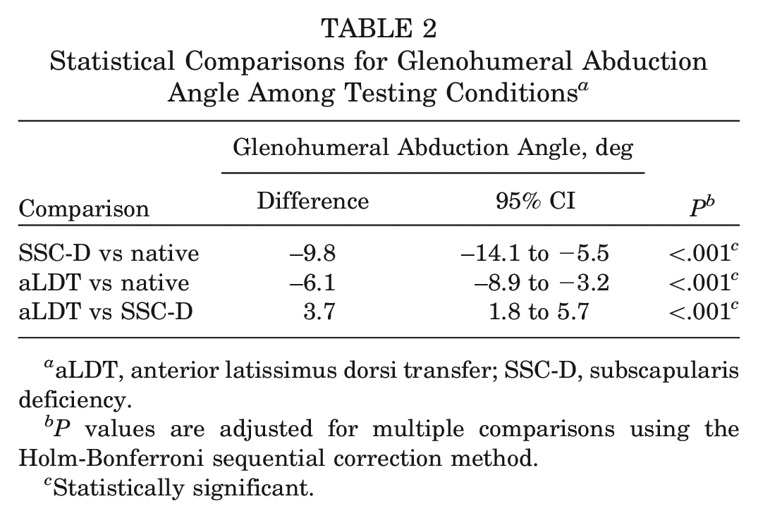
aLDT, anterior latissimus dorsi transfer; SSC-D, subscapularis deficiency.
P values are adjusted for multiple comparisons using the Holm-Bonferroni sequential correction method.
Statistically significant.
Superior Humeral Head Migration
The SSC-D resulted in a significant increase in SM when compared with the native state (Δ2 mm; P = .003) (Tables 1 and 3). There were no differences in SM when comparing the aLDT to the native and SSC-D conditions (P > .05, respectively).
Statistical Comparisons for Superior Humeral Head Migration Among Testing Conditions a
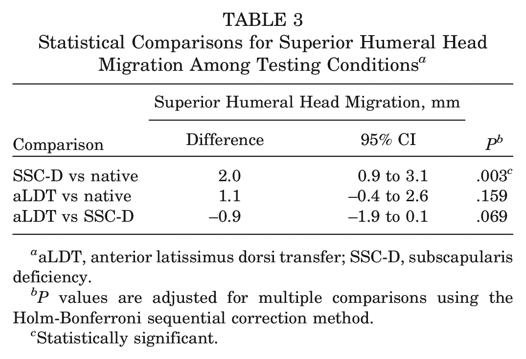
aLDT, anterior latissimus dorsi transfer; SSC-D, subscapularis deficiency.
P values are adjusted for multiple comparisons using the Holm-Bonferroni sequential correction method.
Statistically significant.
Cumulative Deltoid Forces
The aLDT showed a significant decrease in cDF compared with the native shoulder (Δ–28.4°; P < .001) and the SSC-D state (Δ–36.1°; P < .001) (Tables 1 and 4).
Statistical Comparisons for Cumulative Deltoid Force Among Testing Conditions a
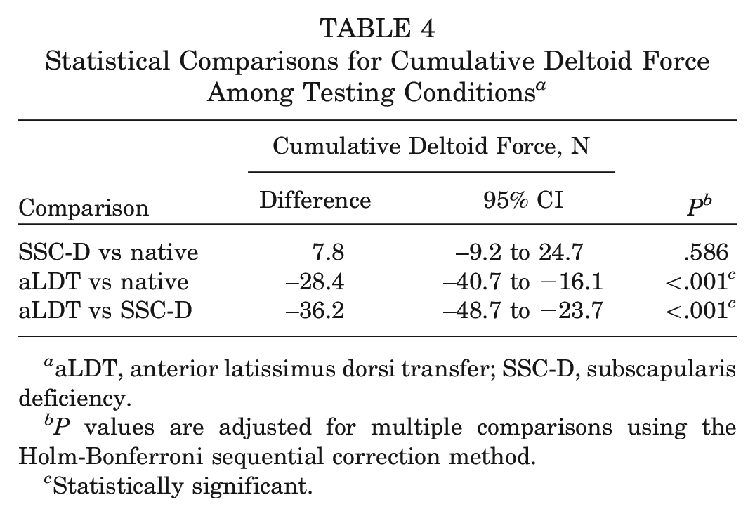
aLDT, anterior latissimus dorsi transfer; SSC-D, subscapularis deficiency.
P values are adjusted for multiple comparisons using the Holm-Bonferroni sequential correction method.
Statistically significant.
Discussion
The most important finding of this study was that performing an aLDT for irreparable SSC insufficiency restored the anteroposterior force couple, prevented superior and anterior humeral head migration, and improved glenohumeral kinematics in a cadaveric model. Additionally, the use of an aLDT can help decrease compensatory deltoid forces that arise due to SSC insufficiency. Given the favorable effect of the aLDT on shoulder kinematics in this dynamic shoulder model, performing an aLDT may be considered as a treatment option in patients with irreparable SSC-D.
As the SSC muscle is one of the most powerful muscles in the shoulder girdle, 19 irreparable SSC tears lead to disruption of the force couple in the transverse or coronal plane, often resulting in pain and loss of function. In this situation, the ultimate goal is to restore the anteroposterior muscular force couple to ensure a centered abduction motion and prevent superior and/or anterior migration of the humeral head.2,6,11,27 Consequently, Elhassan et al12,14 introduced the aLDT as a potential surgical solution, given its ability to replicate the vector of the SSC tendon, its large muscle excursion, and the adequate force required for powerful internal rotation. Besides, the aLDT is thought to counteract the elevation force of the anterior and middle deltoid muscle by pulling down the humeral head. This rationale has led Elhassan et al 12 to conduct an anatomic feasibility study and propose the aLDT for patients with SSC tendon insufficiency. Since then, several surgical techniques have been described, 7 covering open procedures as well as arthroscopically assisted approaches. Clinically, patients show favorable outcomes after aLDT. 20 This may be in part be explained by the aforementioned biomechanical effects, allowing for a recentered glenohumeral motion.
As shown by the data of this study, anterior subluxation and SM did not occur at time-zero testing. This is in accordance with the current literature, in which decentralization was only described in 1 study after performing aLDT.7,20 Interestingly, when anterior subluxation and/or superior humeral head migration are present preoperatively, the ability of an aLDT to fully restore native shoulder kinematics may be limited, as 11% to 26% of the patients have been reported to still show decentralization of the humeral head.7,20 In these cases, performing a tendon transfer should be critically discussed with patients and surgeons.
Biomechanically, there is a substantial body of evidence that insufficiency of the rotator cuff leads to a disruption of the complex interactions between the rotator cuff and the deltoid muscles, with a subsequent increase in deltoid forces to compensate for loss of abduction.1,3-5,10,11,26
As such, based on the results of the present study, the aLDT allows not only for a rebalancing of glenohumeral abduction motion, but also for a reduction of compensatory deltoid forces. This is of clinical importance, as postoperative deltoid fatigue with subsequent pain and loss of glenohumeral abduction motion may be prevented. However, one of the main drawbacks of the aLDT is that this complex surgical technique has a steep learning curve, and attention has to be paid to avoid iatrogenic injuries of major anatomic structures.7,12 More specifically, relevant postoperative complications have been reported to occur in approximately 15% of cases. 20
In general, SSC-D is a debilitating condition, mostly requiring surgical intervention. As shown by the data gained in this dynamic biomechanical investigation, the aLDT may hold the potential to mimic the physiological vector of the SSC tendon, allowing for a promising restoration of glenohumeral kinematics. Even though initial shoulder function may not be fully restored, significant improvements can be expected when compared with a severe SSC insufficiency. Compensatory deltoid forces and superior head migration can be reduced, while improving glenohumeral abduction motion. Functional outcomes after aLDT have been shown to be highly promising, 14 although reliable long-term data are still lacking.
This study has several limitations. First, anterior subluxation could not be directly measured in this dynamic shoulder model. However, this was not deemed of critical necessity, as any disruption in the transverse or coronal force couple would have been reflected in a decreased range of centered glenohumeral abduction motion.2,6,11,27 Second, as this was a biomechanical cadaveric study, only time-zero data are reported without evaluation of biological healing over time. Third, the necessity of securely mounting the specimen to the shoulder simulator with a fixed scapula eliminated any scapulothoracic motion. Additionally, as full torso specimens were not used, the true dynamic lines of pull of the LD and PM may be influenced. However, every attempt was made to place the respective force vectors in a physiological orientation by using several anatomic landmarks. Finally, internal rotation could not be measured in this shoulder model, given the inherent limitations of the dynamic testing setup.
Conclusion
In this cadaveric study, by performing an aLDT for an irreparable SSC insufficiency, we restored the anteroposterior force couple and prevented superior and anterior humeral head migration, thus improving glenohumeral kinematics. Furthermore, compensatory deltoid forces were reduced by performing an aLDT.
Footnotes
Submitted March 17, 2023; accepted November 8, 2023.
One or more of the authors has declared the following potential conflict of interest or source of funding: S.S. has received consulting fees from Arthrex GmbH, Medi GmbH & Co, and KLS Martin Group. A.D.M. has received consulting fees from Arthrex and Astellas Pharma, speaking fees from Kairos Surgical, and honoraria from Arthrosurface. The University of Connecticut Health Center/UConn Musculoskeletal Institute has received direct funding and material support from Arthrex Inc. The company had no influence on study design, data collection, or interpretation of the results or the final manuscript. AOSSM checks author disclosures against the Open Payments Database (OPD). AOSSM has not conducted an independent investigation on the OPD and disclaims any liability or responsibility relating thereto.