
Abstract
Keywords
Trigeminal neuralgia (TN) is a neuropathic pain disorder characterized by recurrent unilateral paroxysmal attacks of severe, brief electric shock–like pain within the distribution of the trigeminal nerve, triggered by innocuous stimuli (1). The International Classification of Headache Disorders (ICHD) distinguishes three forms of TN: classical TN (13.1.1.1), associated with neurovascular compression causing morphological changes of the trigeminal root; secondary TN (13.1.1.2), resulting from an underlying disease such as multiple sclerosis or benign posterior fossa tumors; and idiopathic TN (13.1.1.3), of unknown etiology (1). Increasing evidence, however, points to an additional etiological variant of TN. This recently proposed entity is defined by the presence of a solitary pontine lesion, radiologically characterized by a single T2-hyperintense lesion involving the intraxial course of primary trigeminal afferents, in patients without any other evidence of white matter disease (2).
Clinical and radiological evidence
Published case series and narrative reviews indicate that TN associated with a solitary pontine lesion is rare. To date, only a limited number of patients have been described in the literature. Tohyama et al. (2) identified 24 patients with a solitary pontine lesion in their cohort and noted that only 13 cases had been reported previously in the literature. Earlier publications consist mainly of small case series, as well as several single-case reports (3). Some prior studies reported solitary pontine lesions causing TN with variable interpretations, including pontine infarction or varicella-zoster virus-induced pontine lesion (4). Overall, combining all series and case reports, the total number of documented patients with TN due to a solitary pontine lesion is approximately 40 individuals.
Neuroimaging analyses of patients with solitary pontine lesion–related TN demonstrated that these lesions are uniformly distributed along the trigeminal pontine pathway, with maximal overlap in the region of the trigeminal brainstem sensory nuclear complex (5). Diffusion tensor imaging of solitary pontine lesions showed abnormal white-matter microstructure—specifically reduced fractional anisotropy and increased mean, radial, and axial diffusivity—suggesting neuroinflammation, demyelination, and axonal degeneration (5).
Distinction from multiple sclerosis–related TN
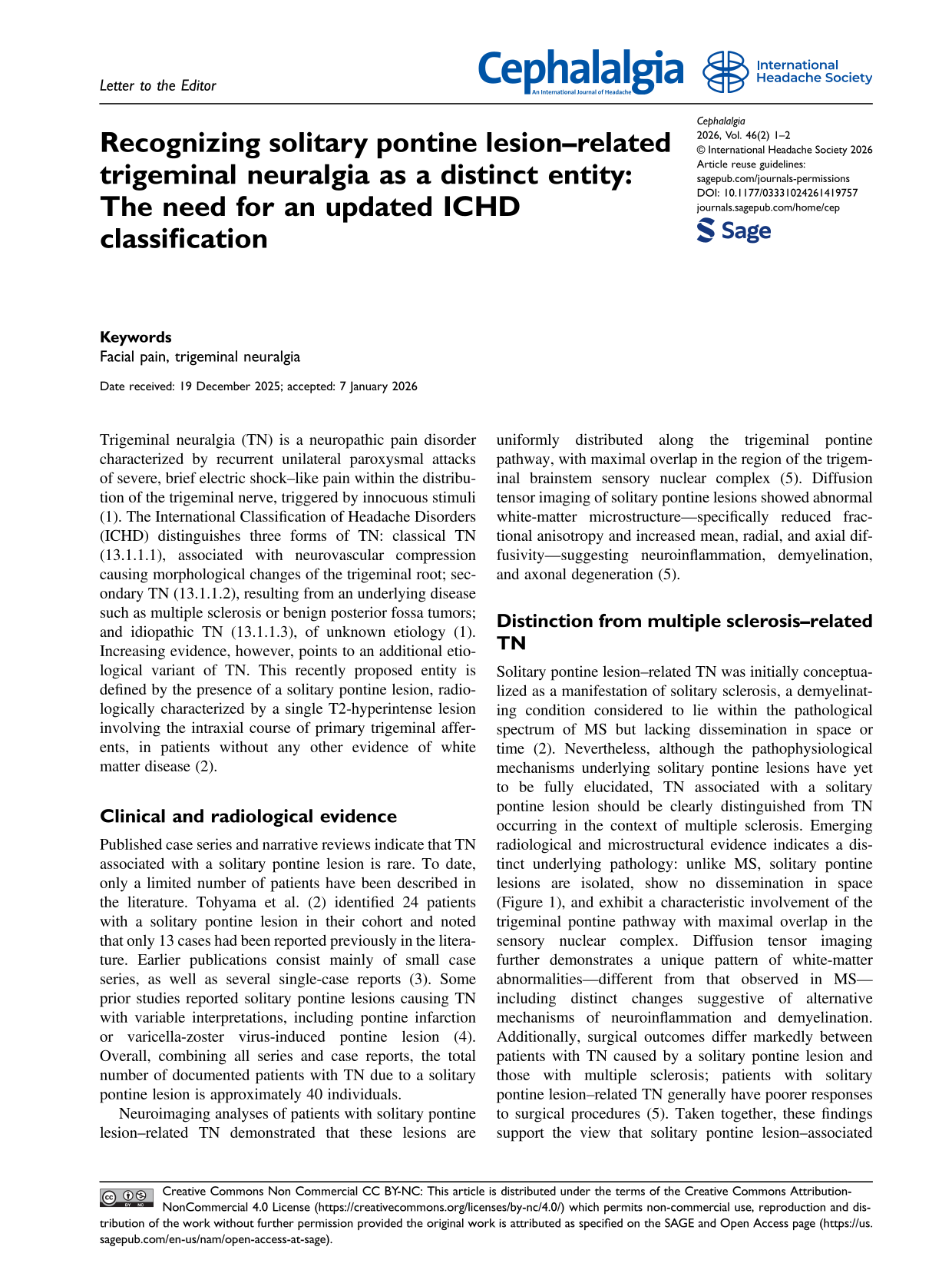
Solitary pontine lesion–related TN was initially conceptualized as a manifestation of solitary sclerosis, a demyelinating condition considered to lie within the pathological spectrum of MS but lacking dissemination in space or time (2). Nevertheless, although the pathophysiological mechanisms underlying solitary pontine lesions have yet to be fully elucidated, TN associated with a solitary pontine lesion should be clearly distinguished from TN occurring in the context of multiple sclerosis. Emerging radiological and microstructural evidence indicates a distinct underlying pathology: unlike MS, solitary pontine lesions are isolated, show no dissemination in space (Figure 1), and exhibit a characteristic involvement of the trigeminal pontine pathway with maximal overlap in the sensory nuclear complex. Diffusion tensor imaging further demonstrates a unique pattern of white-matter abnormalities—different from that observed in MS—including distinct changes suggestive of alternative mechanisms of neuroinflammation and demyelination. Additionally, surgical outcomes differ markedly between patients with TN caused by a solitary pontine lesion and those with multiple sclerosis; patients with solitary pontine lesion–related TN generally have poorer responses to surgical procedures (5). Taken together, these findings support the view that solitary pontine lesion–associated TN represents a separate disease rather than a localized manifestation of multiple sclerosis.

Brain MRI in a representative patient demonstrating a solitary pontine T2 hyperintense lesion without evidence of spatial dissemination. (A): Axial posterior fossa FLAIR image showing a well-defined hyperintense lesion in the pons. (B) and (C): Axial FLAIR and mid-sagittal T2-weighted images showing no white-matter abnormalities.
Rationale for a new ICHD subtype
Inclusion of solitary pontine lesion-related TN in the category 13.1.1.2.3 “Trigeminal neuralgia attributed to other cause” is not appropriate because solitary pontine lesion has a consistent lesion location along the trigeminal pontine sensory pathway and diffusion tensor imaging shows specific microstructural abnormalities. This indicates a specific disease mechanism, rather than a heterogeneous “other cause” category. Unlike many “other causes” of TN, solitary pontine lesions show uniform neuroanatomical patterns, making them recognizable and classifiable. Grouping them with miscellaneous secondary TN cases would obscure this reproducibility.
We therefore believe that incorporating this entity into future editions of the ICHD would improve diagnostic precision, facilitate appropriate etiological stratification, and promote more consistent research on its pathophysiology and treatment outcomes. In this context, we propose that solitary pontine lesion–related trigeminal neuralgia be formally recognized as a new subtype within the category of secondary trigeminal neuralgia.
Footnotes
Consent for publication
All authors approve the publication of the article by Sage.
Author contributions
GDeS: Conceptualization; AT, GDiS: writing original draft; AT, GDeS, KMH, RFSJr: Writing – review & editing.
Funding
The authors received no financial support for the research, authorship, and/or publication of this article.
Declaration of conflicting interests
The authors declared no potential conflicts of interest with respect to the research, authorship, and/or publication of this article.