
Abstract
Background
Surgical management of headache due to anomalies in the cervical spine is uncommon, as most cases improve with drugs and/or physical therapy.
Case
We report two instances of a very uncommon congenital upper cervical spine anomaly due to the presence of a pseudoarthrosis between a unilateral paracondylar process in the base of the skull and an epitransverse process arising from the transverse apophysis (PCP/ETA). The first one corresponds to a male on whom an endoscopic guided puncture was performed, and the second to an adult male from the Neolithic period who showed two cranial trepanations together with the presence of morphine metabolites in both bones and dental calculus.
Discussion
We draw a parallel between the treatment of two individuals separated by a gap of more than 4800 years: contemporary direct vision of the false joint through a small endoscope, which provides an accurate puncture, and ancient double trepanation with clear signs of bone eburnation.
Introduction
The craniovertebral junction (CVJ) is an anatomical area where various bone and soft tissue congenital anomalies may be found.
Although some produce neurological changes to varying degrees, the most common anomalies in the CVJ are usually asymptomatic. However, on certain occasions, these latter ones may cause neck pain and headaches, as in the case of total or partial occipitalization of the atlas (OAC).
One of the rarest OACs is the pseudoarthrosis between a paracondylar process (PCP) of the base of the skull and an epitransverse process (ETP), which consists of a cranial projection of the transverse apophysis of the atlas.
This rare anomaly can cause neck pain, restriction of mobility and headaches, and can require medical or even surgical treatment in some cases.
Its presence in archaeological remains is anecdotal, while its relationship with medical and surgical practices has not been described up until now.
Two similar cases of a PCP/ETP joint that required surgical treatment are presented; these cases are separated by more than 4800 years.
Case 1 (present)
In 2015, a study was carried out on a patient who had been referred from the Neurology Department for assessment by our Orthopedic and Traumatology Department. He was a 41-year-old male with a hard job and had been seen for the study and treatment of pain at the back of the neck with restriction of neck movement and left headache for the previous eight months. Headache was localized in the upper zone of the neck with radiation to parietal and frontal areas. It was marked by continuous daily pain that tended to worsen as the day went on, although there were pain-free periods. Headache was triggered by rotation movements in both sides, but not the flexo-extension or lateralization of the head. Neither aura nor dizziness was present. Pain relief was obtained by dexketoprofen 25 mgplus thiocolchicoside 2 mg plus paracetamol 500 mg, two to four times per day. No other neurological signs were detected.
The initial radiological examination showed a partial fusion between the laminas and the spinal apophyses C2 and C3 of the left side. After analyzing the case, a surgical resection of the fusion by the posterior approach was chosen.
Five months after, the patient showed a slight improvement in neck mobility, with the headaches persisting in the same area as before the intervention.
A new examination was carried out via a CT scan that showed a complete separation of the C2-C3 level, although at the CVJ level there was evidence of an anomaly consistent with a pseudoarthrosis between a unilateral PCP/ETP on the left side (Figure 1(a)). The scintigraphy study showed moderate uptake at the C2-C3 level, compatible with sequelae of the surgical intervention, and a more intense uptake at the level of the left CVJ anomaly. A CT-guided puncture was then performed (Figure 1(b)). At the same time, and for the first time according to the literature, an endoscopic view was achieved using a maxillofacial surgery lens that was able to observe the false joint and compare it with the CT view. It was injected with a 0.2 cc solution of triamcinolone with 0.3 svedocaine.
(a) Coronal slice with the PCP/ETP junction (arrow) in the left side. (b) Transversal shot image of the endoscopic camera in contact with the anomaly (circle).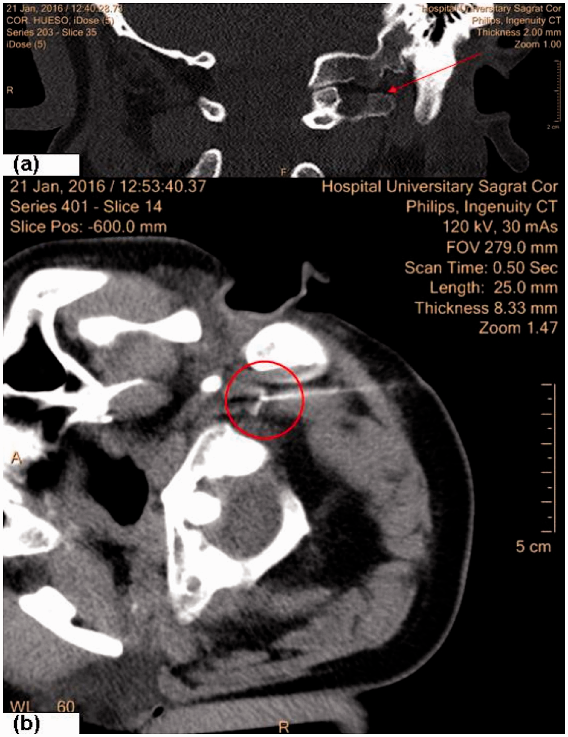
Six months after the injection the patient showed significantly improved neck rigidity, while the headaches had disappeared.
Case 2 (Neolithic period)
During the 1990s, studies were performed in several iron mines in the Can Tintorer archaeological site (Gavá, Barcelona). Mine no. 28 consisted of a primary collective sepulcher of about 5 m2 in which a total of 12 individuals with a good skeletal preservation index were identified.
Dating was performed using 14C on the inhumation and contact layers in two different laboratories (Teledyn Isotopes, New Jersey, USA and the Chemistry Faculty of the University of Barcelona, Spain), with both results coinciding within the limits between 4820 +/−100 BCE.
Individual 10 (M28.10) corresponded to a male individual of about 30 years of age by anthropological criteria, showing marked bone robustness and numerous enthesopathies, mainly in the upper limbs. These were considered to be markers of occupational stress, probably associated with his work in the mine.
However, what made this individual stand out was a double trepanation in the left parietal (f 33 × 23 mm/26 × 17 mm) (Figure 2(a)). The characteristics of these trepanations suggested that they had been performed by the abrasion method, and they showed clear signs of survival. After re-analyzing the skeletal remains of the individual in depth, a unilateral PCP/ETP complex could be detected on the left side (Figure 2(b)).
(a) Posterior view of the 10.M28 skull from the Neolithic period (± 4820 BP) showing two healed parietal trepanations. (b) Image of the 10.M28 atlas (anterior side); an epitransverse process is clear in the left side.
In parallel, with the aim of studying the paleo diet and its relationship with the work practiced, studies were performed on the dental calculus in all the male individuals in mine no. 28 with an optical microscope, as well as with a Scanning Electron Microscopy (SEM) combined with Energy Dispersive Spectroscopy (EDS), at the University of Barcelona. In the specimen from individual 10, fragments of epidermal and parenchymal tissue from the opium poppy seed (Papaver somniferum) were identified. In order to check whether the consumption of the opium poppy was exclusive to individual 10, various paleo-toxicology techniques were performed for the identification of drug metabolites by Radioimmunoassay (RIA) and Gas Chromatography/Mass Spectometry (GC/MS) (1). The results showed that the only individual positive for morphine and codeine was individual 10, in both bone tissue and dental calculus.
Discussion
Congenital anomalies in the CVJ are not rare. The fourth occipital sclerotome contributes to the formation of the foramen magnum, occipital condyles, C1 lateral masses, and the C1 posterior cranial arch area. Any alteration in this state leads to the type of anomalies to which we are referring. Thus the PCP/ETP complex is a vestige of the cranial half of the first cervical sclerotome (2).
The first references to congenital anomalies of the cervical spine were made by Ackerman in 1790 (3). The first description of an occipital-atlas fusion did not occur until a century later (4), whilst the first anatomical description of a PCP/ETP complex was made by McRae in 1960 (5).
The frequencies of these types of anomalies are very variable due to the majority not presenting with symptoms. The OACs vary from 0.08% up to 25%. On the other hand, the PCP frequency varies from 0.125% (6) up to 0.29% (7).
There are few references on the presence of PCP, ETP or PCP/ETP complexes. In current skeleton collections, we can highlight: 1 ETP in 107 individuals (8); 2 PCP in 692 individuals (7); 1 PCP in 728 individuals; and 2 PCP + 1 ETP in 214 individuals (9). Of the studies performed on live subjects, an extensive radiology study should be mentioned, in which 4000 X-rays were reviewed, with five cases of PCP being found (6).
The symptoms arising from the presence of a PCP/ETP complex could be due to mechanical compression of the nerve structures (C1 root), compression of the vascular structures, instability, and rigidity, all of them mainly due to a biomechanical alteration of the osteochondral junction (OCJ). They can range from sporadic headaches to more severe neurological symptoms and most of them appear after a traumatism.
Although there is a higher prevalence in males, the four cases in which this anomaly has required surgical resection have all been in young women.
Partial fusion of C2-C3 associated with OAC has been described previously in four individuals.
The presence of PCP and ETP in archaeological remains is anecdotal. The most extensive study on archaeological remains was carried out in the United Kingdom in 1996 (10), in which 1300 skulls from the medieval period were studied during the excavation of St Gregory’s Priory. In this study there were 1 PCP (9 mm in SK 188 individual) and six paracondylar tubercles (less than 8 mm), which is a frequency of about 0.5%. In no cases did they appear to be associated with an ETP, nor could any update of the cranial anthropic origin be established. One case of right unilateral PCP was described from the archaeological site of Amoxiumqua, New Mexico (NMNH 271804), dated between 1350/1650 CE (11). The oldest case studied until the present one is that described in the case of a female individual from burial 54 from the necropolis of Santa Maria Aprutensis in Teramo, Abruzzi (Italy), which was carbon dated 600-685 AD (12), and only presented with a left PCP.
The coincidence of a PCP/ETP with headaches that required pharmacological treatment and then surgery in our first individual leads us to think of a possible relationship between his case and that of the individual who showed a similar anomaly, with two parietal trepanations practiced on the same left side in his lifetime.
We base this on the fact that in the Neolithic period trepanations were mainly practiced in a ritualistic manner to treat cases of headaches and convulsions, with there being examples of this in different areas of Europe. Opioids, like morphine, codeine, thebaine, papaverine, and noscapine, were used in Neolithic times for their euphorigenic and analgesic proprieties.
Furthermore, individual 10.M28 was the only one showing evidence of trepanation in the 12 individuals of mine no. 28, and in the 38 individuals excavated at the archaeological site.
Although consumption of the opium poppy has been established in different eastern Mediterranean populations, this consumption has never been associated with the practice of trepanation, as appears to be the case for individual 10.M28.
Footnotes
Article highlights
Cephalalgia of the same rare etiology with a gap of more than 4800 years: endoscopic vision versus trepanation in the Neolithic.
First case with endoscopic vision of the craniovertebral joint anomaly (CVJ anomaly).
Relationship between the CVJ anomaly and two trepanations in Neolithic times.
Acknowledgements
The authors would like to thank Dr Adrià Arboix, neurologist, for his expert advice and MJ Sánchez, librarian, for her support in this study.
Declaration of conflicting interests
The authors declared no potential conflicts of interest with respect to the research, authorship, and/or publication of this article.
Funding
The authors received no financial support for the research, authorship, and/or publication of this article.