
Abstract
Background
Successful orthodontic treatment begins with accurate bracket placement. While current tools like Boon’s Gauge and MBT Gauge help guide height and angulation, they often fall short when it comes to aligning the bracket precisely along the tooth’s long axis—especially when working in the mouth with indirect vision. This can lead to misalignments that affect both aesthetics and treatment efficiency.
Objective
To design a simple, chairside device—SAY–AXIS—that helps orthodontists accurately position brackets by clearly identifying both the mesiodistal center and the long axis of each tooth.
Materials and Methods
SAY–AXIS was created by modifying a standard mouth mirror using 22-gauge orthodontic wire. The wire forms clearly marked horizontal and vertical guides, which project outward to assist the clinician in aligning the bracket with the tooth’s natural long axis. The device is removable, rotatable, and adaptable for both front and back teeth, improving visualization even in hard-to-reach areas. A detailed step-by-step fabrication and sterilization protocol was established.
Results
Early in-vitro use and pilot clinical application of SAY–AXIS demonstrated improved bracket placement accuracy, particularly in indirect vision situations. It was found to be easy to use, effective for both anterior and posterior segments, and helped reduce time spent on repositioning brackets during treatment.
Conclusion
SAY–AXIS offers a practical and innovative solution to a common clinical challenge. By enabling better visualization and alignment, this tool supports more accurate bracket placement from the start—leading to better treatment results, fewer adjustments, and greater efficiency for both clinicians and patients.
Introduction
Background and Significance
Accurate bracket positioning is fundamental to the success of fixed orthodontic therapy, particularly in systems utilizing the preadjusted edgewise appliance (PEA). According to McLaughlin, Bennett, and Trevisi (MBT), the effectiveness of the built-in prescription is heavily reliant on the precision with which brackets are placed on individual teeth.
Conventional Methodology
Traditionally, bracket positioning is performed using gauges and standard vertical measurements from the incisal or occlusal edges. For maxillary and mandibular central incisors, ideal bracket placement involves aligning the bracket slot at the facial axis of the clinical crown (FACC)—the most prominent vertical line on the facial surface, representing the long axis of the tooth. The FA point, located at the midpoint of this axis, serves as the intended site for bracket placement.1, 2
Clinical Challenges
Despite the availability of various bracket positioning instruments, many fail to address axial accuracy, particularly in terms of aligning the bracket along the true vertical axis of the clinical crown. Visual distortions, intraoral constraints, and limited angulation control can lead to suboptimal placement, affecting treatment efficacy and necessitating mid-treatment adjustments. 3
Need for Innovation
There is a pressing clinical need for a simple, accurate, and reproducible method to visualize and align brackets along both vertical and horizontal axes during placement. An ideal device would aid in locating the FA point accurately, while also serving as a reference for long-axis orientation.
The SAY-AXIS Solution
To meet this need, we introduce the SAY-AXIS innovatively derived from a standard mouth mirror. SAY-AXIS integrates vertical and horizontal axis guidance into a single tool, enhancing bracket positioning accuracy. By enabling precise visualization of the FACC and FA point, this device facilitates improved alignment of the bracket slot with the tooth’s true anatomical axis.
Clinical Relevance
This device offers orthodontists a reliable, non-invasive, and time-efficient method to enhance the accuracy of bracket positioning, ultimately contributing to better treatment outcomes, fewer mid-course corrections, and improved long-term stability.
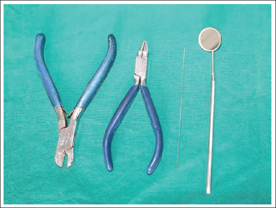
Armamentarium (Figure 1)
Universal plier
Hard-wire cutter
22-gauge stainless-steel wire
Mouth mirror
Armamentarium.
Fabrication Protocol
Wire preparation: Take a segment of 22-gauge stainless steel orthodontic wire and straighten it using a universal plier.
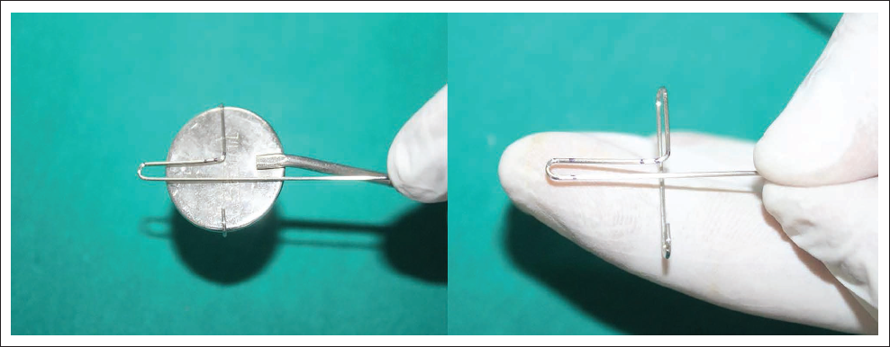
Initial engagement: Create a J-bend at one terminal end of the wire and insert it securely into the shaft of a standard mouth mirror to achieve initial stabilization (Figure 2).
Opposing fixation: At the opposite end of the mirror handle, mark the engagement point on the wire and form a U-bend to secure the mirror between the two anchorage bends (Figure 3).
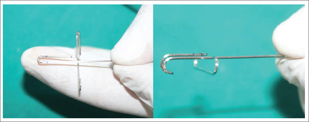
Axis reference marking: Identify and mark the midline on the posterior aspect of the mirror handle. Place two additional reference points 2 mm mesial and distal to the central mark. These serve as vertical-axis guides for bracket positioning (Figure 4).
Axial alignment arms: At each lateral mark, execute a precise 90° bend perpendicular to the long axis of the mirror handle, projecting anteriorly (Figure 5).
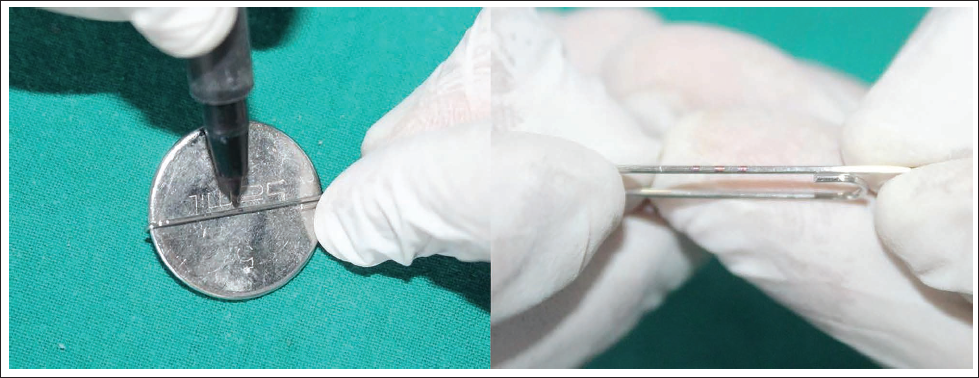
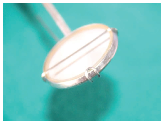
Horizontal guide loop: Measure 4 mm beyond the transverse width of the mirror and construct a U-shaped loop that encircles the mirror anteriorly. This loop acts as a horizontal positioning guide (Figure 6).
Locking mechanism: Rotate the free end of the wire across the reflective surface of the mirror and engage it into the contralateral end via a terminal locking bend, ensuring rotational and positional stability (Figure 7).
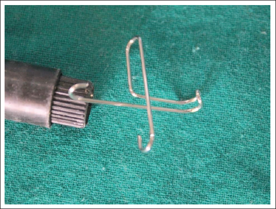
Final assembly: Ensure that the final configuration allows removable, rotatable manipulation of the wire around the mirror, while maintaining the stability of all axial and transverse guides. The assembled device should be visually symmetrical and functionally stable (Figure 8).
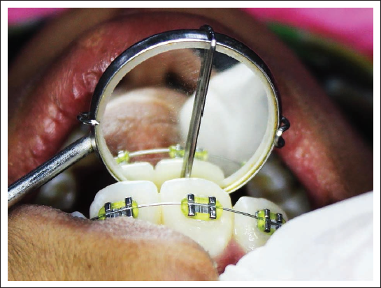
Anterior segment application: Use the device to assess and align the bracket slot precisely with the FACC on maxillary and mandibular anterior teeth, ensuring accurate vertical and mesiodistal bracket placement (Figure 9).
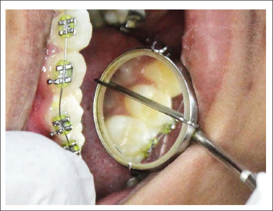
Posterior segment adaptation: Apply the device to posterior teeth by angling the mirror for indirect visualization, maintaining FACC guidance and FA point referencing even in restricted buccal corridors (Figure 10).
J-bend Was Given at One End of the Wire and Engage It to the Mouth Mirror.
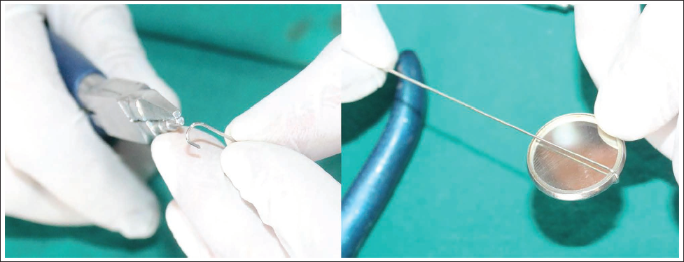
Mark the Opposite End of the Mouth Mirror on the Wire and Give a U-bend.
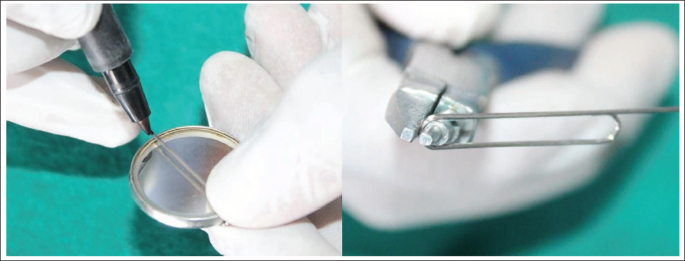
Mark the Midpoint on the Wire at the Back Side of the Mouth Mirror. Also, Add Two Marks 2 mm Away from the Central Mark.
On Proximal Marking Give a 90° Bend.
Take 4 mm Extra than the Width of the Mouth Mirror and Bend a U-loop Encircling It.
Free End will Turn Around and Above the Mouth Mirror.
Final Design of SAY-AXIS.
SAY-AXIS Used in Anterior Teeth.
SAY-AXIS Used in Posterior Teeth.
Sterilization Protocol
Cleaning: Rinse under water, soak in an enzymatic solution, or use an ultrasonic cleaner. Scrub gently and rinse again.
Drying: Air dry or wipe with lint-free gauze.
Sterilization:
Autoclave at 121°C for 15-20 min or 134°C for 3-4 min (preferred).
Chemical: Soak in 2% glutaraldehyde for 10 h if an autoclave is unavailable.
Storage: Keep in sterile pouch until use.
Pre-use check: Inspect for damage, corrosion, or clarity issues before use.
Advantages
Enhanced accuracy: Ensures precise alignment of the mesiodistal center and long axis of the tooth for optimal bracket placement.
Improved visualization: Facilitates better indirect visualization, overcoming challenges in assessing bracket position along the long axis.
Three-dimensional alignment: Enables precise 3D alignment of brackets, improving overall treatment outcomes.
Ease of use: Simple to use, and it integrates seamlessly into routine orthodontic procedures.
Versatility: Suitable for both anterior and posterior teeth.
Minimizes human error: Reduces the risk of errors in bracket placement, ensuring consistent and predictable results.
Time efficiency: Reduces time spent on adjustments and refinements during treatment.
Non-invasive: No modifications are needed to existing equipment, making it easy to adopt.
Improved treatment stability: Promotes long-term orthodontic stability by ensuring accurate initial bracket positioning.
Limitations and Future Scope
The SAY-AXIS device is in the early clinical innovation phase, with successful in vitro testing and pilot clinical application. A formal clinical trial will strengthen the evidence base. Accordingly, a protocol is being developed for a prospective, randomized clinical trial. Lack of clinical data, mirror access in posterior segments, and a need for operator calibration.
Conclusion
SAY-AXIS is a novel, removable, rotating, bracket positioning device, which is modified from a simple mouth mirror to provide vertical as well as horizontal accuracy.
Footnotes
Declaration of Conflicting Interests
The authors declared no potential conflicts of interest with respect to the research, authorship, and/or publication of this article.
Ethical Approval
Ethical consent has been obtained for research involving human subjects.
Funding
The authors received no financial support for the research, authorship, and/or publication of this article.
Informed Consent
The authors certifies that they have obtained all the appropriate patient consent forms. The patients are aware that their initials and names will not be disclosed.