
Abstract
Objective
To synthesize selenium nanoparticles (SeNPs) from plant extract, characterize them, coat them on clear aligners, and evaluate their cytotoxicity and antimicrobial activity.
Materials and Methods
The green synthesis of SeNPs utilized the leaves of Withania somnifera. The biosynthesized nanoparticles are incorporated in clear aligners using sol-gel thin film dip coating method to check for antimicrobial effect against Streptococcus mutans and Lactobacillus species using time kill curve assay. The cytotoxicity of the material was checked using brine shrimp lethality assay.
Results
The optical density values indicated that the SeNPs-coated clear aligners effectively inhibited the growth of Lactobacillus sp. and S. mutans. Time kill curve assessed in varying concentration of 25 µg/mL, 50 µg/mL, and 100 µg/mL revealed greater antibacterial activity at a lower concentration with increasing time.
Conclusion
Clear aligners coated with SeNPs had positive antibacterial effects on suspensions of Lactobacillus sp. and S. mutans and are biocompatible.
Introduction
There is a felt need for an enormous change to make advances in orthodontics to enhance the aesthetic sense and accelerate the comfort of the patient. Thus, there rose an invaluable technique of focusing on the perspective of clear aligners. Eliminating the hostile method that prevailed, an incredible approach is being facilitated to elevate the confidence, comfort, and contentment of people for a millennium.
Clear aligners are superior when compared to conventional fixed appliance in certain aspects as they reduce the treatment time, root resorption, and prevalence of white spot lesions.1–3 It was seen that the incidence of developing white spot lesions was less in patients treated with aligners due to shorter treatment duration. 1 As aligners are removable devices, they help in maintaining the oral hygiene to a certain extent, thus reducing the possibility of greater plaque accumulation. 4 Though clear aligners are known to be more hygienic, incidence of white spot lesions is known to occur with this kind of treatment also, which may be due to the composite attachments that cover a portion of the tooth surface. Thus, patient motivation is an important criterion in maintaining a better oral hygiene, hence preventing the incidence of white spot lesions. 5
However, this does not completely eliminate the bacteria present. 3 The proposal of incorporating a nanoparticle has an appealing effect due to their increased surface area and proven antibacterial action. 6
Gold, zirconium, copper, zinc, silver, and titanium were some of the metals used to evaluate their antimicrobial properties. 7 Copper nanoparticles have an excellent antibacterial property but an increased risk of toxicity. 8 Studies have shown that usage of silver nanoparticles has been known to cause cytotoxicity.6, 9 Gold nanoparticles have been proven to have antibacterial actions but with a limited effect, 4 zirconium nanoparticles exhibit considerable antimicrobial effect at higher concentrations, 10 but the cost of zirconium is a major drawback. 11
All these nanoparticles used have been reported to possess some amount of toxicity. Among them, copper and zinc were considered to be most cytotoxic nanoparticles. The cytotoxicity is mainly attributed to synthesizing process. Chemical and physical methods released toxic byproducts. 12
Hence, green synthesis is a method of choice especially to wield off the toxic byproducts. Therefore, as proven, biosynthesis of nanoparticles is being done as it has good biocompatibility, reactivity, and no cytotoxic effects. 13 Also, in this method of green synthesis, biomolecules play the role of reducing and stabilizing agents of selenium nanoparticles (SeNPs). 14
SeNPs are gaining importance in the field of medicine due to their antibacterial property. SeNPs are biocompatible, nontoxic, and have the ability to disrupt microbial biofilms. 5
Various methods were used to coat nanoparticles onto the clear aligners. Carboxymethyl cellulose and chitosan were used to fabricate polysaccharide-based antibacterial coating on polyethylene terephthalate glycomodified, which is a routine clear aligner raw material. Polysilsequioxane (PSQ) was used for the modification of clear aligners. Clear aligners were coated with a ladder-like PSQ containing ammonium cations and long alkyl chains, and the antibacterial ability was investigated. Xie et al modified clear aligners with quaternary ammonium-modified gold nanoclusters (QA-GNCs). Gold nanocomposites, 4,6-diamino-2-pyrimidinerhiol-modified gold nanoparticles (AuDAPT), was also used for coating clear aligners. 15
Since clear aligners are being worn almost the entire day, and they cover the teeth and gingiva, incorporating biosynthesized nanoparticles onto them would bring about positive antimicrobial effects. 4
Hence, the aim of our study is to assess the antimicrobial activity of biosynthesized SeNPs coated on to clear aligners and evaluate their cytotoxicity.
Materials and Methods
Study Design
This is an in vitro nanolaboratory-based study to assess the cytotoxicity and antimicrobial activity of clear aligners coated with SeNPs.
Sampling Method and Sample Size Calculation
Conventional sampling method was used for the selection of the samples. Sample size was derived to be 10.
t tests – Correlation: Point biserial model
Analysis: A priori: Compute required sample size
Input: Tail(s) = Two
Effect size |ρ| = 0.8
α err prob = 0.05
Power (1-β err prob) = 0.95
Output: Noncentrality parameter δ = 4.2163702
Critical t = 2.3060041
Df = 8
Total sample size = 10
Actual power = 0.9566452
And data is analyzed by means of one-way and two-way analysis of variance and post hoc tests (α = 0.05).
Preparation of Plant Extract
Withania somnifera leaves were washed with sterile distilled water to remove the dust particles and then shade dried. W. somnifera leaves extract was prepared by placing 10 g of dried fine cut in a 500-ml beaker along with 500 mL of deionized water. The mixture was boiled for 15 min until the colorless aqueous solution changed to yellow, subjected to cooling at room temperature and filtered with Whatman no. 1 filter paper.
Synthesis of Selenium Nanoparticle
Based on the previous studies, sodium selenite salt used as a precursor was mixed with the appropriate concentration, 30 nm of SeNPs was dissolved in 50 mL of distilled water. 16 To that, 50 mL of the prepared plant extract was added slowly. Hydrothermal, microwave irradiation, ultrasonication, UV radiation, self-assembling, and conventional heating were the various methods used in the green synthesis of SeNPs. The reaction mixture was kept on a magnetic stirrer at 650 to 700 rpm for 48 to 72 h. The collected SeNPs were subjected to centrifugation at 8,000 rpm for 10 min. The material was transferred to a beaker and magnetron sputtering method was done for the production of nanoparticles.
Incorporation of Selenium Nanoparticle in Clear Aligners
The green synthesized SeNPs are incorporated in clear aligners using sol-gel thin film dip coating method. First, the aligners were treated with oxygen plasma for 3 min and then they were immersed immediately in the prepared solution for 12 h. Then the sample was washed with water and unbound nanoparticles were removed by washing them with phosphate-buffered saline (PBS) with ultrasonic treatment for 1 h. The selenium nanoparticle-coated clear aligners were dried and stored at around 20ºC.
Characterization of SeNPs-coated Clear Aligners
The morphology and distribution of the SeNPs were analyzed by SEM-EDX as shown in Figures 1 and 2, respectively. The bacterial cells were incubated for 24 h and the samples were gently washed 3 times with PBS and fixed with 2% glutaraldehyde for 2 h at 4ºC. The fixed samples were dehydrated through a gradient ethanol series. The samples coated with SeNPs were observed by Scanning Electron Microscopy (SEM). SEM images were viewed at different magnifications 20,000 X (scale bar 1 µm).
Analysis of Selenium Nanoparticles-coated Clear Aligner With SEM.

Analysis of Selenium Nanoparticle-coated Clear Aligner With EDX.
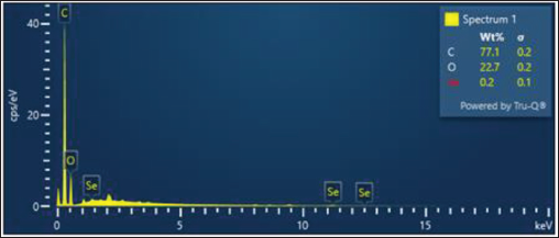
The particles were spherical in shape with approximately 60 to 70 nm in size. Most of the particles were aggregates with only a few of them scattered seen under this microscope which confirms the presence of SeNPs.
Brine Shrimp Lethality Assay
Salt Water Preparation
Two grams of iodine free salt was weighed and dissolved in 200 mL of distilled water. Six-well ELISA plates were taken and 10 to 12 mL of saline water was filled. To that, 10 nauplii were slowly added to each well. Then the nanoparticles-coated clear aligners were added according to the concentration level. The plates were incubated for 24 h. After 24 h, the ELISA plates were observed and noted for number of live nauplii’s present and calculated by using the following formula,
Number of dead nauplii/Number of dead nauplii + Number of live nauplii × 100
Antibacterial Activity
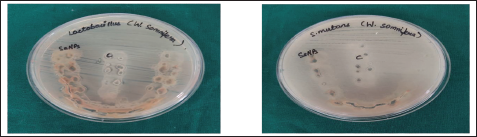
Antibacterial activity of the nanoparticles against the strain Streptococcus mutans and Lactobacillus was tested. Mueller Hinton agar (MHA) was utilized for this activity to determine the zone of inhibition. MHA was prepared and sterilized for 45 min at 120 lbs. Media was poured into the sterilized plates and was let stable for solidification. The test organisms were swabbed and then the SeNPs-coated clear aligners were placed on the surface of the agar plates, and the plates were incubated for 24 h at 37 ° C. After the incubation time, the zone of inhibition was measured.
Nutrient broth was prepared, sterilized, and 6 mL was added to all 3 test tubes.
Bacterial suspension (S. mutans and Lactobacillus) was added to all 3 test tubes in the range of 5 × 10 5 CFU/mL.
The first and second tubes contain the Acorus calamus extracts (aqueous, ethanolic, methanolic) tested usually at final concentrations of 0.25× MIC and 1× MIC and the third tube is considered as the growth control.
The incubation is done under suitable conditions for varied time intervals (0, 4, 6, 8, 10, 12, and 24 h).
Then the percentage of dead cells is calculated at wavelength of 600 nm at regular time intervals.
Results
Cytotoxicity Analysis
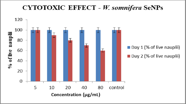
Graph 1 shows the cytotoxicity analysis of SeNPs assessed by brine shrimp lethality assay in varying concentrations of 5 (μg/mL), 10 (μg/mL), 20 (μg/mL), 40 (μg/mL), and 80 (μg/mL). Among 100 live nauplii taken as control, it was seen that 100%, 90%, 80%, 70%, and 60% of nauplii survived till 48 h. It is seen that there is reduced cytotoxicity at a lower concentration.
Antibacterial Activity of SeNPs-coated Clear Aligners
As depicted in Figure 3, to investigate the antibacterial activity of SeNPs-coated clear aligner, the samples were placed in bacterial suspension of Lactobacillus sp. and S. mutans. Further, we performed time kill curve assay to assess the concentration, which shows better antibacterial activity (Graphs 2 and 3).
Petri Dish Containing Muller Hilton Agar With Selenium Nanoparticles-coated Clear Aligners and an Uncoated Aligner Taken as Control to Assess the Antibacterial Activity.
Graphical Representation Showing Cytotoxicity of Selenium Nanoparticles in 48 H.
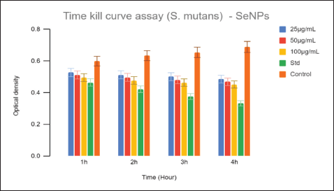
Graphical Representation of Streptococcus Mutans Growth in 4 H.
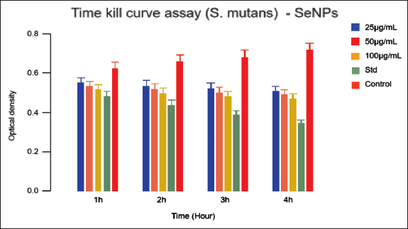
The optical density values indicated that the SeNPs-coated clear aligners effectively inhibited the growth of Lactobacillus sp. and S. mutans. Time kill curve was assessed in varying concentration of 25 µg/mL, 50 µg/mL, and 100 µg/mL along with a standard and negative control at time intervals between 1, 2, 3, and 4 h. The negative control did not contain any antibiotics while antibiotics were added in the standard.
Taking the standard value into consideration, it is seen that as the time increases the optical density also decreases, which is 0.3 indicating that there is more antibacterial activity at a lower concentration. Similarly, with concentrations of 25 µg/mL, 50 µg/mL, and 100 µg/mL, the optical density was 0.47, 0.46, and 0.44, respectively, after 4 h while the negative control keeps increasing as concentration decreases. This shows that better antibacterial activity can be achieved at low concentrations.
Discussion
Clear aligners being the recent trend in orthodontics are being used as an alternative to traditional fixed appliance which is beneficial in many ways, especially in preventing white spot lesions that frequently occurs after orthodontic treatment. 1
The invaluable use of nanoparticles has lucrative manifestation in the field of dentistry in the recent years. Various studies are being done in the field of nanobiomedicine due to their unique properties. Wenjing Song and Shaohua Ge proved that biosynthesized nanoparticles did not possess cytotoxicity under certain concentration ranges. 6
Previous studies have been done by incorporating gold nanoparticle in clear aligner that prevents Porphyromonas gingivalis biofilm formation where it was proven that it had antimicrobial effect to a certain extent. 4
A plant-mediated selenium nanoparticle study has been done by Johnson et al which stated that SeNPs, when biosynthesized using Withania somnifera, could be used in many medical applications. 17 It was also seen that SeNPs act as a suppressing agent for biofilm formation. Oxidative stress is a main reason for selenite nanoparticles to exhibit antibacterial activity. Selenium nanoparticles adhere to the bacterial cell wall and penetrate the cell membrane, causing disruption of the DNA thus acting as an antibacterial agent.6, 14
Hence, in our study we have incorporated SeNPs as they have been proven to have antibacterial property. Green synthesis was the method chosen to prepare the nanoparticle as they would have a better biocompatibility with reduced cytotoxic effect. 13 The organisms chosen here are S. mutans and Lactobacillus due to their high prevalence in plaque and salivary samples of orthodontic patients. 18
The various methods used for coating the nanoparticles are top-down or bottom-up approach, sol-gel method, electro explosion, laser pyrolysis, mechanical milling, laser ablation sputtering, thermal decomposition, spinning method, and so on. The green synthesis of nanoparticles was done by using sputtering method. 19 Extracts of Withania somnifera were used in the process of green synthesis of SeNPs.
The morphology and distribution of the SeNPs were analyzed by SEM-EDX. SEM magnifies the images and the particle size of the coated sample can be measured. It was seen that the particles were spherical in shape which was about 60 to 70 nm in size. Presence of the coated nanoparticle can be assessed by EDX. Most of the particles were clumped together due to the plant extract responsible for biosynthesis of nanoparticles while only a few of them were seen scattered under this microscope.
Nanoparticles have an increased surface area, thus increasing its biological reactivity when compared to larger particles. The increased activity can be either positive and desirable or negative and undesirable or a combination of both. 20 Hence, cytotoxicity analysis was done here by brine shrimp lethality assay which proved that there was reduced cytotoxicity at a lower concentration observed within a period of 48 h.
On evaluating the antimicrobial effect of SeNPs, it was seen that it was a potent antibacterial agent against gram-positive and gram-negative organisms. 21
On the other hand, by performing the time kill curve assay, taking the standard and negative control into consideration, we could infer that as time increases, a better antibacterial effect is observed at a lower concentration. On the contrary, the negative control keeps increasing with decreasing concentration as no antibiotics have been added to it. 22
Limitations and future recommendations:
It was an in vitro study whereas under in vivo conditions the results may vary.
Helping patients achieve better oral health while undergoing treatment might be a better choice in the future as clear aligners are in high demand. Further, studies can be conducted by incorporating different nanoparticles in clear aligners.
Conclusion
In conclusion, by evaluating the SeNPs-coated clear aligners, it was seen that they had a favorable antibacterial effect on suspension of Lactobacillus sp. and S. mutans and reduced cytotoxicity at a low concentration. Green synthesis of SeNPs would be one of the positive attributes to the reduced toxicity and increased antimicrobial activity.
Footnotes
Declaration of Conflicting Interests
The authors declared no potential conflicts of interest with respect to the research, authorship and/or publication of this article.
Ethical Clearance Number
Ethical approval from Institutional Review Boards (IRBs) was obtained (275/IRB-IBSEC/SIST).
Funding
The authors received no financial support for the research, authorship and/or publication of this article.
Informed Consent
The participant has consented to the submission of the article to the journal.