
Abstract
Introduction:
Force is applied through brackets in fixed mechanotherapy which generates stress in periodontal ligament and root of the teeth and is difficult to measure directly. Finite element analysis is used as a solution for understanding this biomechanical response. So the aims and objectives of this study were to evaluate the stress magnitude induced in the labial and lingual surface of mandibular incisors using superelastic NiTi arch wires of different cross-section at different inclination through finite element analysis.
Materials and Methods:
Finite element model of all mandibular teeth including periodontal ligament, alveolar bone, buccal tubes, brackets, and arch wire were constructed using mechanical elastic properties of the materials. Three different NiTi arch wires, round (0.016″), square (0.016″ × 0.016″), and rectangular (0.016″ × 0.022″), were placed into the bracket slots and models were constructed which were analyzed for stress distribution on root surface and periodontal ligament with 3 different cross-sections at 90°, 100°, and 110° with Ansys Version 14 software.
Conclusion:
Labial stress was found highest in round arch wire as compared to square and rectangular wire. Lingual stress was found to be slightly more in rectangular wire as compared to round and square arch wire.
Keywords
Introduction
When the orthodontic force is applied, areas of compression and tension will be created that lead to the remodeling of bone. During fixed mechanotherapy, when the force is applied through the brackets, it results in tipping of tooth as force is applied away from the center of resistance. 1
The curve of Spee is one among the Andrews Six Keys of occlusion. The key to successful orthodontic treatment is flat curve of Spee. The arch wire will deflect vertically by superiorly positioned mandibular incisor teeth if the curve of Spee exists.
The lower incisor position in orthodontic treatment planning is important because of its effect on aesthetics, periodontal health, and long-term stability.2, 3 The mandibular region is more prone to dehiscence and gingival recession because of its anatomical limitations in anterioposterior region. 4 6 The stress induced by biomechanical forces is difficult to measure directly, a precise method for numerical analysis should be used.
The finite element analysis has been suggested as a solution for understanding the biomechanical response.7, 8 Thus, the objective of our study was to evaluate the stress magnitude induced in the labial and lingual surface of mandibular incisors using superelastic NiTi arch wires of different cross-section at different inclination through finite element analysis method.
Materials and Methods
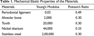
Mechanical Elastic Properties of the Materials.
Finite Element Model of Mandible With All Mandibular Teeth, Periodontal Ligament, Alveolar Bone, Buccal Tubes, Brackets, and Arch Wires.
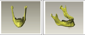
To determine the stress magnitude, a 3-dimensional model of mandibular central incisor was constructed along with periodontal ligament and alveolar bone and the interface between the alveolar bone socket and root surface was 0.2 mm as the width of periodontal ligament (PDL) is 0.25 to 0.35 mm.9, 10 The final meshwork consisted of 6,197 nodes and 3,115 elements. Standard edgewise prescriptions with no torque and no angulation brackets were used for all models based on computer-aided design with slot dimension of 0.018 × 0.025 inch.
The finite element model of mandibular teeth revealed a good alignment with deep curve of Spee. Three different NiTi arch wires (0.016″ round-shaped wire, 0.016″ × 0.016″ square-shaped wire, and 0.016″ × 0.022″ rectangular-shaped wire) were passively placed into the bracket slots and 3 models were constructed using computer-aided design as follows:
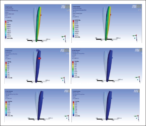
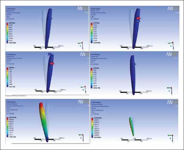
Showing the Stress Generated and Incisal Edge Displacement by 0.016″ NiTi Arch Wire at Different Inclination of Mandibular Incisors.
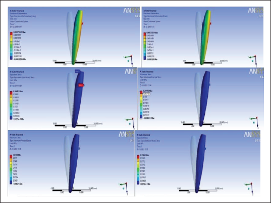
Showing the Stress Generated and Incisal Edge Displacement by 0.016″ × 0.016″ NiTi Arch Wire at Different Inclination of Mandibular Incisor.
Showing the Stress Generated and Incisal Edge Displacement by 0.016″ × 0.022″ NiTi Arch Wire at Different Inclination of Mandibular Incisors.
These models were then analyzed for stress distribution on root surfaces and PDL and were then evaluated for different types of mandibular incisor movement caused by NiTi arch wires with different cross-sections at 90°, 100°, and 110° inclinations. An intrusive movement in the apical direction of 0.2 mm from arch wire deflection was applied to simulate the initial displacement of lower incisor.
The von mises stress was generated at the root surface and PDL from arch wire on the bracket. To know the pattern of tooth movement, the Z axis displacement in each model was investigated as movement in anterioposterior direction.
Results
Showing the Stress Generated and Incisal Edge Displacement by 0.016″ NiTi Arch Wire at Different Inclination of Mandibular Incisors.
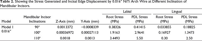
Showing the Stress Generated and Incisal Edge Displacement by 0.016″ × 0.016″ NiTi Arch Wire at Different Inclination of Mandibular Incisors.
Showing the Stress Generated and Incisal Edge Displacement by 0.016″ × 0.022″ NiTi Arch Wire at Different Inclination of Mandibular Incisors.
Discussion
In the present study for the evaluation of the stress magnitude in roots and periodontal ligament, finite element model of mandibular teeth was constructed with continuous arch wire system and force was applied to simulate the real clinical situation at different inclinations of 90°, 100°, and 110°.
The results of the present study showed that when NiTi arch wire with round cross-section (0.016″) was passively placed in the bracket slots (FEM Model 1), the labial stress was found to be 0.38 at 90° inclination, 1.91 at 100° inclination, and 3.44 at 110° inclination. Lingual stress in Model 1 was found to be 0.03 at 90° inclination, 0.16 at 100° inclination, and 0.30 at 110° inclination as shown in Table 2 and Figure 2. This suggests that as inclination increases, both labial and lingual stress increase. The reason for increase in both labial and lingual stress in round wires may be due to the single contact point of wire with bracket. When force is applied, it causes labial tipping due to which stress generated at labial side is more. The stress distribution was found to be more on labial side as compared to lingual side which represented the tooth movement in labial side. This might be due to the fact that the biomechanical force did not pass through the center of resistance of teeth.
During the initial levelling, the round wire-shaped arch wire (Model 1) showed more displacement in Y-axis due to relative intrusion of the mandibular incisors as shown in Table 2 and Figure 2. The results of the present study were in accordance with the study conducted by Yan et al, 11 they found highest von Mises stress at the labial root area. The results of our study were also in favor of the study conducted by Lombardo et al 1 where they found more stress at the labial side during displacement of lower incisors.
The results of present study showed that when NiTi arch wire with square cross-section (0.016″ × 0.016″) was passively placed in the bracket slots (FEM Model 2), the labial stress was found to be 0.038 at 90° inclination, 0.19 at 100° inclination, and 0.344 at 110° inclination. Lingual stress in Model 2 was found to be 0.03 at 90° inclination, 0.19 at 100° inclination, and 0.35 at 110° inclination as shown in Table 3 and Figure 3.
The results of present study showed that when NiTi arch wire with rectangular (0.016″ × 0.022″) cross-section was passively placed in the bracket slots (FEM Model 3), the labial stress was found to be 0.04 at 90° inclination, 0.21 at 100° inclination, and 0.38 at 110° inclination. Lingual stress in Model 3 was found to be 0.003 at 90° inclination, 0.02 at 100° inclination, and 0.40 at 110° inclination as shown in Table 4 and Figure 4.
In Models 2 and 3, the square (0.016″ × 0.016″) and rectangular (0.016″ × 0.022″) cross-section of NiTi arch wires showed lower Von Mises stress than the round arch wire (Model 1) as shown in Tables 2 to 4 and Figures 2 to 4. The reason for lesser stress with these wires might be because the square and rectangular wires have 2-point contact with the bracket which caused the torque expression and reduced their tipping. The results of our study was in accordance with the study done by Theerasopon et al, 12 they found that the labial stress with square and rectangular wires was less as compared to the stress on labial side with round wire. Further, they concluded that there is slightly increased level of maximum stress at lingual site in rectangular arch wire and both square and rectangular arch wires expressed lower stress at labial site.
The results of our study showed that the displacement shown by square (0.016″ × 0.016″) and rectangular wire (0.016″ × 0.022″) in Z direction was less than that of displacement produced by the round wire (0.016″) as shown in Tables 2 to 4 and Figures 2 to 4. This might be due to the pure intrusion and less labial crown tipping. The displacement in Y axis was found to be highest in round arch wire model (NiTi 0.016″) as compared to the square (0.016″ × 0.016″) and rectangular (0.016″ × 0.022″) cross-sectional arch wires which suggest the relative intrusion of mandibular incisors due to the labial tipping movement along with levelling of curve of Spee.
Stress analysis obtained from finite element model showed that the stress distribution was found at both the labial and lingual surfaces of mandibular incisor in all the 3 models shown in Tables 2 to 4 and Figures 2 to 4. This stress distribution pattern showed intrusive movement which might be due to conical shape of the mandibular incisors roots. However, it was found that the stress magnitude was maximum at the labial surface than that of the lingual surface in finite element model of round wire cross-section which confirms the labial tipping. Further, it was found that the lingual stress magnitude was slightly increased in rectangular arch wire as compared to square and round wire because of torque effect from square- and rectangular-shaped arch wire which reduced the flaring of the mandibular incisors.
The finite element method used in the study was a virtual model of the mandibular jaw with all the teeth and periodontal tissue to simulate the clinical situation in which orthodontic force made to act on the brackets is reproducible and needs further evaluation. As our study has certain limitations, the continuous arch wire was used and the amount of force was not known.
Though this study provides the magnitude of stress distribution on roots of mandibular incisors at labial and lingual surface by different cross-sections of NiTi arch wires at different inclinations using finite element analysis, but it requires further clinical investigation.
Conclusion
The stress magnitude on labial surface at 110° was found to be highest in round wire whereas the stress magnitude was found to be slightly more on lingual surface by rectangular wire at 110°.
Labial stress was found highest in round arch wire as compared to square and rectangular wire.
Lingual stress was found to be slightly more in rectangular wire as compared to round and square arch wire.
Footnotes
Declaration of Conflicting Interests
The authors declared no potential conflicts of interest with respect to the research, authorship, and/or publication of this article.
Funding
The authors received no financial support for the research, authorship, and/or publication of this article.
Statement of Informed Consent and Ethical Approval
Necessary ethical clearances and informed consent was received and obtained respectively before initiating the study from all participants.