
Abstract
Objective:
Studying torque expression and biomechanical effects of various wires on giving palatal root torque, using finite element (FE) method (FEM).
Conclusion:
TMA wires are most favourable for torquing, in terms of torque expression and susceptibility to root resorption.
Materials and Method:
Geometric model of maxillary right central incisor was developed, using computed tomography (CT) scan. 0.022" × 0.028" Standard edgewise brackets and 10-mm-long stainless steel (SS), titanium molybdenum (TMA) and titanium niobium (TiNb) archwires of dimension 0.019" × 0.025" were modeled and a palatal root torque of 25 degrees was applied on all wires. The angular displacement of the crown and root and nodal displacement at the incisal edge and root apex in y and z axis were analyzed along with stresses on periodontal ligament (PDL), bone, cementum, enamel, and bracket.
Results:
Buccal crown and palatal root movement was seen, which was maximum for SS and least for TMA. Angular displacement was also highest for SS. Compressive stresses were concentrated at the bucco-cervical and linguo-apical regions in the PDL, cementum, and bone and tensile stresses were concentrated in the linguo-cervical regions. In the enamel, the bracket attachment site showed maximum stresses, and slot base showed higher values of stresses than wings. All stress values were highest for SS and least for TMA.
Introduction
For correction of certain malocclusions associated with irregular axial inclination of teeth, a controlled root movement is required, which is commonly called the root torque. The expression of torque depends on various factors such as material and dimension of archwire, bevelling and angle of twist, ligation, position of bracket, its dimensions and material. 1
Nowadays, several kinds of alloy wires are used for torque application. There are various methods for predicting applied torque, 2 like torsionmeter, which is designed for measuring and studying torque moments. 3 Thus, it is a commonly used apparatus to study the effective torque. However, torquing is one of the most important and potent forces of the edgewise mechanism, which may cause root resorption, 4 so stresses produced on different biological structures by torquing need to be evaluated. But till date, none of the studies have been carried out for evaluating the overall biomechanical effects of torquing forces on biological structures.
Thus, the aim of the present study was to compare the effective torque and the stresses produced on the various biological structures by stainless steel (SS), titanium molybdenum alloy (TMA) and titanium niobium (TiNb) wires, with the help of finite element (FE) analysis (FEA).
Materials and Method
The setup consisted of workstation (P4 HP) having a processor (Intel Pentium 4) with primary storage (8 GB RAM) and secondary storage (500 GB). HyperMesh version 13.0 (Hyperworks CAE Software, Altair Engineering, Troy, MI) was used for initial modelling and preprocessing, and ANSYS version 12.1 was used for post-processing and FEA.
Material Characteristics Used in the Study.
MIMICS Processing CT-Scan Images.
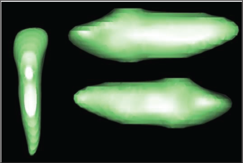
Model in HyperMesh.
Total Number of Nodes and Elements in the Model.
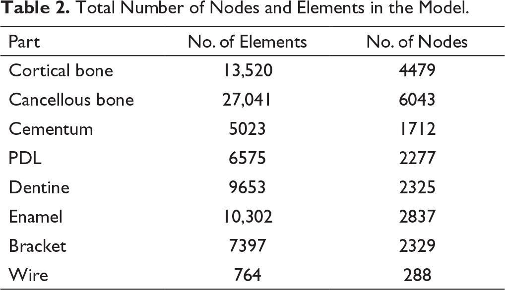
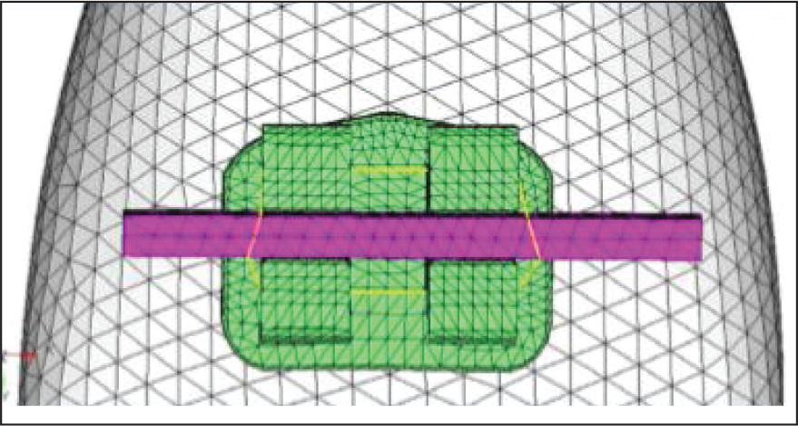
A three-dimensional model of 0.022 × 0.028-inch slot Standard edgewise maxillary right central incisor SS bracket—Mini Diamond Twin bracket (ORMCO, CA)—was modeled by reverse engineering technique. The bracket was placed on the buccal aspect of the tooth, at the center of the clinical crown, and the tooth and bracket were connected with a 0.2-mm-thick adhesive layer of composite resin. The archwires were modeled using the computer-aided three-dimensional interactive application (CATIA) software, and they were 10 mm long and of the dimension 0.019 × 0.025 inches. They were modeled in the xy-plane of global coordinate system, and then material properties were assigned to every wire. The archwire was then inserted passively in the bracket and was ligated with a 0.010-inch SS ligature (G&H Wire Company) (Figure 2). Based on these 3D solid models, a coarsening factor of 1.5 was taken (as it was seen to be reliable) and a node-to-node connection was made by creating an FE mesh among alveolar bone, PDL, tooth, bracket, and adhesive. Wire and ligatures were meshed separately from the bracket to allow contact analyses and bracket–wire frictional coefficient was taken as 0.2. Mesh refinement was carried out, which verifies convergence of results, and leads to an ideal meshing of FEs. Table 2 presents total number of elements and nodes in models.
Torque was applied along the plane of the archwire as forced rotational displacement, and the axis of rotation coincided with the plane of the wire. Twisting of wire after inserting in bracket is carried out to simulate the clinical situation, where a palatal root torque of 25 degrees is given. The rectangular wire was inserted in the bracket slot keeping it parallel and passive, and the displacements were applied to rotate the wire. This method takes into account bracket–wire play, which is 10 degrees in this case. The boundary conditions were applied, which included tight ligation with the help of a spring nodal tie, as well as restricting movement of the apical bone (outer bone surface). The geometry of the tooth and alveolus was kept constant during movement.
The various models created were:
Model 1: TMA wire with 25 degrees of palatal root torque; Model 2: TiNb wire with 25 degrees of palatal root torque; and Model 3: SS wire with 25 degrees of palatal root torque.
Angular displacement of the crown and root along with the von Mises stresses and the principal stresses were evaluated for various structures. The displacement of nodes at the root apex and incisal edge were analyzed in y and z axes. All analyses were performed using ANSYS software after applying 25 degrees of palatal root torque in the wires.
Results
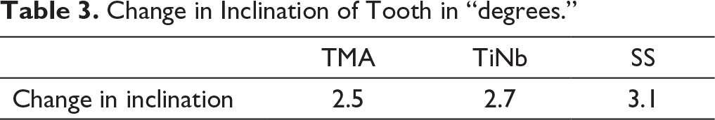
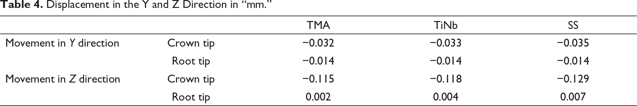
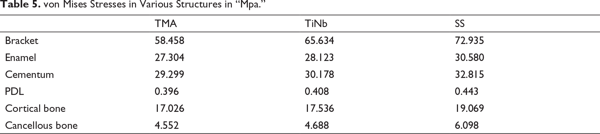
Results consisted of angular displacement (Table 3), linear displacements of the crown and root tips in vertical and buccolingual planes (Table 4), von Mises stresses (Table 5), and the principal stresses (Table 6), which were determined by the ANSYS software.
FEM Model of Bracket with Wire.
Change in Inclination of Tooth in “degrees.”
Displacement in the Y and Z Direction in “mm.”
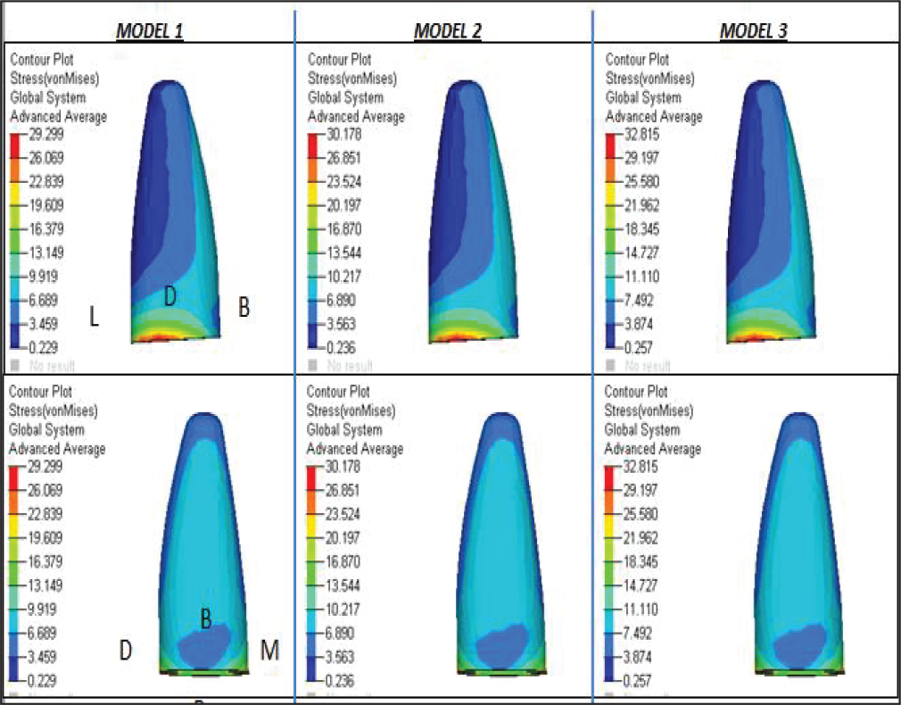
von Mises Stresses in Various Structures in “Mpa.”
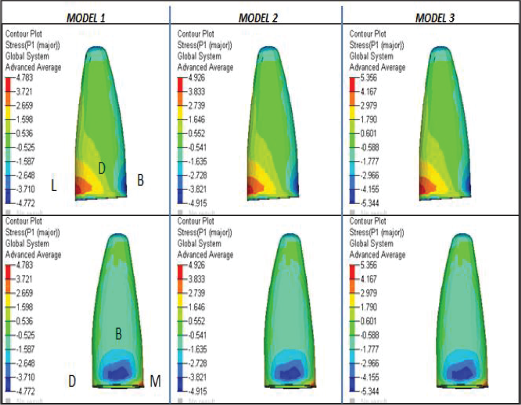
Principal Stresses in Various Structures in “Mpa.”
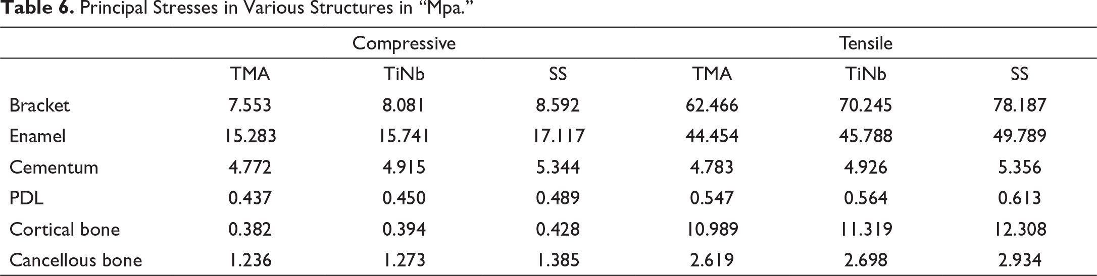
An evaluation of the displacement was as follows: overall buccal crown and palatal root movement was seen along with relative intrusion, which was maximum for SS wire, followed by TiNb and TMA wires. The movement of crown was more than the root in all the three cases. The angular displacement values were highest for SS wire and the least for TMA (Figure 3).
Displacement of the Crown and Root, and Change in Inclination of Tooth.
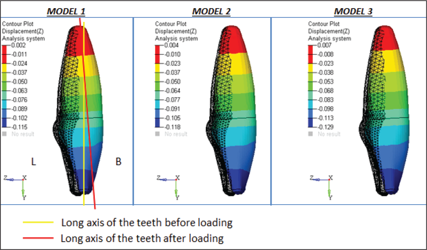
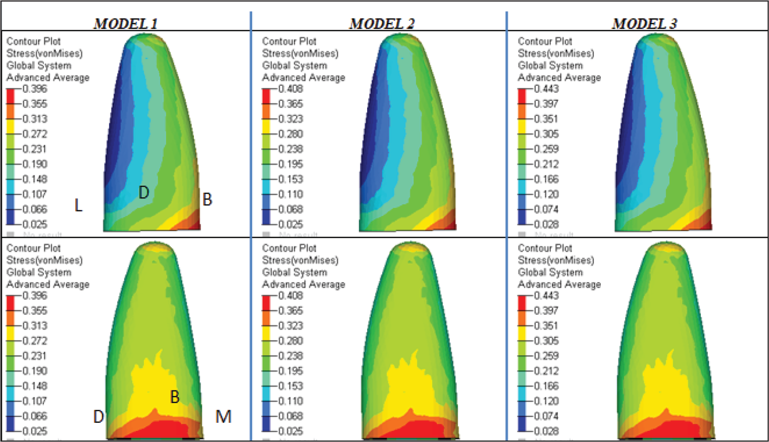
On studying stress patterns on the various structures, all the stress values were the highest for SS wire, followed by TiNb, and the least for TMA wire.
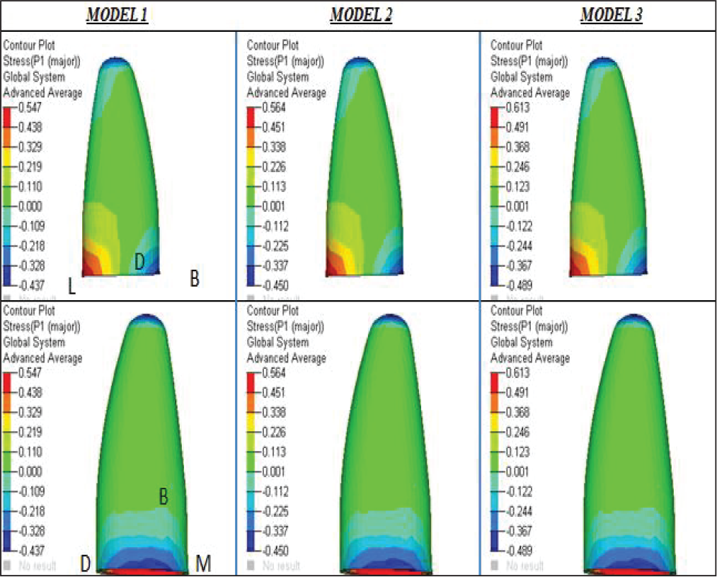
In the PDL, peak von Mises stresses were obtained on the cervical region of buccal surface and apical region. The maximum compressive stresses were obtained at the cervical region of the buccal surface and apical region. The maximum tensile stresses were seen at the linguo-cervical region. On the buccal aspect, the pattern of stresses was from compressive to tensile, from cervical to apical region, and, on the lingual aspect, stresses varied from tensile to compressive, from cervical to apical (Figures 4(a) and 4(b)).
von Mises Stress in PDL “Mpa.”
The peak von Mises stresses in the cementum were obtained on the cervical region. The maximum compressive stresses were seen at the bucco-cervical region and apical region, and the maximum tensile stresses were seen at linguo-cervical region. On the buccal aspect, the compressive stresses were decreasing from cervical to apical region, and on the lingual aspect, the pattern of stresses were from tensile to compressive from cervical to apical (Figures 5(a) and 5(b)).
In the bone, the peak von Mises stresses and maximum tensile stresses were obtained in the cortical bone, whereas maximum compressive stresses were obtained in the cancellous bone. Peak von Mises stresses were at the linguo-cervical region, and the maximum compressive stresses were at the bucco-cervical region.
The peak von Mises stresses in the enamel were obtained at the slot base, which were decreasing toward the bracket base and wings. The maximum compressive stresses were obtained at the cervical region of bracket base and wings, and the maximum tensile stresses were obtained at the slot base incisally.
Principal Stress in PDL “Mpa.”
von Mises Stress in Cementum “Mpa.”
Principal Stress in the Cementum “Mpa.”
Discussion
Torque is a moment, which is generated by twisting a wire of rectangular cross section in the bracket slot. Torque of maxillary incisors is critical in establishing an esthetic smile line, proper anterior guidance, and a solid class I relationship, and under-torqued anteriors can preclude the retraction of the anterior maxillary teeth. Torque expression depends on various factors, of which archwire material is one. SS has been the wire alloy of choice for torque application traditionally; however, β-titanium is also being employed for this purpose at present. 5
Root resorption is one of the undesirable side effects encountered during orthodontic tooth movement, and torque moment is probably one of the most important and potent forces of edgewise arch mechanism, which may be an immediate cause of external apical root resorption. 4 Till date, no studies have been carried out to compare susceptibility to root resorption caused by torquing of various archwires. Thus, the present study was conducted to compare the torque expression, as well as the susceptibility to root resorption of various archwires.
In the present study, maxillary central incisor was chosen for the study, as per the studies conducted by various authors,5, 6 since the maxillary central incisors are usually subjected to orthodontic forces for longer durations, and also as apical root resorption is observed mainly in the anterior dentition with the maxillary teeth being more severely affected than the mandibular teeth. Standard edgewise bracket was chosen for the study, as per the study carried out by Papageorgiou et al, 7 and 10 mm length of 0.019 × 0.025-inch wires were modeled according to the average crown size, as conducted in a study by Jayade et al, 8 and the dimensions were chosen since the largest archwire normally used is 0.019 × 0.025 inch in the 0.022 inch slot, as stated by Pandurangan et al 9 A palatal root torque of 25 degrees was given, as the clinical range of torque angles is from 5 degrees to 40 degrees.
SS archwires are relatively stiff, exhibit low coefficient of friction, and have a steep load deflection, that is, the forces delivered by the SS wires dissipate over a very short amount of deactivation, thereby, requiring more frequent activations. In comparison with SS, TMA produced gentle, linear forces with greater elastic range, which made it an ideal archwire in many ways. The stiffness of TiNb in torsion is only 36% of steel, yet, the springback of TiNb in torsion is slightly higher than SS due to which it is possible to utilize this wire for even major third-order corrections. 10
Angular displacement expressed by SS wire was the maximum, followed by TiNb and TMA. Similar results were obtained in a study conducted by Archambault et al, 11 who suggested that alloys like NiTi and beta-titanium (β-Ti), with reduced modulus, and with only a fraction of the stiffness of SS wire, may be ineffective in transmitting torque within a bracket slot. Thus, archwire stiffness can modulate torque expression. 12 Wire stiffness depends on the composition and structure of the wire alloy, and geometry of wire, that is, the cross section, shape, and the size of the wire. Torsional stiffness determines how a material will behave in torsion, and it depends on the shear modulus of the material (which depends on the alloy), the polar moment of inertia, and the beam length. The shear modulus of an archwire depends on the alloy, and SS is seen to have almost two times the torsional stiffness of β-Ti. 13
In all the cases, intrusion was seen, which was relative, and not actual. The incisal edge showed labial displacement, and the root tip showed palatal displacement, which was in accordance with study carried out by Papageorgiou et al, 7 and the amount of root movement was slight, with more crown movement, because FEM runs for milliseconds, and only the initial movement is seen. The movement of crown was more in the case of SS wire, but as torquing is the movement of root, with little or no crown movement, in terms of effective torquing, SS wire was not found to be very effective for torque expression.
The quantification of PDL stress is important, as this stress is transmitted to alveolus with subsequent bone remodeling and tooth movement. 14 In this study, stresses in the PDL were maximum for SS wire, and minimum for TMA wire, in accordance with various studies,7, 15 and the pattern of stresses obtained is in agreement with various studies.16, 17 It is due to the formation of zones of compression at the bucco-cervical and linguo-apical regions, and zones of tension at the linguo-cervical region. The different blood sources for PDL vascularization may also play a part, depending on their position in the PDL, as seen in the case of the cervical third, which is connected with the supraperiosteal arteries, and the apical third, which is near the arteria dentalis, which makes the apical region most susceptible to necrosis. The distribution of capillary blood pressure along the PDL is not known, but it might also contribute to the differences in stresses observed.
The stress distribution within the cementum should be considered rather than the stress within the PDL, when we need to correlate root resorption and force magnitude during various tooth movements. 6 In this study, like various studies, stresses were maximum for SS wire and minimum for TMA wire, and also the obtained pattern of stresses was in accordance with various studies.4, 18-20 According to Malek et al, 21 susceptibility of apical region to resorption is because the apical cementum is softer than cervical cementum and has fewer Sharpey’s fibers. Henry and Weinmann 22 discovered that the cervical acellular cementum is more mineralized than the apical cellular cementum, and the mineral content influences susceptibility to resorption; thus, there is more susceptibility of the apical region to root resorption as compared to the cervical region.
The analysis of stress on bone is an important aspect in FEM studies as it helps to identify the pattern in which the stresses are dissipated on the bone during the application of forces. In the current study, the stress pattern obtained was in agreement with the study by Puente et al 18 Also, highest stresses were obtained for SS wire and least for TMA wire. As cortical bone has Young’s Modulus values higher than cancellous bone, it is able to resist greater deformation and sustain greater loads, due to which the stresses in the cortical bone were more than cancellous bone.14, 23 All the stress values were far below 135 MPa, which is the ultimate tensile strength of the alveolar bone, thus concluding that the bone can resist torquing forces in clinically acceptable ranges.
In fixed mechanotherapy, force is transferred from wire to bracket, and finally to teeth, and as a result, high amounts of stresses are produced, especially during torque, which increase the possibility of bracket deformation, as well as debonding. In order to understand the effective torque, bracket deformation needs to be studied. According to Pandurangan et al, 9 applying a palatal root torque creates forces, which are usually translated through the gingival wall of the bracket slot to the gingival wings that produce more deformation of the gingival slot wall than the incisal slot wall. The stresses produced by SS wire are higher than TiNb and TMA, due to which the chances of deformation of slot, as well as debonding of the bracket, are more in the case of SS wire.
Clinical Implications of the Study: Choice of material for the orthodontic appliance should be carried out carefully. When considering torque expression, most of the studies have considered only angular displacement, which was highest for SS; thus, most authors of various studies concluded that SS wire is the most effective in terms of torque expression. But on considering linear displacement of the crown and the root separately, as done in our study, it was seen that the crown showed minimum displacement in the case of TMA, followed by TiNb, and maximum for SS. Though root displacement showed slight difference in the three cases, crown displacement was more in SS wire. Thus, from a mechanical point of view, TMA wire is most favorable for torquing, followed by TiNb wire. Also, from a biological aspect, using a TMA wire would be favorable over both SS and TiNb wires, in terms of reducing the stresses produced on the various biological structures. Though, in terms of cost-effectiveness, TiNb wire is cheaper than TMA, it can also be considered in certain cases for torquing.
Limitations
FEM is a study of approximation. In our study, the tooth model was created from CT scan data of a patient having particular characteristics such as bone density, root length, root angulation, and crown size. So, the results of this study are valid with patients exhibiting similar features. During the orthodontic treatment, various forces act continuously on the maxillary dentition (from the mandibular teeth, the tongue, and the lips and cheek). The direction and amount of these forces are not determined, and their effect on orthodontic tooth movement is thus not clear. Therefore, the results obtained by FEM studies need to be correlated with similar preclinical and long-term clinical studies in order to validate the research models. Another drawback of our study was the inability to quantitatively predict through simulation, the long-term tooth movement. Clinically, during the torque application, there is dissipation of the applied force due to tooth movement. It is difficult to assess the amount of time-related torque force in the bracket slot with the relevant tooth movement, and it was not possible to incorporate this factor in this FE analysis. Also, the deformational changes in the slot, due to direct contact of torqued wire with slot, were not studied in detail.
Conclusion
An overall buccal crown and palatal root movement was seen in all the three cases, with more crown movement. The highest values were obtained for SS wire, followed by TiNb, and least for TMA.
The stresses in PDL, cementum, and bone were concentrated in the bucco-cervical and apical regions, with the nature of stresses being compressive. Thus, these areas were more susceptible to resorption. The highest values were obtained for SS wire, followed by TiNb, and least for TMA.
For torquing, TMA wires are the most favorable, in terms of both torque expression and susceptibility to root resorption, and TiNb wires can also be considered in terms of cost-effectiveness.
Footnotes
Acknowledgments
The authors would like to thank Mr Rupesh Kumar, from Ruthvi Tech Solutions Pvt. Ltd. for his technical assistance.
Declaration of Conflicting Interests
Funding
The authors received no financial support for the research, authorship, and/or publication of this article.
Statement of Informed Consent and Ethical Approval
Necessary ethical clearances and informed consent was received and obtained respectively before initiating the study from all participants.