
Abstract
Abstract
In orthodontics, the impaction of maxillary canine is a common problem. Treatment for this clinical hitch usually requires surgical exposure of the impacted tooth, followed by orthodontic traction to align the tooth in the arch. To expose labially impacted canine, several methods are used in literature. In this article, we throw light on an innovative technique showing the surgical procedure for uncovering impacted canine by using a punch biopsy instrument.
Introduction
Maxillary canine is the most frequently impacted tooth after the third molar. In orthodontic patients 1% to 2% labial tooth impactions are seen.1,2 The exposure of the impacted canine allows for bonding, which could advance treatment competence rather than merely waiting for the tooth to erupt.
Depending on the impacted tooth position, various surgical techniques have been used for their exposure. Excisional gingivectomy (simple window excision) and the placement of an apically positioned flap are the most common methods used for uncovering labial impaction.3-6 The disadvantage of these techniques is that orthodontists need to depend on a surgeon for exposing the impacted canine.
Impacted right mandibular canine
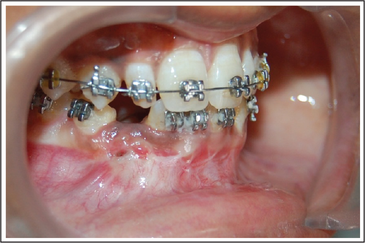
Here, we presented a novel technique of surgically uncovering the mandibular right impacted canine (Figure 1), which can be performed by an orthodontist himself/herself by using a punch biopsy instrument.
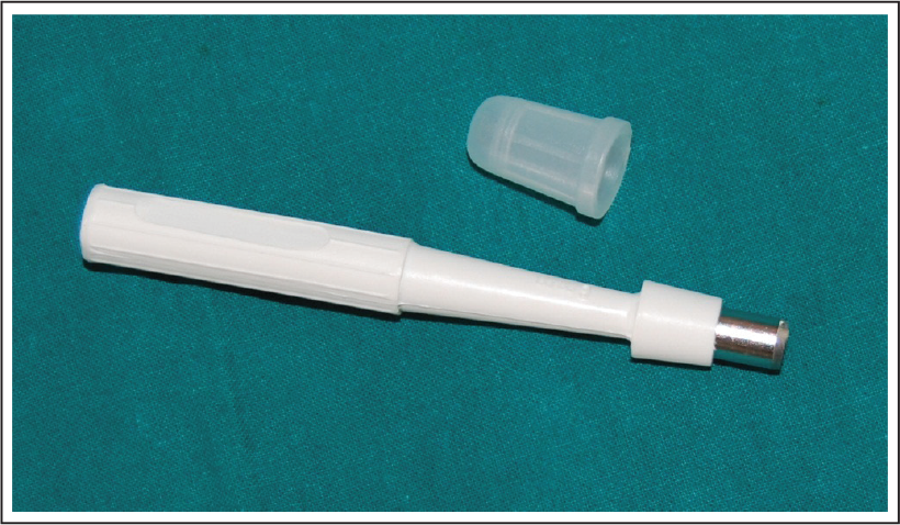
A punch biopsy instrument is a sharp, hollow, circular instrument. It is available in various sizes ranging from 1 to 8 mm. The blade is directly attached to the handle.7 This instrument is mainly used in the punch biopsy of mucosa (Figure 2).
Procedure
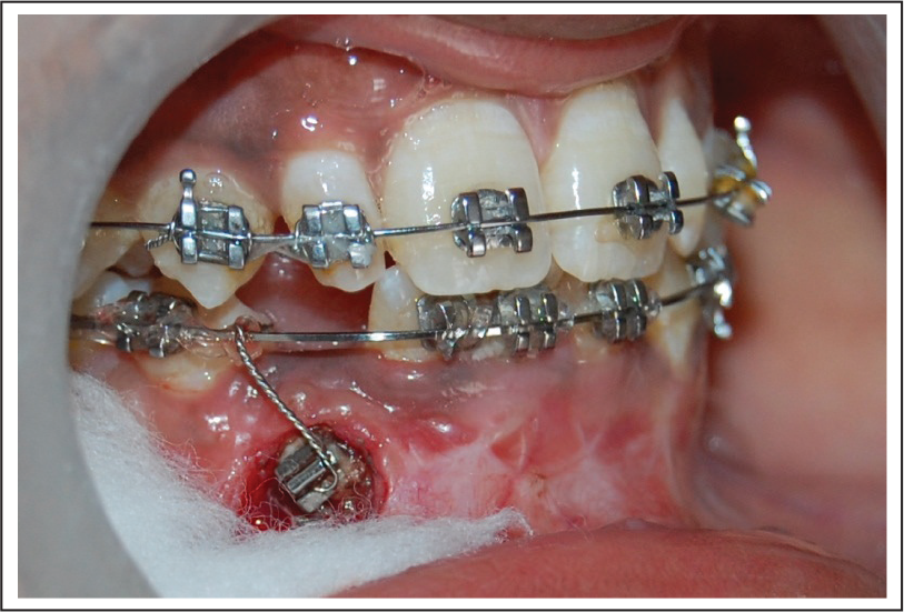
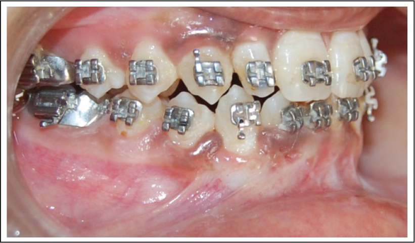
This procedure was carried out by cleaning the mucosa around the impacted right mandibular canine, and then, local anesthesia was administered. A punch biopsy instrument with 6 mm size was selected for this procedure. To expose the impacted right mandibular canine, the punch biopsy instrument was placed vertically over the gingival covering of right mandibular canine, and the smooth twisting motion was used. Once the instrument reached the impacted tooth, the instrument was removed. The tissue was lifted out with tissue-holding forceps. The excision of the tissue on the impacted tooth was enough to bond a bracket. Curved Begg bracket with 0.009″ twisted ligature wire was attached on the exposed canine. Traction for the impacted right mandibular canine was done with 0.009″ ligature wire (Figure 3). The canine fully aligned in the arch after 8 months (Figure 4).
Punch biopsy instrument
Curved Begg bracket with 0.009” ligature wire attached
Canine in occlusion after 8 months
Indications for This Technique
Following are the indications for this technique:
Adequate zone of attached gingival present for erupting canine Labially placed canine Canine is palpable with blanching of tissue on finger pressure Panoramic radiograph showing crown eruption beyond the dento-alveolus.
Advantages of This Technique
The technique is easy, cost-effective, requires a disposable instrument, and an orthodontist can do this simple procedure. Further studies are required to evaluate the long-term efficiency of such a procedure.
Declaration of Conflicting Interests
The authors declared no potential conflicts of interest with respect to the research, authorship, and/or publication of this article.
Funding
The authors received no financial support for the research, authorship, and/or publication of this article.