
Abstract
People often look at objects that they are holding in their hands. It is therefore reasonable to expect them to be able to direct their gaze precisely with respect to their fingers. However, we know that people make reproducible idiosyncratic errors of up to a few centimetres when they try to align a visible cursor to their own finger hidden below a surface. To find out whether they also make idiosyncratic errors when they try to look at their finger, we asked participants to hold their finger in front of their head in the dark, and look at it. Participants made idiosyncratic errors of a similar magnitude to those previously found when matching a visual cursor to their hidden finger. This shows that proprioceptive position sense of finger and gaze are not aligned, suggesting that people rely on vision to guide their gaze to their own finger.
Keywords
People direct their gaze to objects that they intend to interact with and keep their gaze on such objects while interacting with them. When performing tasks that involve manipulating several objects with their hands, people typically look at key positions for the next step of the task rather than keeping their gaze on the object in their hands (e.g., Hayhoe, 2000; Johansson et al., 2001; Land & Hayhoe, 2001). Presumably, looking at an object helps them guide the digits of their hand to appropriate positions for the impending interaction. If the sense of position based on proprioception of the finger is well aligned with the extraretinal sense of gaze position, looking at an object could be useful in guiding the finger because the relevant position on the object can be judged with respect to gaze. If the two are not aligned, looking at the object can only be helpful by decreasing the retinal eccentricity of the finger so that it can be guided to the relevant position using more precise visual information (Cámara et al., 2018).
One reason to suspect that people may not be able to accurately look at their finger is that they make idiosyncratic errors of up to a few centimetres when attempting to align a visible cursor with their own finger hidden below a surface (Kuling et al., 2017; see also van Beers et al., 1998). Presumably, they fixate the cursor when doing so, suggesting that they systematically misperceive the alignment between their gaze and their finger. This is somewhat surprising considering how often we direct our gaze at objects that we are manipulating with our visible fingers. However, we know that small differences in how the alignment task is executed can give rise to quite different patterns of errors (Kuling et al., 2017). Are such misalignments of experimental artefacts caused by factors such as the unusual posture of the arm below the surface or the procedure that is used for guiding the cursor to the finger? Or are the senses of position of the finger and gaze misaligned when the finger is not visible? In the latter case, some people may even be unable to direct their gaze at their own finger in complete darkness.
Methods
Participants
Ten young adults (one female, two left-handed) volunteered to participate in the experiment. The experiment was approved by the Vaste Commissie Wetenschap en Ethiek van de Faculteit der Gedrags- en Bewegingswetenschappen. All participants gave their written informed consent prior to the experiment.
Experimental Setup
Eye movements were recorded with a head-mounted video-based eye tracker (Eyelink II). Participants were fitted with individual dental impression bite-boards that were fixed rigidly in space so that the head could not move. A wooden board was placed vertically, facing the participant, at a distance at which the participant could comfortably hold his or her finger (about 40 cm from the eyes). A single lamp above the board provided dim illumination whenever required. Four light-emitting diodes (LEDs) were embedded within holes in the board through which they could emit dim light. They were arranged at the corners of a 25 by 25 cm square. When the lamp and LEDs were off, the room was completely dark. Four infrared emitting Optotrak markers were used to determine the position of the board. An additional Optotrak marker was attached to the nail of the index finger of the participant’s dominant hand to check for unwanted finger movements.
Procedure
Each participant took part in two sessions, with a short break between the sessions. There were 10 trials in each session, so each participant did 20 trials. Before the first session, the procedure was explained to the participants, and they were told that the marker on their finger was the part of the finger to look at when asked to do so. They were told that they were free to choose any position on the board and that they should vary this position across trials but that they should keep the finger at the same position during the trial. A trial started by the light turning off and a recorded instruction asking the participant to move their finger to a new position on the board and to press a button with their nondominant hand when they were ready. The remainder of the trial consisted of two looking periods, one in the dark and one in the light. Additional recorded instructions were automatically presented at the appropriate moments during these periods to remind participants that they had to switch between looking at the LEDs and looking at their visible or invisible finger. From the moment the participant pressed the button, the timing of the sequence of events was fixed (Figure 1A).

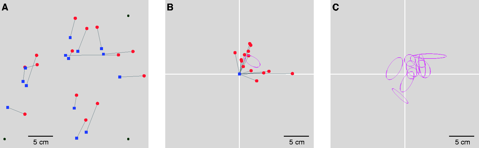
Procedure and Results. A: Summary of the sequence of events within each trial. The trial started with the light turning off and the participant placing his or her index finger somewhere on the board in front of them. The participant then fixated a sequence of LEDs, looked at the finger, and fixated the sequence of LEDs again. The light then turned on, and the sequence of looking at the LEDs, finger, and then LEDs again was repeated with the finger visible (still at the same position on the board). The text in this panel indicates when the corresponding recorded instruction was played, with a black background indicating that the light was off at the time, and a grey background indicating that it was on. B: All trials of one participant. For each trial, the median horizontal and vertical gaze locations in the dark ((red (round) symbols) and in the light (blue (square) symbols) are connected with a thin line. The black symbols indicate the positions of the four LEDs. C: The positions in the dark with respect to those in the light for the same participant as in B. The positions are biased upwards and to the right. The purple ellipse is the 95% confidence ellipse for the mean bias. D: The confidence ellipses for all 10 participants’ biases. The position of gaze in the light (origin) is outside the ellipse for all participants.
The sequence started with the instruction to look at the LEDs and then each of the four LEDs was illuminated for 2 s in a fixed order in the dark (clockwise, starting at the top left). Then, there was a 2-s dark interval with the instruction to look at the finger, followed by 3 s in the dark for looking at the finger. After that there was a 2-s interval in which the instruction was given to look at the LEDs, and the same sequence of LEDs being illuminated was repeated. Then, the light was turned on, and after 2 s to adjust to the new illumination, the same procedure was repeated in the light. Thus, after having positioned the index finger of their dominant hand and pressed the button, each trial lasted 48 s, yielding 3 s of fixating the finger and 16 s of fixating the LEDs both in the dark and in the light. The fixed LED sequences were used to make the measurements as similar as possible both between measurements made in the dark and in the light, and between trials.
Data Handling
Each trial provided us with two sets of four-point LED fixation data in the dark and another two such sets in the light. Using LED fixations before as well as after looking at the finger for calibrating gaze, and spreading the calibration throughout the experiment, makes it very unlikely that drift in the eye tracking will introduce systematic errors. To convert eye-tracking data to gaze locations, we first determined the median horizontal and vertical eye tracker coordinates during each 2-s interval of fixation on the LEDs. We then determined the median of these horizontal and vertical values for each LED during each session. This was done separately for the parts of the trials with the light on and off. These values were then used to determine the gaze position of the participants when looking at their finger at that position with the light on and off (based on the median horizontal and vertical eye-tracker positions during the 3-s intervals in which they were looking at the finger). For each eye, we determined the translation, scaling, rotation, and shear that relates the Eyelink camera coordinates to positions on the board. Finally, we averaged the gaze positions of the two eyes.
It is certainly conceivable that the offsets of gaze in the dark from (the presumably perfect) gaze in the light would depend systematically on the finger’s position on the board. For instance, gaze positions might be compressed or expanded due to misjudging the distance to the finger in the dark. However, looking at plots of where gaze was directed (as shown for one participant in Figure 1B) suggested that within this area the main difference between gaze in the light and gaze in the dark was a uniform shift. In the further analysis, we therefore only looked for systematic global shifts of relative gaze positions in the dark. Note that ignoring other potential systematic effects does not affect the average shift; it might result in an increase of the variability in the shift across trials.
Trials were excluded if the finger marker data were missing when looking at the finger. This sometimes happened if the participant placed their finger at a position that was hidden from the Optotrak camera by their own body. Trials were also excluded if gaze data were incomplete (due to the eye tracker failing to detect the pupil). In addition, we present the analysis of a subset of the data that comply with additional selection criteria with regard to the confidence of the measurements.
Results
Ten participants each performed 20 trials. In the analysis, we excluded 11 of the 200 trials (0–7 per participant; finger marker data were missing for 2 trials, and gaze data were incomplete for 9 trials). Participants generally held their finger very still: The median displacement from when they looked at the finger in the dark to when they did so in the light was only 1.0 ± 0.8 mm. As could be expected, the area of the pupil was 26 ± 12% larger in the dark than in the light (mean ± standard deviation across participants).
Before presenting the main results, we present some characteristics of the fixations. The median two-dimensional distance between the estimated gaze locations of the two eyes (on the board) was 1.7 ± 0.5 cm when looking at the finger in the light and 1.3 ± 0.3 cm when doing so in the dark. A small horizontal difference between the gaze locations of the two eyes is to be expected if participants looked at the marker, as instructed, rather than at a position on the board. The marker was attached to the nail of the finger and was therefore slightly closer than the board. Nine of the 10 participants looked further away in the dark than in the light. Seven of them even did so significantly (t test across trials). In the dark, the marker was not visible, so participants might have tried to look at the position at which the finger was touching the board instead of the marker. With a viewing distance of 40 cm and an average distance between the pupils of the two eyes of 6.4 cm, a difference in fixation distance in depth of 1 cm (a reasonable thickness for a finger) would result in a distance between the gaze of the two eyes on the board of 1.6 mm. In the vertical direction, four participants had systematic differences between the two eyes’ relative gaze in the light and in the dark, but the direction of the effects was not consistent across participants.
To visualize any systematic global shifts of gaze positions in the dark, we compared the gaze coordinates when looking at the fingernail in the light and in the dark (the trials of one participant are shown in Figure 1B). Typically, gaze in the dark was not evenly distributed around gaze in the light. This is clearly seen when the positions in the light are superimposed so that only the differences between the coordinates in the dark and in the light are visible (see Figure 1C for the data of the example participant). We determined 95% confidence ellipses for the mean of such differences (purple ellipse in Figure 1C). All 10 participants had a significant difference between gaze position in the light and in the dark: The intersection of the white lines is not within the ellipse (Figure 1D). The mean absolute value of the 10 participants’ biases was 3.4 cm (with a standard deviation across participants of 1.8 cm). The mean area of the 95% confidence ellipses was 4.5 cm2 (with a standard deviation across participants of 2.0 cm2).
Reanalysing the Data With Strict Inclusion Criteria
In our analysis, we considered all measurements equally, although our confidence in the measurements differed between trials. We therefore also analysed the data after applying strict criteria for including trials. In addition to the obvious criteria that the data had to be complete, we now excluded trials if the finger marker moved more than 5 mm during the trial or if the measured gaze positions of the two eyes were more than 2.5 cm apart. Forty additional trials were excluded because the finger moved more than 5 mm during the trial. Thirty-five additional trials were excluded because the measured eye positions were more than 2.5 cm apart on the board. This left us with 114 of the 200 trials (5–16 per participant).
The remaining data of the example participant and the confidence ellipses of all participants can be seen in Figure 2. Also, when only analysing the most reliable data, all 10 participants’ gaze was significantly different in the dark than in the light: The intersection of the white lines is not within the ellipse (Panel C of Figure 2). The mean absolute value of the 10 participants’ biases was 3.3 cm (with a standard deviation across participants of 1.4 cm). The mean area of the 95% confidence ellipses was 5.5 cm2 (with a standard deviation across participants of 2.0 cm2). The fact that the strict selection hardly made a difference to the biases makes it unlikely that the biases were caused by the finger moving, the participant not looking at the finger when asked to do so, or gaze having been measured inaccurately.
Analysis With Stricter Trial Selection (Details as in Figure 1). A and B: Data for the remaining trials (14 of the original 20) of the selected participant. The purple ellipse is similar to that in Figure 1 despite being based on fewer points. C: The confidence ellipses for all 10 participants’ biases.Note. Please refer to the online version of the article to view the figure in colour.
Discussion
To investigate whether it is possible to look at your finger in the dark, we asked participants to place their finger on a frontal surface and then to look at the finger, first in the dark and then under dim illumination. We found that they made idiosyncratic errors of several centimetres when they could not see their finger, similar to those that have previously been found in other alignment tasks (e.g., Kuling et al., 2013, 2016, 2019; Kuling, Brenner, et al., 2014; Kuling, van der Graaff, et al., 2014; Rincon-Gonzalez et al., 2011; Smeets et al., 2006; Sousa et al., 2010; Tillery et al., 1991; van Beers et al., 1996, 1998; van der Kooij et al., 2013). The fact that none of the participants’ confidence ellipses included an error of zero and that 7 of our 10 participants have a bias to the top right is probably a coincidence. The main finding is that there are idiosyncratic mismatches and that these mismatches are of a similar magnitude to those found in previous studies comparing vision and proprioception.
Finding that people even make idiosyncratic errors when trying to look at their own finger that they have actively positioned in front of their own eyes in the dark is in line with our earlier arguments (Kuling et al., 2017) that similar biases that we found in related tasks (e.g., Kuling, van der Graaff, et al. 2014, Kuling et al., 2017) were not only the result of the unusual postures that were used in those studies. Although the idiosyncratic errors in directing gaze are not huge, they are orders of magnitude larger than the spatial resolution at similar distances from the fovea (Woog & Legras, 2018). Thus, it is likely that retinal information is normally used to guide the digits to their goals (Cámara et al., 2018). Where the eyes are oriented for guiding the digits to their goals is presumably determined by where one expects it to be useful to have a high retinal resolution (Voudouris et al., 2018), rather than by it being easier to move a digit to where one is looking (Neggers & Bekkering, 2001). We conclude that people rely on vision to direct their gaze, even when directing gaze to their own finger.
Footnotes
Declaration of Conflicting Interests
The author(s) declared no potential conflicts of interest with respect to the research, authorship, and/or publication of this article.
Funding
The author(s) received no financial support for the research, authorship, and/or publication of this article.