
Abstract
Three donkeys were presented with progressive lameness and distal suspensory ligament breakdown in multiple limbs. Treatment with nonsteroidal anti-inflammatory drugs was only partially effective and eventually the donkeys were euthanized due to further progression of the lameness and concerns for their welfare. At necropsy, the distal part of the suspensory ligaments in multiple limbs, including the suspensory ligament branches, was markedly thickened, enlarged, and mottled white and brown on cut section. In one case, adult Onchocerca sp. nematodes were grossly identified embedded within the suspensory ligaments. Histopathologic examination revealed chronic, multifocal to coalescing, moderate to severe, lymphoplasmacytic, eosinophilic, and fibrosing desmitis and tendinitis with intralesional, coiled adult nematodes of Onchocerca sp., accompanied by osseous and cartilaginous metaplasia. To the authors’ knowledge, this is the first histopathologic description of suspensory ligament desmitis and tendinitis associated with Onchocerca sp. in donkeys.
Equine onchocercosis is a parasitic infection caused by filarial nematode parasites of the genus Onchocerca affecting donkeys, horses, and other equids with a worldwide distribution. 19,20 Cutaneous and connective tissue onchocercosis is a common problem in working donkeys in most developing countries. 10,16 It is widely known that the skin-dwelling microfilariae of equine Onchocerca spp. induce cutaneous and ocular lesions of varying degrees of severity. 10,11,14,19 O. cervicalis (“neck threadworm”) and O. railletie are the 2 common species often reported from the connective tissue of the nuchal ligament of working donkeys. 1,7,16 O. cervicalis is the frequently reported species in horses. 2,8,11 –13 The cattle filariid, O. gutturosa, has not been reported in donkeys but rare reports exist in the nuchal ligament of the horse. 15 The only Onchocerca sp. reported from the suspensory ligaments (SLs) and tendons of the limbs of donkeys and horses is O. reticulata. Infection by this parasite is often characterized by the presence of subcutaneous nodules over or within the flexor tendons and suspensory ligaments, where it is potentially associated with swelling and lameness. 6,10,16,19 O. cervicalis has a worldwide distribution but O. reticulata seems to be restricted to Europe, Africa, and Asia. 7,16,19,20 Onchocercosis caused by O. cervicalis was recently reported in horses from some European countries. 8,9 Reports of equine onchocercosis in the United Kingdom are rare, and date back to the early 1970s and 1980s. 2,13 There is no report of onchocercosis in donkeys in the United Kingdom or the rest of Europe.
Three donkeys (Table 1), 1 gelding and 2 females, aged 18, 21, and 23 years old, respectively, were presented to the Veterinary Hospital of The Donkey Sanctuary, Devon, United Kingdom. All 3 donkeys had similar clinical signs, namely, chronic multilimb lameness, stiffness, and dropped fetlocks on the affected limbs. Both forelimbs were affected in case 1, all 4 limbs were affected in case 2, and only the hindlimbs were affected in case 3. All 3 donkeys were treated with nonsteroidal anti-inflammatory drugs (phenylbutazone [Equipalazone], carprofen [Carprieve]). All 3 donkeys were routinely dewormed with moxidectin (Equest) yearly at a dose rate of 0.4 mg/kg body weight and had further monitoring for strongyles via fecal worm egg counts and targeted anthelmintic treatments. Further deterioration of the clinical signs resulted in humane euthanasia.
Gross Postmortem and Histopathological Findings in the Suspensory Ligaments and Tendons Examined in 3 Cases of Connective Tissue Onchocercosis in Donkeys.
Abbreviations: SL, suspensory ligament; LF, left fore; RF, right fore; RH, right hind; LH, left hind; SDFT, superficial digital flexor tendon.
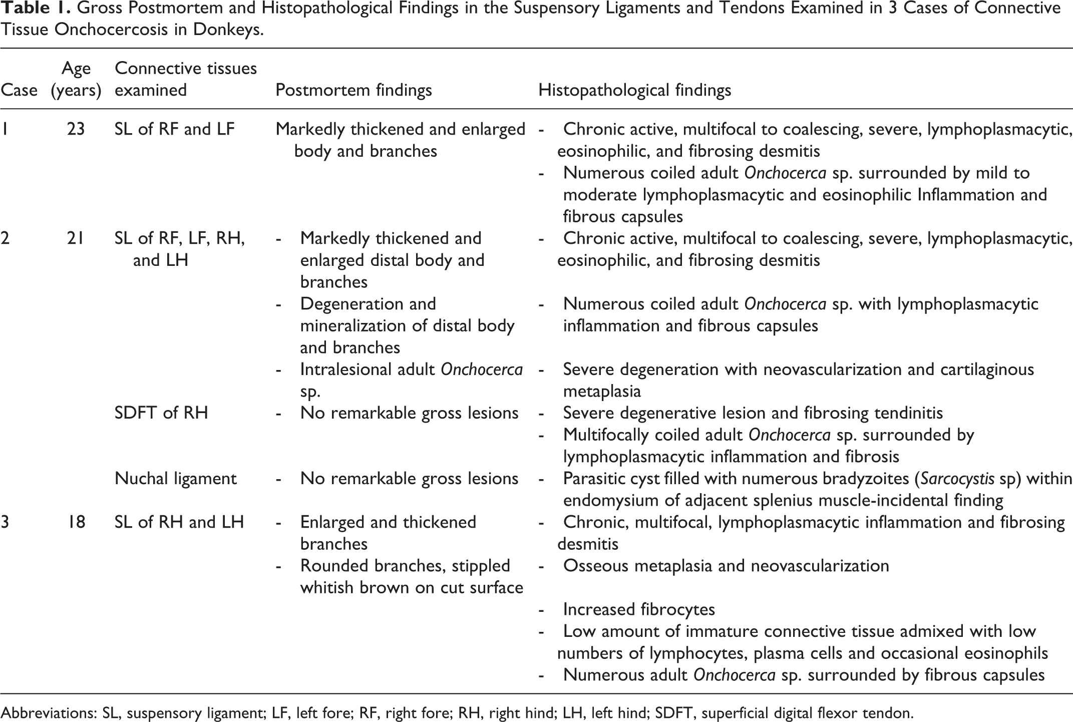
At necropsy, the distal third of the SLs (body and branches) of the affected limbs were markedly enlarged, firm, rounded, and mottled white and brown (Table 1). No other significant gross abnormalities were observed. In case 2, numerous coiled adult nematodes were identified embedded within the branches of the affected SLs (Figs. 1, 2). In cases 1 and 3, the nematodes could not be seen grossly. There were no visible cutaneous lesions.
Onchocercosis, suspensory ligament (SL), donkey, case 2.
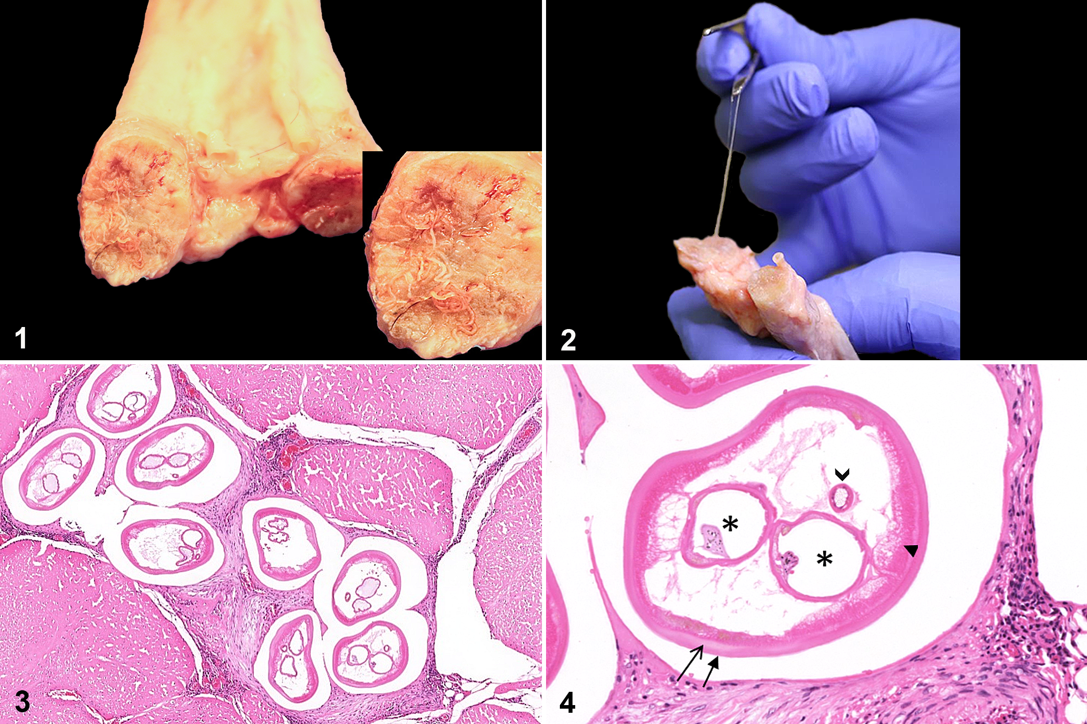
Histopathological findings were similar in all 3 cases with different degrees of severity (Table 1). Multifocally within the fascicles and interfascicular matrix of the SLs of all cases and SDFT of case 2 there were multiple cross sections of coiled, viable, and degenerate adult nematodes with an outer cuticular layer, subcuticular hypodermal layer with characteristic ridges, coelomyarian-polymyarian musculature, paired uteri (sometimes filled with microfilariae), and small gastrointestinal tract (Fig. 4), consistent with Onchocerca sp. 5 Parasites were surrounded by moderate to high numbers of lymphocytes and plasma cells, moderate numbers of eosinophils and a moderate amount of immature and mature fibrous connective tissue (Fig. 3). The inflammatory reaction was more intense around nonviable degenerated nematodes (Fig. 3). In addition, there was moderate to severe degeneration of SLs with cartilaginous metaplasia and neovascularization in case 2, and osseous metaplasia in case 3. Paired gravid uteri with intrauterine microfilariae indicated patent infections in all 3 cases and was one of the most characteristic features of the cross sections of adult female Onchocerca sp. in most of the affected SLs and tendons (Fig. 4).
Histological examination of the nuchal ligament in case 2 revealed no Onchocerca sp. but focally within the endomysium of the adjacent to the nuchal ligament splenius muscle there was a parasitic cyst filled with numerous bradyzoites, presumed to be Sarcocystis sp. Individual Sarcocystis sp. cysts in skeletal muscle are considered incidental.
Records of SL and tendon infections by Onchocerca spp. in equids from Africa and associated gross lesions date back to the 1950s and 1960s as cited in Ottley et al. 15 These studies showed the association of O. reticulata with suspensory ligament desmitis, tendinitis, sesamoiditis, and navicular bone disease in horses. The desmitis and tendinitis in the 3 cases described here are similar to their macroscopic findings. Similar gross pathological changes in the SLs and tendons associated with O. reticulata infection were more recently reported in donkeys from Africa, 16 although histopathologic examination was not performed. Consistent with the cases described here, infiltration of the nuchal ligament by lymphocytes, eosinophils, plasma cells, and cartilaginous metaplasia were reported in donkeys infected by O. cervicalis and O. railliete and horses infested by O. cervicalis. 1,17 The presence of inflammation likely demonstrates a reaction to the Onchocerca spp. parasites. Whether the parasites are the direct cause of the inflammatory infiltrates or it is due to hypersensitivity reaction to the presence of the parasites cannot be determined. 10,16 Chronic inflammation in the SLs, accompanied by mineralization, cartilaginous metaplasia, and ossification in cases 2 and 3 illustrates the severity of pathological changes which appeared to be irreversible. This is likely associated with the protracted course of infection with progressive pathological changes as the parasites can live and reproduce for several years in the connective tissues. 18 These lesions were likely responsible for the lameness and marked pain that were unresponsive to analgesics.
Microfilariae of Onchocerca spp. are transmitted by blood-sucking insects in which an obligatory stage of development occurs. Several different insects may be suitable hosts, but only Culicoides spp. midges and Anopheles spp. mosquitoes are known to play a significant role. 3 The time and location of infection of the 3 donkeys in this case report is unknown. Cases 1 and 3 which originated from northwest and southwest England, had been at the sanctuary for over 6 years. However, case 2 had only been relinquished to the sanctuary from Scotland 6 months previously. The prepatent period of Onchocerca spp. is over 1 year. 4 The clinical history of prolonged and progressive lameness along with the histopathologic finding of many adult gravid female Onchocerca sp. strongly suggests that case 2 had been infected at least 6 months before its relinquishment. Furthermore, adult Onchocerca spp. may survive for several years in connective tissue. 18 The deworming history of these donkeys is not fully known before they were relinquished. The donkeys were routinely dewormed with moxidectin (Equest) every year while at the sanctuary. Insensitivity of the adult stage to the microfilaricides has been previously reported. 19 Repeated and periodic retreatment is required even to eliminate the cutaneous microfilariae. 19 The sanctuary practice of selective anthelmintic treatment in donkeys to prevent anthelmintic resistance might have played a role in sustaining the infection through incomplete elimination. Another possible explanation is that the Onchocerca sp. in donkeys might have developed resistance due to increased exposure to macrocyclic lactone treatment during life. However, this needs to be substantiated through well-designed studies and anthelmintic efficacy trials.
The characteristic histomorphological features enabled identification of the nematodes as species of the Genus Onchocerca. It is not possible to distinguish between the different equine Onchocerca spp. in tissue sections where only parts of the nematode can be seen. 15 Based on its suggested geographical distribution in Europe, and its common predilection site in the connective tissues of SLs and tendons of the lower limbs, 15,19 it is most likely that the species recovered was O. reticulata. However, molecular identification would be required to confirm this.
To the authors’ knowledge, this is the first histopathologic description of SL lesions associated with this nematode, which has similarities to nuchal ligament onchocercosis in equids. Differential diagnosis during lameness evaluation of donkeys with dropped fetlocks should include SL desmitis and SDFT tendinitis associated with Onchocerca sp.
Supplemental Material
Supplemental Material, Combined_supplemental_materials-Paraschou_et_al - Suspensory Ligament Desmitis Caused by Onchocerca sp. in Three Donkeys
Supplemental Material, Combined_supplemental_materials-Paraschou_et_al for Suspensory Ligament Desmitis Caused by Onchocerca sp. in Three Donkeys by Georgios Paraschou, Getachew M. Adako, Simon L. Priestnall and Faith A. Burden in Veterinary Pathology
Footnotes
Acknowledgements
The authors would like to thank Chris Platts and Edo Santangelo, laboratory manager and photographer at The Donkey Sanctuary, respectively, and Dr Alejandro Suárez-Bonnet, lecturer in comparative pathology at Royal Veterinary College, for their constructive advice and assistance with image processing; Allan Emmett, post mortem assistant, for the invaluable help collecting the tissue samples; and the histology technicians at The Nationwide Laboratories-Abbey veterinary services, Devon, UK, for their help processing the histopathology slides.
Declaration of Conflicting Interests
The author(s) declared no potential conflicts of interest with respect to the research, authorship, and/or publication of this article.
Funding
The author(s) received no financial support for the research, authorship, and/or publication of this article.
Supplemental material for this article is available online.
References
Supplementary Material
Please find the following supplemental material available below.
For Open Access articles published under a Creative Commons License, all supplemental material carries the same license as the article it is associated with.
For non-Open Access articles published, all supplemental material carries a non-exclusive license, and permission requests for re-use of supplemental material or any part of supplemental material shall be sent directly to the copyright owner as specified in the copyright notice associated with the article.