
Abstract
T-zone lymphoma (TZL) is an indolent nodal T-cell lymphoma most commonly observed in submandibular lymph nodes in dogs. The diagnosis is based on its distinct morphology and expression of CD3. TZL has been reported to have a low Ki67 index and to lack expression of CD45. The latter feature has been used to diagnose this type of lymphoma via fine needle aspirate and flow cytometry without confirmation of the characteristic tissue architecture. The goal of this study was to characterize the immunophenotype of canine nodal TZL in greater detail. Twenty-seven TZLs were selected based on their characteristic morphology. A tissue microarray was generated, and immunohistochemical expression of CD3, CD5, CD20, CD21, CD25, CD45, Bcl-6, and Ki67 was evaluated. Neoplastic T cells in all cases were positive for CD3, CD5, and CD25, and negative for CD20, CD21, and Bcl-6. Positive labelling for CD45 was detected in 2 of the 27 cases with the remaining 25 cases being negative. All cases had a low Ki67 index with an average index of 19.56%. For the CD45-positive TZLs, clonality of the T-cell antigen receptor gamma gene was confirmed in only one of these cases. The observed immunophenotype of canine TZL is similar to previous publications with the exception that 2 cases expressed CD45. Expression of CD45 in TZLs in this study emphasizes the importance of interpreting immunophenotypic findings in conjunction with histopathology to reach an accurate diagnosis and not to use lack of expression of a particular antigen as the sole diagnostic criterion.
T-zone lymphoma (TZL) is a type of nodal T-cell lymphoma that has been well characterized in dogs. 17 –20 In humans, TZL is considered a morphologic variant of unspecified peripheral T-cell lymphoma, while in dogs it is considered a distinct entity. 13 In contrast to other nodal T-cell lymphomas in dogs (ie, anaplastic large T-cell lymphoma, angioimmunoblastic T-cell lymphoma, and unspecified peripheral T-cell lymphoma), TZL follows an indolent course.
Diagnosis of TZL is based on characteristic paracortical expansion by neoplastic T cells that cause peripheralization and compression of lymphoid follicles against the capsule. 18 –20 Neoplastic T cells are uniform, small to intermediate sized, and have moderate amounts of pale, often water-clear cytoplasm. Nuclei are densely stained and often have sharp indentations and inapparent nucleoli. The mitotic count and Ki67 proliferation index are low. In one study, the average Ki67 proliferation index was reported to be 17.6% with a standard deviation of 12.1%. 4
To assist in distinguishing TZL from other types of lymphomas, immunophenotyping in combination with histopathology is recommended. 15,19 TZL has an immunophenotype typical of all mature T-cell neoplasms, in that it is positive for both CD3 and CD5, and negative for B-cell antigens CD20, CD79a, and Pax-5. 4,9,10,13,17,19,20 Furthermore, TZLs are typically positive for CD21 and CD25. 8,10
Interestingly, loss of expression of leukocyte common antigen CD45 has been reported as a characteristic feature of canine TZLs. 9,13,14 This is in contrast to most T- or B-cell lymphomas that are CD45-positive. The loss of CD45 expression in canine TZLs has been linked to impaired gene transcription in one study, though the exact mechanism is not understood. 8 This lack of CD45 expression has been proposed as the primary diagnostic feature of TZL allowing for a diagnosis with fine needle aspirate and flow cytometry without confirmation of the characteristic tissue architecture. 1,5,14 As histopathologic examination represents a critical component to accurately subtype lymphomas, and loss of CD45 expression has been described in other T-cell malignancies, 3,5,11 such an approach may result in potential diagnostic errors.
The goal of this study was to characterize the immunophenotype of canine nodal TZL in greater detail with special emphasis on the expression of CD45.
Materials and Methods
Case Selection
TZL cases were retrospectively selected from cases submitted between 2010 and 2015 to the Veterinary Diagnostic Laboratory at Michigan State University. A total of 60 cases were selected, some of which were excluded due to unavailability of paraffin-embedded blocks, and 35 remained in the study. Cases had been previously diagnosed based on morphologic characteristics, immunophenotyping for CD3 and CD20, and, in some cases, PARR testing for clonality of the T-cell antigen receptor gene. 4,6,7,16 –20
Histopathology and Immunohistochemistry
For each case, sections stained with hematoxylin and eosin (HE) and serial sections immunohistochemically labeled for CD3 and CD20 slides were evaluated to confirm the previous diagnosis. Eight cases were omitted based on large nuclear size and high mitotic activity that was inconsistent with a diagnosis of TZL. A tissue microarray (TMA) was generated with five 1-cm diameter punches for each of the 27 remaining cases. Punches were taken from the original paraffin block from multiple regions. Specifically, we sampled paracortical T cells, which were easily identified in HE-stained slides based on their characteristic histologic features especially when also combined with immunophenotyping for CD3 and CD20 (Figs. 1–3). The completed TMA block was stained with HE to confirm the proper selection of neoplastic cells. Punches that missed the targeted areas or had poor representation of the neoplastic cells were omitted. Immunohistochemistry (IHC) for CD3, CD5, CD20, CD21, CD25, CD45, Bcl-6, and Ki67 was performed on serial sections of the tissue microarray. The choice of these antibodies was based on previous studies of canine TZL. 2,4,9,10,13,16,18 –20 B-cell markers CD20 and Bcl-6 were included to better highlight nodal follicular architecture and to aid in differentiating T and B cells on the TMA punches. Immunohistochemistry was carried out in accordance with the guidelines set forth by the American Association of Veterinary Diagnosticians Subcommittee on Standardization of Immunohistochemistry. 12 IHC was performed on a BOND-MAX Automated Staining System (Vision BioSystems; Leica) using the Bond Polymer Detection System (Vision BioSystems; Leica) with 3,3′-diaminobenzidine (DAB) as the chromogen for CD3, CD20, CD25, and CD45; on the Dako link 48 Automated Staining System (Agilent Technologies) using the peroxidase conjugated EnVision Polymer Detection System (Agilent Technologies) with DAB as the chromogen for CD5, CD21, and Bcl-6; and with the Discovery Ultra staining platform (Ventana Medical Systems) using the Vetana medium cell conditioner protocol (Roche) and detection with DISCOVERY UltraMap alkaline phosphatase system with the ChromoMap AP Red Staining kit (Roche) for Ki67. Details of the antibodies, retrieval methods, and detection are shown in Table 1. Normal canine lymph node was used as a positive control for all antibodies. IHC-labeled microarray slides were blindly reviewed by 2 authors (LS, MK) and immunoreactivity was subjectively scored as an average percent of positive neoplastic cells per punch per case. A case was called positive for CD3, CD20, CD45, and Bcl-6 if the majority of neoplastic cells showed strong, diffuse, marker-specific labeling. For CD5, CD21, and CD25 a case was called positive if at least 10% of the neoplastic cells showed positive labeling. This was based on the limited reports of CD5, CD21, and CD25 use for immunohistochemistry in canine neoplasms. 2,10,16,18 The Ki67 index was determined by first identifying the area of each punch with the highest proportion of Ki67 labeling neoplastic cells. 4 In these areas, 100 neoplastic cells were counted, as well as the number of those 100 cells that had strong nuclear labeling for Ki67. The percentage of Ki67-positive cells was calculated to yield the Ki67 index. For each individual case, the indexes of all 5 punches were averaged to determine the Ki67 index. The raw IHC data analyzed in this study are available in Supplemental Table S1.
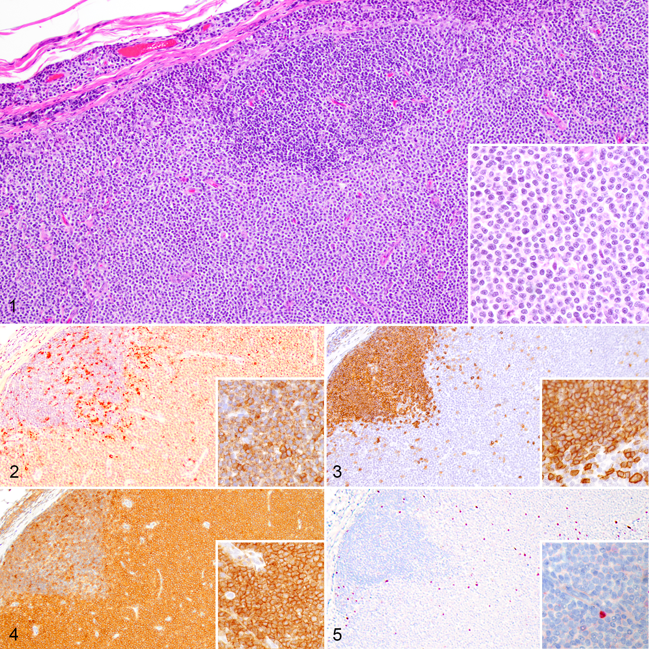
T-zone lymphoma (TZL), lymph node, dog.
Immunohistochemistry Methods Used in This Study.
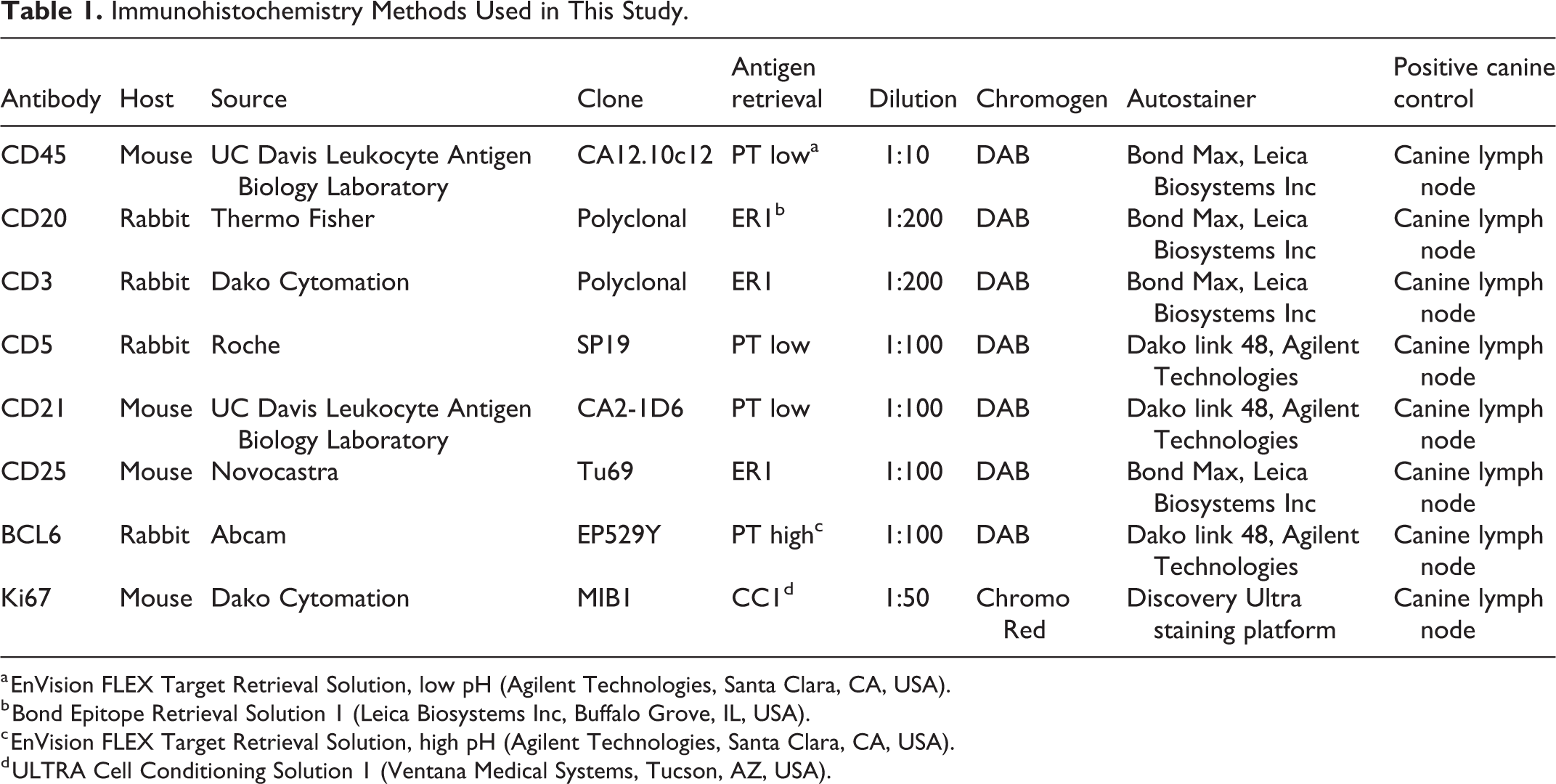
a EnVision FLEX Target Retrieval Solution, low pH (Agilent Technologies, Santa Clara, CA, USA).
b Bond Epitope Retrieval Solution 1 (Leica Biosystems Inc, Buffalo Grove, IL, USA).
c EnVision FLEX Target Retrieval Solution, high pH (Agilent Technologies, Santa Clara, CA, USA).
d ULTRA Cell Conditioning Solution 1 (Ventana Medical Systems, Tucson, AZ, USA).
Results
TZLs were characterized by neoplastic small to intermediate sized T cells with moderate to abundant water-clear cytoplasm and densely stained nuclei (Fig. 1). Cells expanded the paracortex and compressed lymphoid follicles against the capsule. The mitotic count ranged from 1 to 5 mitotic figures in 10 high power fields (2.37 mm2) across all cases.
Immunolabeling with all antibodies was positive in canine control tissues. Neoplastic cells in all TZLs showed diffuse immunoreactivity for CD3 (Fig. 2). Neoplastic cells were not immunoreactive for CD20 (Fig. 3), CD21, or Bcl-6. Immunoreactivity for CD5 ranged from 10% to 100% across all cases, with 24/27 TZLs having at least 80% labeling of neoplastic cells and 3/27 having 10% to 20% labeling. Immunoreactivity for CD25 ranged from 20% to 100% across all cases, with 25/27 TZLs having at least 50% labeling of neoplastic cells and 2/27 having 20% to 40% labeling. Two of the 27 TZLs had strong immunoreactivity for CD45 in all neoplastic cells (Fig. 4). The number of cells expressing the different markers in a given punch were either very high or very low depending on the antibody, and therefore, the distinction between positive and negative cases was straightforward based on the described criteria. There was minimal variation in the percentage of cells expressing a specific antigen among punches of the same case.
Neoplastic cells in all TZLs had infrequent nuclear labeling for Ki67 (Fig. 5). The average Ki67 index across all 27 TZLs was 19.5%. Individual case averages ranged from 7.67% to 26.67%. The average Ki67 index in the 2 CD45-positive cases was 21.8% and 22.5%, respectively.
Discussion
The observed immunophenotype of canine nodal TZL was consistent with previous reports, with the exception that 2 of 27 TZLs expressed CD45. While a diagnosis of TZL was confirmed in these 2 cases based on the characteristic morphology and immunophenotype, as well as a low Ki67 proliferation index that was within one standard deviation of previous reports, subsequent PARR testing confirmed clonal rearrangement of the T-cell antigen receptor gene in only one of these 2 cases. Failure to detect clonality in the other CD45-expressing TZL may be due to the rearrangement not being covered by our primers. Alternatively, it could be because the proliferation in that case was focal (ie, early in the disease) so the polyclonal population in the remainder of the node may have masked the smaller clonal population. 6,7
There are a number of studies that have reported the loss of expression of CD45 as a specific diagnostic feature of TZL. 9,13,14 However, for the majority of TZLs reported in the literature, the diagnosis was based entirely on cytology and an absence of CD45 expression on flow cytometry. The number of published cases that were diagnosed as TZL based on the characteristic histomorphology followed by detailed immunophenotyping, as is the gold standard, is limited. The initial hypothesis that canine TZLs have lost expression of CD45 was based on a series of only 6 CD4+/CD45− T-cell lymphomas that exhibited a less aggressive behavior for 3 cases. 1 In a follow-up study, only those cases were selected that were composed of a CD45− T-cell population comprising more than 30% of all T cells on flow cytometry. 14 Cases were excluded from that study if expression of CD45 represented a continuum from CD45 low to high within the T-cell population. Based on those selection criteria, only 20 cases were identified where the lymph node had been examined histologically, and paraffin blocks were only available for 13 of these cases. Immunohistochemistry for CD45 was not performed on any of these paraffin blocks and lack of expression was based entirely on the previous selection criterion of being CD45 negative on flow cytometry. In another study, 51 canine TZLs were examined histologically; however, lack of CD45 expression was again used as a selection criterion and only CD45-negative lymphomas were included in that study. 8 It is also important to recognize that CD45 expression in the initial flow cytometry study was detected by using a rat anti-dog CD45 antibody, clone YKIX716.13 (AbD Serotec), whereas in studies of formalin-fixed paraffin-embedded tissues, including ours, a mouse anti-dog CD45 antibody, clone CA12.10c12 (UC Davis Leukocyte Antigen Biology Laboratory), has been utilized. 8,9,14
While the data from these previous studies document that a significant subset of TZLs lack expression of CD45, the design of these studies may result in a vast overestimation of the percentage of TZLs that do not express CD45. As such, lack of CD45 expression cannot be confidently used as the primary diagnostic feature of TZL. It is important to recognize that all cases in our study were selected based on the characteristic histomorphologic appearance and low proliferation index of TZL, regardless of their expression of CD45. It is also worth noting that the anti-CD45 antibody used in our study was the same that was used in a study by Martini et al for immunohistochemical evaluation of TZL. 8 However, in that study, cases were initially selected based on cytologic appearance and CD45-negative immunophenotype using flow cytometry. Histopathology and immunohistochemistry were only performed on those preselected CD45-negative cases, if available, and, as expected, showed complete lack of CD45 expression. It is unlikely that the positive labeling for CD45 of some TZLs in our study was caused by differences in antibody selection. It is more likely that we were able to identify these CD45-positive cases by using unbiased selection criteria when designing our study, which is crucial when evaluating the diagnostic significance of single-antigen expression or lack thereof in a neoplasm. Future, large-scale studies that use similar selection criteria and compare immunohistochemistry and flow cytometry are essential to accurately assess the diagnostic value of CD45 expression in TZL.
Loss of CD45 expression in TZL has been linked to impaired gene transcription. 8 Martini et al reported significantly lower than normal CD45 mRNA transcript levels in canine TZLs compared to nonneoplastic T cells or aggressive T-cell lymphomas. 8 The small amounts of CD45 transcript that were detected in TZLs were assumed to be related to contamination or residual nonneoplastic lymphocytes, suggesting the virtual absence of CD45 transcript in TZLs. 8 However, our results suggest that in at least some cases of TZL, the CD45 transcript is not completely lost or diminished and may warrant further consideration of the underlying genetic alterations leading to the development of TZL.
From a diagnostic perspective, our study emphasizes the importance of interpreting immunophenotypic findings in conjunction with histopathology. Diagnosing TZL solely by the expression of CD3 and loss of CD45 expression without considering the histomorphology of the neoplasm may result in misdiagnosis of a more aggressive T-cell lymphoma and, by extension, inaccurate communication of prognosis and therapeutic options. Loss of CD45 expression has been identified in other T-cell lymphomas, such as peripheral T-cell lymphoma (PTCL). In one case report, PTCL was misdiagnosed as TZL based on fine needle aspirate and CD45-negative flow cytometry results. 11 Ultimately, a PTCL was confirmed histologically based on the lymph node architecture, cellular features, and high mitotic activity. It can be argued that the interpretation of the fine needle aspirate may have differed among pathologists, as it is subjective, but the misdiagnosis demonstrates the confidence that was erroneously placed on the CD45-negative immunophenotype. Additionally, there is always the risk that a fine needle aspirate could miss the heterogeneity of a cell population and clusters of larger cells with increased mitotic activity that would suggest malignant transformation. The combination of cytology, flow cytometry, and clinical features can be useful but remains insufficient to definitively diagnose this entity. While cytology and flow cytometry are less invasive and less expensive diagnostic techniques, histopathology in combination with immunophenotyping remains the gold standard for diagnosing and subtyping lymphomas.
The results of this study suggest that an accurate diagnosis of TZL should be made by histopathologic examination; immunophenotyping for CD3, CD20, and CD45; analysis of the Ki67 proliferation index; and, if needed, clonality testing. If histopathology is not utilized, then a diagnosis of TZL must be made with caution and through the use of more than just a CD45-negative immunophenotype.
Supplemental Material
Supplemental Material, Supplemental_materials-Stein_et_al - Immunophenotypic Characterization of Canine Nodal T-Zone Lymphoma
Supplemental Material, Supplemental_materials-Stein_et_al for Immunophenotypic Characterization of Canine Nodal T-Zone Lymphoma by Leah Stein, Cynthia Bacmeister and Matti Kiupel in Veterinary Pathology
Footnotes
Declaration of Conflicting Interests
The author(s) declared no potential conflicts of interest with respect to the research, authorship, and/or publication of this article.
Funding
The author(s) received no financial support for the research, authorship, and/or publication of this article.
Supplemental material for this article is available online.
References
Supplementary Material
Please find the following supplemental material available below.
For Open Access articles published under a Creative Commons License, all supplemental material carries the same license as the article it is associated with.
For non-Open Access articles published, all supplemental material carries a non-exclusive license, and permission requests for re-use of supplemental material or any part of supplemental material shall be sent directly to the copyright owner as specified in the copyright notice associated with the article.