
Abstract
Equine intervertebral disc degeneration is thought to be rare and of limited clinical relevance, although research is lacking. To objectively assess pathological changes of the equine intervertebral disc and their clinical relevance, description of the normal morphology and a practical, biologically credible grading scheme are needed. The objectives of this study are to describe the gross and histological appearance of the equine intervertebral discs and to propose a grading scheme for macroscopic degeneration. Spinal units from 33 warmblood horses were grossly analyzed and scored. Of the 286 intervertebral discs analyzed, 107 (37%) were assigned grade 1 and grade 2 (considered normal) and were analyzed histologically. A nucleus pulposus and an annulus fibrosus could be identified macroscopically and histologically. Histologically, the nucleus pulposus was composed of a cartilaginous matrix and the annulus fibrosus of parallel collagenous bands. A transition zone was also histologically visible. Intra- and inter-observer reliability scores were high for all observers. Higher grades were associated with greater age. Gross changes associated with equine intervertebral disc degeneration (grades 3–5)—that is, yellow discoloration, cleft formation (tearing), and changes in consistency of the nucleus pulposus—were largely similar to those in humans and dogs and were most prevalent in the caudal cervical spine. Equine intervertebral disc degeneration was not associated with osteophyte formation. Changes of the vertebral bone were most common in the thoracolumbar spine but were not correlated with higher grades of intervertebral disc degeneration. Thus, changes of the vertebral bone should be excluded from grading for equine intervertebral disc degeneration.
Neurological conditions originating from the equine neck are common. 57 Noninfectious, nontraumatic causes have mostly been attributed to space-occupying changes of the facet joints, vertebral dorsal laminae, and ligamentum flavum, leading to static compression of the spinal cord and nerves or to cervical vertebral instability characterized by dynamic narrowing of the spinal canal during movement of the neck. 12,28,35 Although cervical neurological abnormalities in horses have been related to intervertebral discs (IVDs) in case reports,* degeneration of the IVD is thought to be clinically less relevant, 7,26,33,36,53 and a detailed morphological description is missing. However, numerous studies in humans and dogs do show a compelling relationship between IVD degeneration (IVDD) and clinical signs, 2,9,29,31,46 but systematic studies of equine IVD disease seem to be lacking.
Moreover, there is absence of consensus among authors about the anatomical characteristics of the equine IVD. Some publications claim the lack of a nucleus pulposus (NP), 7,53 while in others, a fibrous 33,36,52,53 or fibrocartilagenous 26,58 NP is acknowledged. Nonetheless, like humans and dogs, 5,8 it is agreed that there is also a peripheral annulus fibrosus (AF) in horses, 17,24,44,56,58 which is composed of collagenous bands. 58
To be able to determine the clinical importance of equine IVDD, a precise definition of the anatomical structures and the morphological characteristics of degeneration is required. Therefore, this study had 3 objectives: (1) to describe the gross and histological appearance of the normal equine IVD, as well as facilitate the development of a grading scheme for gross changes of the IVDs and the vertebral bone, including changes of the subchondral bone (vertebral endplate) and changes of the ventral part of the vertebral body since these anatomical structures are also included in grading of human and canine IVDs; 5,51 (2) to develop a grading scheme of the gross pathological changes of the equine IVD; and (3) to evaluate the prevalence of IVDD within different spinal regions.
Materials and Methods
Animals
Vertebral columns were harvested postmortem from 33 warmblood horses that either died unexpectedly or were humanly euthanized for reasons unrelated to the current study and referred for necropsy to the Department of Pathobiology, Faculty of Veterinary Medicine, Utrecht University (Suppl. Table S1). All but 2 of the horses used in this study were privately owned. One horse was used for police work. Necropsy was performed with the owner’s informed consent to investigate the cause of death or the cause for the clinical signs. One of the horses was owned by the Faculty of Veterinary Medicine, Department of Equine Sciences of the Utrecht University and used for teaching purposes, which included obtaining surgical skills with subsequent euthanasia with approval of the local ethics committee (DEC number 2013.III.01.012). Horses were of different ages, varying from 8 months to 21 years (mean age, 8.8 ± 6.1 years), different breeds (28 Royal Dutch Sport horses, 2 Zangersheide horses, 1 Trakehner, 1 Holsteiner, 1 Westphalian horse), and different sexes (15 mares, 6 stallions, 12 geldings).
Sampling
The vertebral columns were dissected and cut mid-sagittally using a K430 band saw (Kolbe, Elchingen, Germany; blades Munkfors, Värmland, Sweden). The cut surfaces of the IVDs were photographed with a Nikon D80 digital single-lens reflex camera (Nikon, Tokyo, Japan), and the analysis was based on the spinal unit (subchondral bone–IVD–subchondral bone). Only 1 IVD was visible per photograph to allow unbiased evaluation. Only pictures of good quality to evaluate subtle changes were used, resulting in a total of 286 spinal units (86% of the number photographed) being analyzed. Four different regions were selected for examination, based on common sites of IVDD and related diseases in humans and in nonchondrodystrophic dogs, the 2 most commonly studied species affected by IVDD. 3,23 The IVDs of the cervical spine (C2–T1) were selected because in humans, this is the second most common region showing IVDD after the lumbar region. 22,49 In addition, in nonchondrodystrophic dogs, the caudal cervical spine is predisposed to develop IVDD-related diseases. 9 The IVDs of the cervical spine were further subdivided into 2 regions—(1) cranial to mid-cervical region (IVDs between the cervical vertebrae C2 and C5 10 ) and (2) the caudal cervical region (IVDs between C5 and the thoracic vertebra T1 11,28 )—to identify possible differences in the distribution of IVDD in the equine cervical spine as seen in the dog. 9 In humans and dogs, severe IVDD can lead to osteophyte formation (spondylosis). 3,51 To investigate whether IVDD is also important in the development of osteophytes (spondylosis) in the horse, the intervertebral discs between thoracic vertebrae T11 and T13, the most common region for horses to develop spondylosis, 25,53 were selected. Finally, the lumbosacral region (IVDs between the lumbar vertebrae L4 and the sacral vertebrae S1) was chosen, as this is the most common location for IVDD in humans and for diseases associated with IVDD in nonchondrodystrophic dogs. 2,22,49 To make sure not to damage the IVD between C7 and T1 during necropsy, the cervical and thoracic parts of the spine were separated by sawing through vertebra T2. Consequently, the IVD between T1 and T2 was readily available and therefore also used to develop and validate the grading scheme.
Development of a Macroscopic Grading Scheme for IVDD and the Vertebral Bone
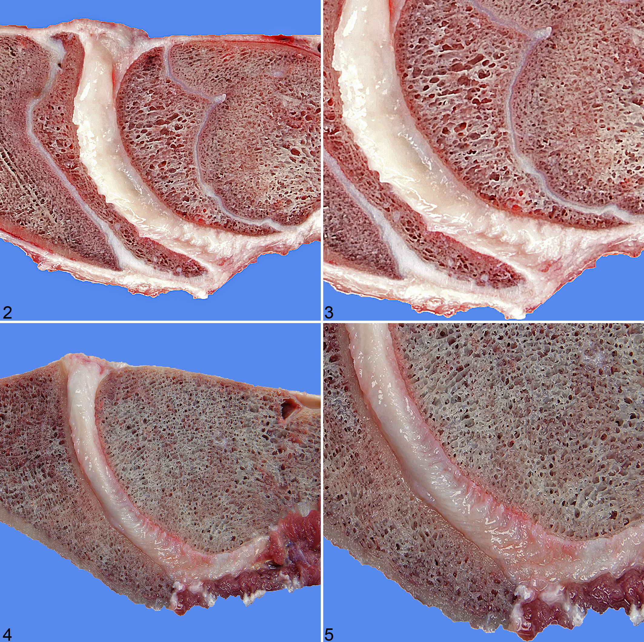
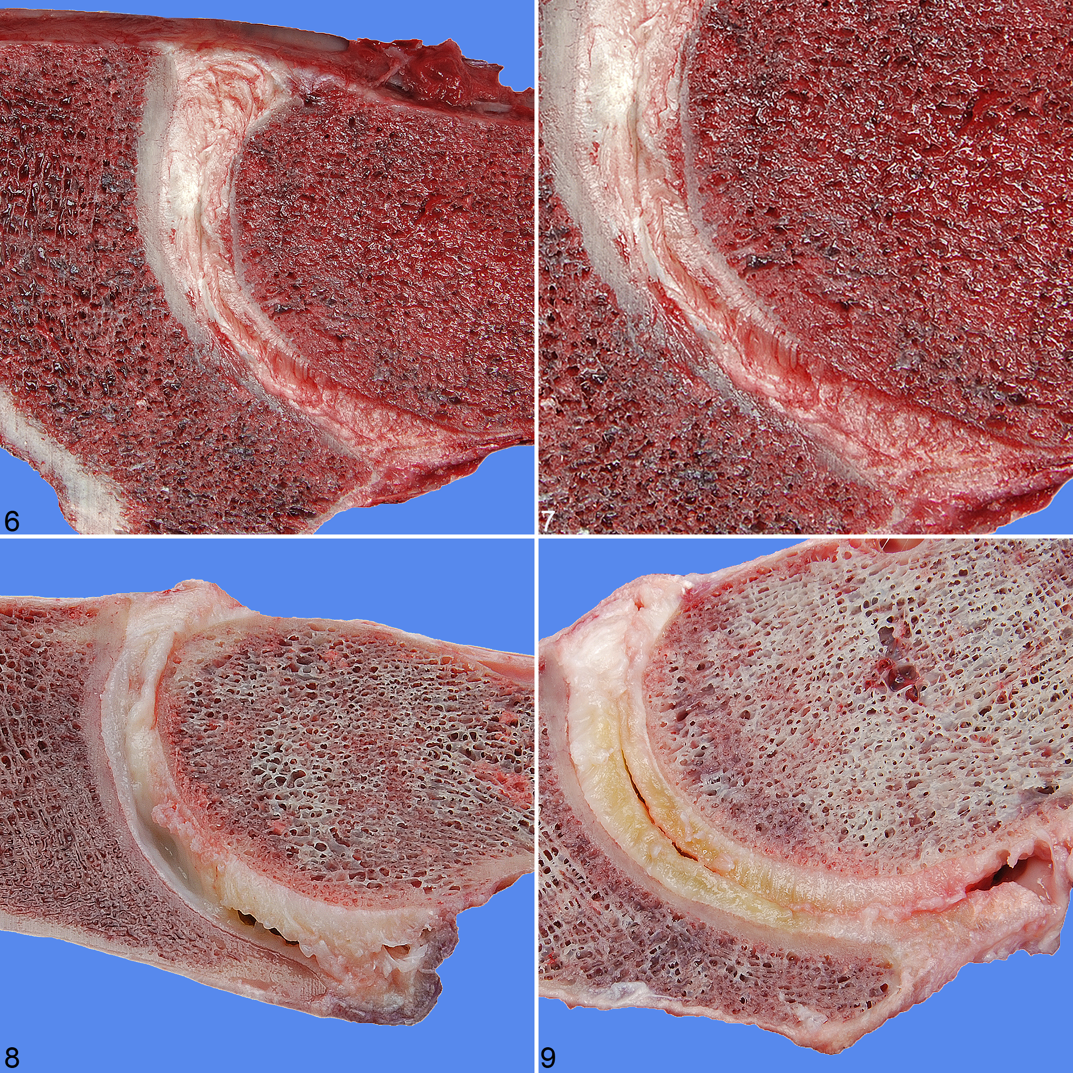
The photographs of the IVDs were analyzed for morphologic changes of the NP, AF, and vertebral bone using a human grading scheme, 51 which is also validated for the dog 3 and was subsequently adjusted for the horse as deemed necessary. In short, for grading of the IVD, the annulus fibrosus and the nucleus pulposus were evaluated for its texture, color, and the presence of clefts. IVD considered normal showed a gelatinous, semi-translucent nucleus and white shiny lamellae. Degenerated discs were characterized by a fibrillary nucleus pulposus, with yellow (-whitish) discoloration of both the nucleus and the annulus with or without clefts (i.e. tears) (Table 1, Figs. 1–9). For grading of the vertebral body, both the subchondral bone and the ventral part of the vertebral body were evaluated. With increasing grades, the subchondral bone changed from trabecular to more compact. The ventral part of the vertebral bone was being reviewed for the loss of rounded margins and the development of osteophytes (Table 2, Fig. 1 and Figs. 10–14). For human and canine discs, grades 1 and 2 are considered normal and grades 4 and 5 degenerated, with grade 3 representing a transitional stage. 6,50 In a preceding pilot study (results not shown), the grading of morphological changes of the subchondral bone as well as osteophyte formation and spondylosis of the ventral part of the vertebral body did not correlate with the grades for changes of the AF and NP, and therefore these morphological aspects were graded in 2 separate schemes.
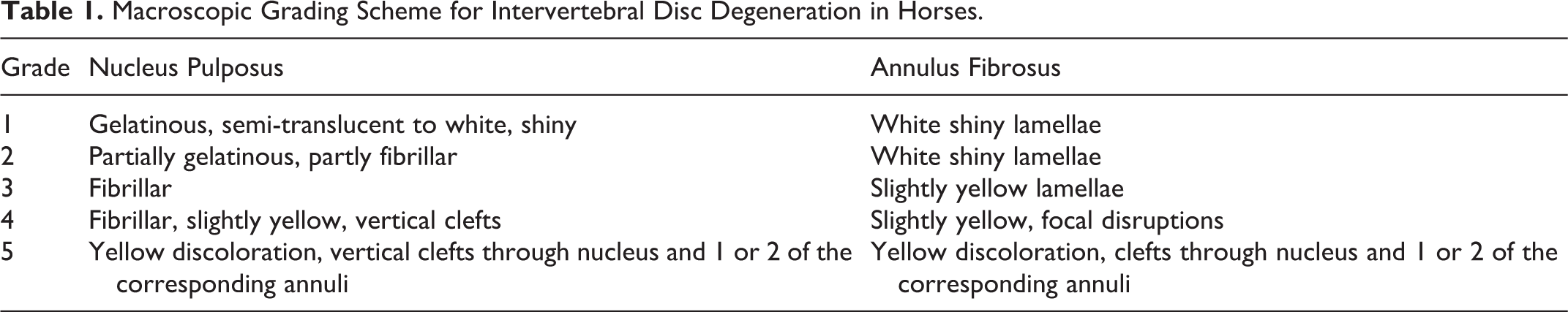
Macroscopic Grading Scheme for Intervertebral Disc Degeneration in Horses.
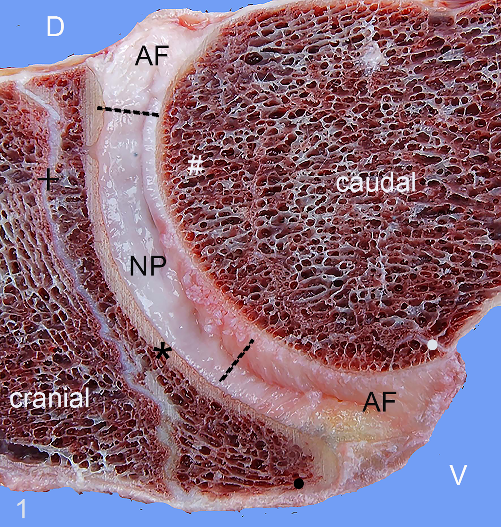
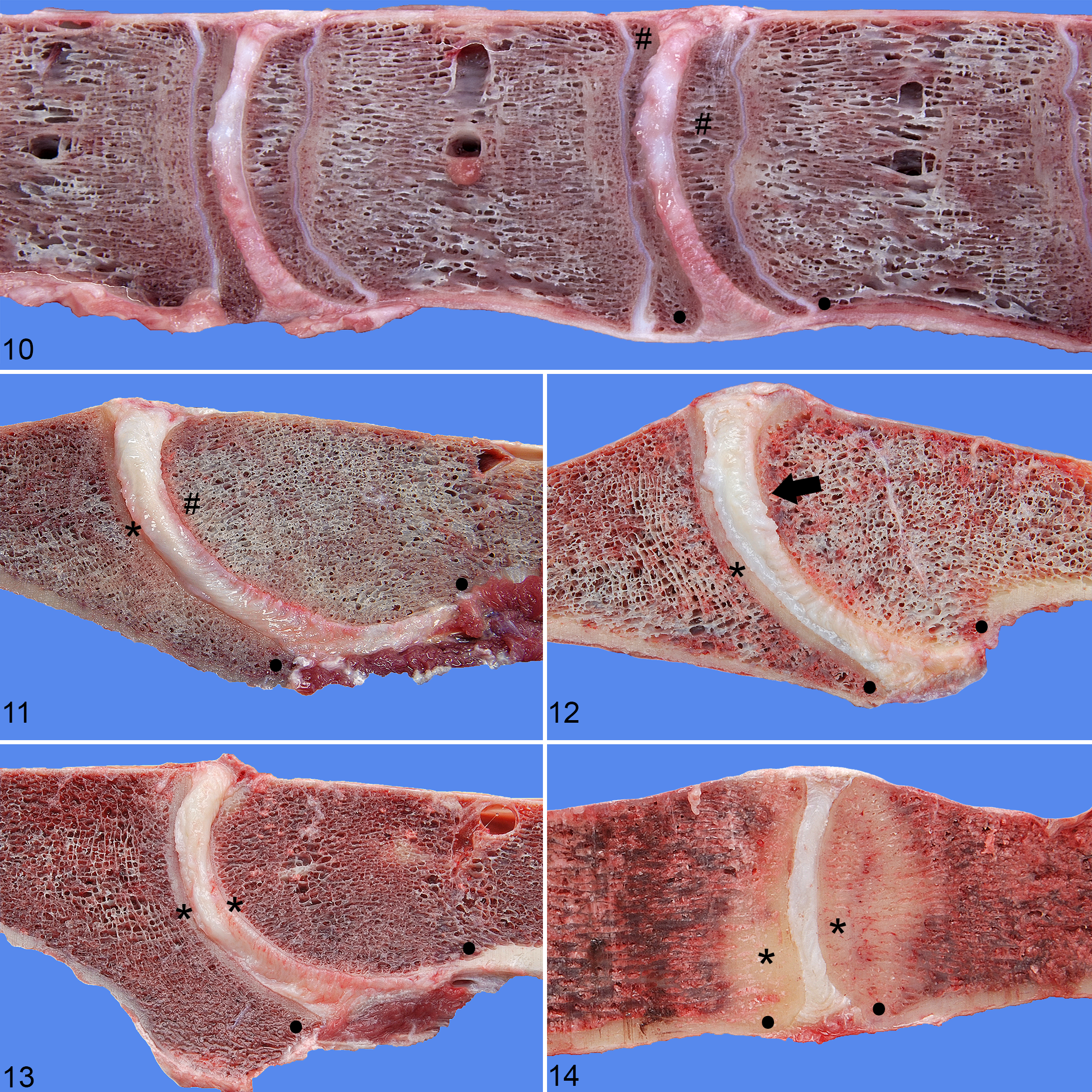
Normal intervertebral disc, C7 to T1 (intervertebral disc between cervical vertebra 7 and thoracic vertebra 1), mid-sagittal spinal section, 2-year-old horse. Grade 1 (normal) for intervertebral disc degeneration and grade 2 for changes of the vertebral bone. The gelatinous, semi-translucent, white, and shiny nucleus pulposus (NP) is visible between the dotted lines. The annulus fibrosus (AF) is white and contains horizontal lamellae. The dorsal AF shows minimal dorsal bulging. Cranially, there is a rim of subchondral compact bone (*), whereas the caudal subchondral bone is trabecular. The ventral part of the vertebral body (•) is rounded. D, dorsal; V, ventral; +, growth plate; *, compact subchondral bone/vertebral endplate; #, subchondral trabecular bone/vertebral endplate; •, ventral part of the vertebral body.
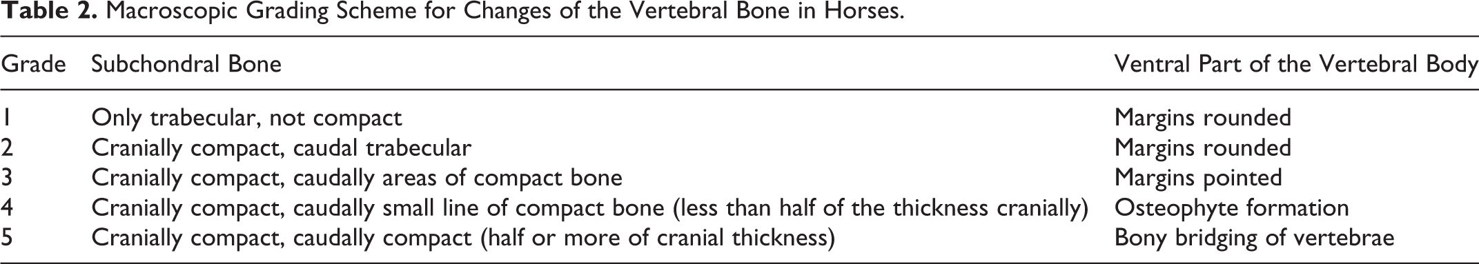
Macroscopic Grading Scheme for Changes of the Vertebral Bone in Horses.
Validation of the Macroscopic Grading Schemes
The photographs were assigned a random number using an online randomization program and graded twice each by 3 independent observers with a time lapse of at least a week between grading sessions of each observer. Inter- and intraobserver reliabilities were calculated by using a Cohen’s weighted κ analysis (quadratic weights of 1, 0.9375, 0.75, 0.4375, and 0). A κ value of less than 0.00 is considered poor, a value between 0.00 and 0.20 as slight, between 0.21 and 0.40 as fair, between 0.41 and 0.60 as moderate, between 0.61 and 0.80 as substantial, and a value between 0.81 and 1.00 as almost perfect. 3,27
The grades assigned during the second round of grading by each observer were added and averaged for a total averaged final grade per spinal unit. If the assigned grade from the second grading differed by more than 1 between the observers, the image was reviewed by the 3 observers and a consensus grade was allotted.
Statistical Analysis
To evaluate the association of the grade for IVDD and the grade for changes of the vertebral bone with age and distribution, the final grades were log-transformed to meet the model assumptions for normality and constant variance. The outcomes were analyzed using a linear mixed model with explanatory continuous variable age and factor variable region, and horse was added to the model as random effect to take the correlated observations within horse into account. The Akaike information criterion was used to select the best model, and Bonferroni correction was applied to correct for multiple comparisons. Residuals were used to investigate the correctness of the model assumptions.
A scatterplot showed a linear association between the grades for IVDD and the grades for the changes of the vertebral bone within the same spinal unit only up to grade 3 (Fig. 15). Therefore, correlation was determined with a Spearman’s rank-order correlation test.
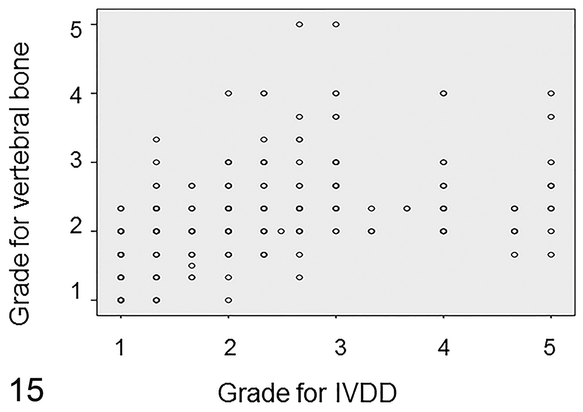
Correlation between the total averaged final grade for intervertebral disc degeneration (IVDD) and for changes of the vertebral bone. Changes in the vertebral bone are linearly associated with grades 1 to 3 for IVDD but are not associated with grades 4 to 5 for IVDD.
All statistical analyses were performed using SPSS version 22 (SPSS, Inc, an IBM Company, Chicago, IL).
Normal Histology
For analysis of the histological characteristics of the normal equine IVD, 107 discs assigned grade 1 and grade 2 (37%) were cut into 0.5-cm-thick slices and fixed in 10% neutral-buffered formalin and then decalcified in 10% EDTA before routine processing into 4-μm sections that were stained with hematoxylin and eosin (HE) and with Alcian blue/picrosirius red (AB/PSR). 4,18,37 Depending on the size of the IVD, 2 to 5 histological sections were necessary to encompass the entire IVD.
The data analyzed in this study will be used for further research and are therefore not available as supplemental materials in the near future.
Results
Macroscopically, the AF and the NP could be identified using mid-sagittal sections in all dissected regions of the vertebral column (Figs. 1–9). The IVD was convex on the cranial aspect and concave at the caudal aspect, and this shape was most prominent in the cervical region. The NP was characterized by a gelatinous, nonlamellar texture compared to the AF and was placed slightly eccentrically toward the dorsal part of the IVD (Fig. 1). The NP of a normal disc was defined as shiny, blue-white, semi-translucent, and pulpy. A firmer, more solid NP with a fibrillar texture and often with yellow-white discoloration was interpreted as mild, early degeneration. Cleft formation of the NP was interpreted as severe degeneration occurring in later stages. The normal AF was defined as a lamellar structure with the ventral AF mildly broader and about twice the height of the dorsal AF. Cleft formation of the lamellae in a dorsoventral direction, nearly always accompanied by a yellow discoloration, was interpreted as degeneration. Bulging of a few millimeters of the dorsal AF into the spinal canal was seen commonly in all grades (Figs. 1, 2, 8, 9). However, in none of the IVDs was this deemed extensive enough to have caused pressure on the spinal cord as visible on mid-sagittal sections of the vertebral column. Herniation of the nucleus pulposus was not seen in any of the IVDs examined.
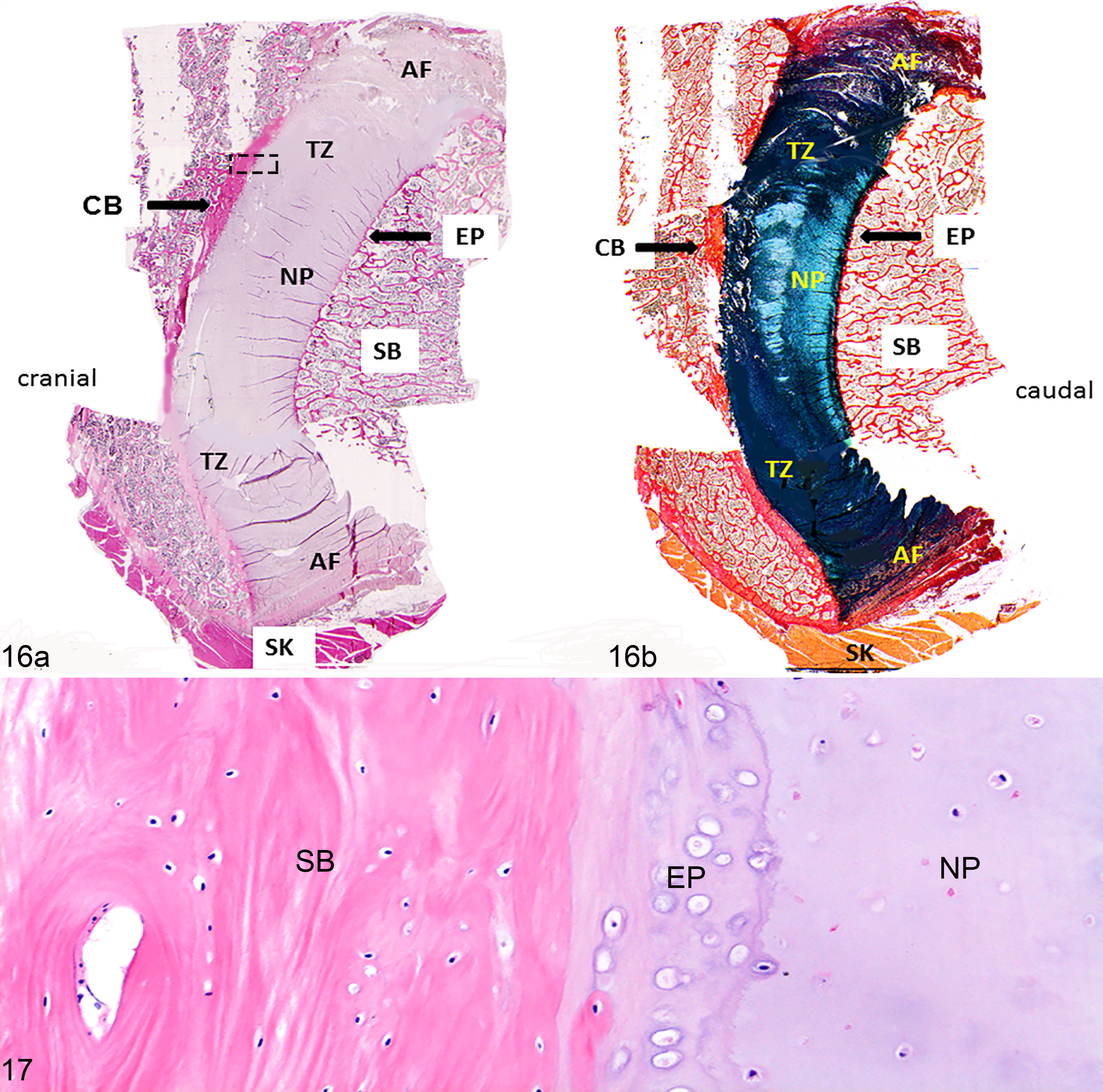
Histologically, the normal IVD was dorsally and ventrally composed of approximately 10 parallel bands that stained brightly red with the AB/PSR stain, consistent with an AF that is rich in collagen type I. The more centrally located part of the IVD consisted of a blue intercellular matrix, compatible with a proteoglycan-rich (mucinous) cartilaginous NP. Between these 2 parts, a rather poorly demarcated transition zone was visible in which there was a gradual transition of the staining characteristics of the intercellular matrix (Fig. 16). This histological difference was colocalized with the grossly visible different morphological characteristics of the peripheral annulus region and the central nucleus region of the equine IVD. At the cranial and caudal margins, the NP and AF were bordered by a rim of hyaline cartilage of approximately 8 chondrocytes thick, compatible with a cartilaginous endplate (Figs. 16, 17).
IVDD was significantly more severe in the caudal cervical region (C5–T1) with a mean ± standard deviation (SD) grade of 2.69 ± 1.64 compared with the cranial cervical region (2.00 ± 0.89), thoracic region (2.02 ± 0.86), and lumbosacral region (2.08 ± 0.87) (Fig. 18, Table 3; P < .05). The grade for IVDD increased significantly with age: grade 1, 4.12 ± 4.15 years of age (mean ± SD); grade 2, 8.10 ± 4.91 years; grade 3, 11.52 ± 5.84 years; grade 4, 11.13 ± 5.35 years; and grade 5, 14.20 ± 3.31 years (P < .05). The grades for changes of the vertebral bone also increased significantly with age: grade 1, 2.05 ± 1.99 years of age (mean ± SD); grade 2, 8.12 ± 5.2 years; grade 3, 11.79 ± 6.08 years; and grade 4, 14.5 ± 3.8 years (P < .05). The formation of subchondral compact bone was significantly more extensive in the thoracic and lumbosacral regions (mean ± SD grade of 2.34 ± 0.75) compared to the cervical segments (2.01 ± 0.67) (Fig. 19, Table 4; P < .05). The cartilaginous endplate was not visible without microscopic magnification in any of the IVDs examined. There was a significant correlation between the grade for IVDD and the grade for changes of the vertebral bone (rs = 0.56, P < .01), which was almost exclusively due to the changes of the subchondral bone. Changes of the ventral part of the vertebral body were only present at the 2 thoracic IVDs of the oldest horse and were characterized by a partially bony, partially fibrous bridge with involvement of the outer layers of the ventral AF between the corresponding vertebrae. These IVDs were both assigned a grade 3.
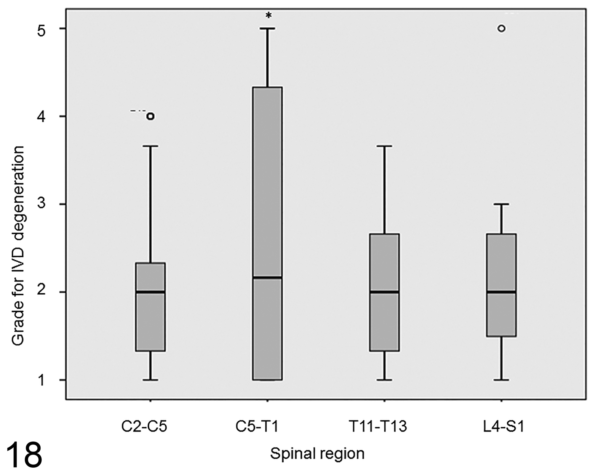
Distribution of grades for intervertebral disc degeneration (IVDD) within the different spinal regions. IVDD was significantly more severe in the caudal cervical region (C5–T1) compared to the cranial cervical, thoracic, and lumbosacral regions. *P < 0.05. The box spans the first to the third quartiles (interquartile range). The line inside the box represents the median. The whiskers show the minimum and maximum. o, outliers.
Grades for Intervertebral Disc Degeneration Distributed Among the Different Spinal Regions.a
aThe data show the number (and percentage) of discs with each grade for each of the anatomic regions.
bIntervertebral disc degeneration was significantly more severe in this region than in the other regions.
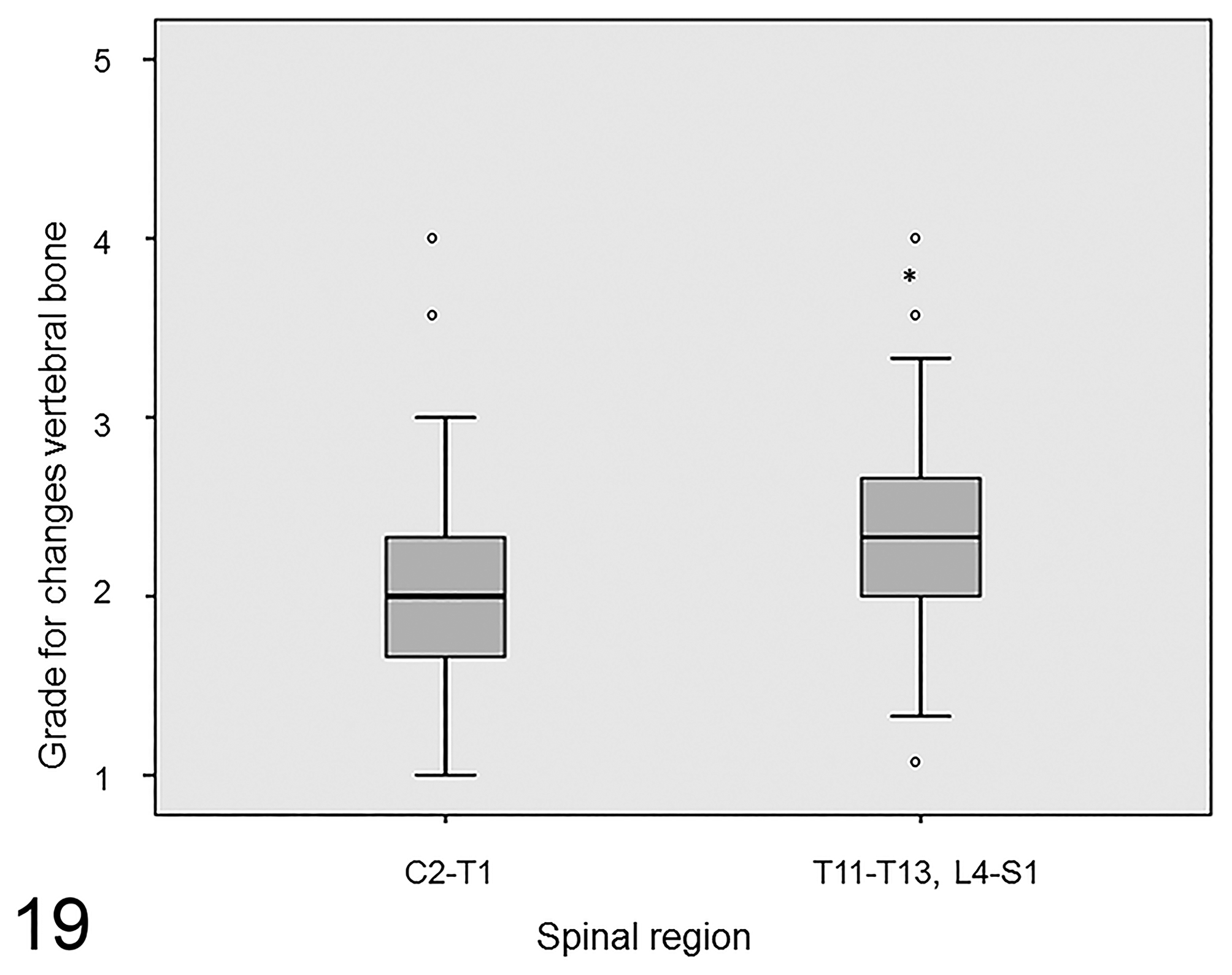
Distribution of grades for changes of the subchondral bone within the different spinal regions. Increased density of subchondral bone was significantly more extensive in the thoracic and lumbosacral regions compared to the cervical segments. *P < 0.05. The box spans the first to the third quartiles (interquartile range). The line inside the box represents the median. The whiskers show the minimum and maximum. o, outliers.
Grades for Changes of the Vertebral Bone Distributed Among the Different Spinal Regions.a
aThe data show the number (and percentage) of discs with each grade for each of the anatomic regions.
bThe changes of the vertebral bone were significantly more severe in these 2 regions combined, compared to the other 2 regions combined.
The intraobserver reliability was substantial to almost perfect, with mean κ scores of 0.78, 0.87, and 0.87 for the 3 observers when using the grading scheme for IVDD. The interobserver reliability for this grading scheme was also substantial to almost perfect, with κ scores of 0.75 and 0.82.
The intraobserver reliability for the scoring scheme for changes of the vertebral bone was moderate to substantial, with mean κ scores of 0.50, 0.70, and 0.77. The interobserver reliability for this grading scheme was fair to moderate, with mean κ scores of 0.39 and 0.56.
Discussion
In contrast to previous publications, 7,53 this study showed that the equine IVD does have a grossly and histologically discernible proteoglycan-rich NP that is distinct from the lamellar collagenous AF. This is similar to the situation in humans and dogs (Fig. 1). 5,6,8,37,51 Furthermore, cranially and caudally located cartilaginous endplates separated the IVD from the bony vertebral endplates, and a transition zone was present between the AF and the NP, which is also comparable to the situation in humans and dogs. 6,37 In literature on the human and canine intervertebral disc, the bony vertebral endplate adjacent to the cartilaginous endplate of the IVD is commonly referred to as subchondral bone. As shown in this study and described previously, 44,58 a rim of cartilage between the IVD and the vertebral bone is also present in the horse. To be consistent with the vast majority of literature, the vertebral endplate is called subchondral bone in this study. The distinction between the AF and the NP was much less clear, both macroscopically and histologically (in the HE stain), in horses compared to human and dogs (Figs. 1, 16). Grossly, in horses of all ages, the NP was never as translucent as in normal human and canine IVDs, making it more difficult to distinguish the NP from the AF. Presumably, this is why in previous publications, 7,53 the equine IVD was said to lack a NP. Nevertheless, the AB/PSR stain clearly showed a morphological and biochemical difference between the AF and NP, and this histological difference was colocalized with the different gross morphological characteristics of the peripheral annulus region and the central nucleus region of the equine IVD. Bulging of a few millimeters of the dorsal AF into the spinal canal was commonly seen in all grades and was considered too small to have caused pressure on the spinal cord, suggesting that minimal bulging of the dorsal AF occurs in normal horses.
Gross changes that were in accordance with degeneration as described for human and canine IVDs could be identified in all parts of the IVD. 1,5,6,51 The presence of yellow discoloration and cleft formation are the only features previously described for equine IVDD, 7,53 whereas the present study identified additional morphological characteristics like a reduced gelatinous and an increased fibrillary appearance of the NP. This interpretation is likely to also apply to horses.
Although the correlation between grades for IVDD and changes of the vertebral bone was statistically significant, this significance seemed to be present for grades 1 to 3 only (Fig. 15). The more severe grades for IVDD (grades 4 and 5) were not associated with a substantial increase in changes of the vertebral bone, suggesting that age is a more dominant factor for these changes than IVDD. Furthermore, grades for IVDD were significantly associated with localization in the caudal cervical spine, whereas grades for changes of the vertebral bone were significantly associated with the thoracolumbar spine. Therefore, macroscopic grading of equine IVDD should only include the morphology of the NP and the AF to prevent under- or overinterpretation of the severity of IVDD.
It has been suggested that osteophyte formation in the horse is not secondary to IVDD. 7 The findings in the present study support this assumption. Of the 39 IVDs assigned a grade 4 or a grade 5, none showed changes of the ventral part of the vertebral body. In addition, ventral bridging was seen only between 3 thoracic vertebrae of 1 horse, and in that case, both associated IVDs were assigned a grade 3 for IVDD.
An age-related increase in the bone density of the equine cervical subchondral bone, directly adjacent to the cartilaginous endplate, was found in this and a previous study. 58 The increased density of the subchondral bone is probably a common feature of aging in horses, as we have found no correlation with pronounced IVDD (grades 4 and 5). The density of the subchondral trabecular bone was significantly less in the cervical than in the thoracic and lumbosacral segments, perhaps because the former trabeculae are oriented less horizontally, possibly leading to less axial compression stress and subsequently lower bone density as dictated by Wolff’s law. 42
The equine grading scheme presented here was based on a commonly used human grading scheme that does not include the parameters’ joint space width (considered indicative of IVDD on radiographs in both humans and dogs 40,46 ) or dorsal bulging and herniation of the IVD. These parameters were not included in the human grading scheme because they are considered consequences of and not part of the degenerative process, 20 and therefore these parameters were also not incorporated in the equine scheme.
When investigating the pathogenesis and the clinical relevance of equine IVDD, a standardized method for evaluation and grading is necessary to assess the gross characteristics and the severity of degeneration. Such a method will allow reliable comparison of results in future research. A good grading scheme is reproducible, which is reflected in high κ scores for intra- and interobserver reliability, 51 as was the case in the adapted grading scheme used in the present publication. A good grading scheme should also be biologically credible. 51 In humans, dogs, and also horses, IVDD is correlated with age. 1,7,36,54 The grades for IVDD assigned in the present study were also significantly associated with age, supporting its biological credibility.
In accordance with the existing literature, 7,36 IVDD was significantly more prevalent in the caudal cervical segment compared to the other segments examined. In addition to the age-related component, IVDD in humans and dogs is thought to be influenced by genetic and mechanical factors, as well as reduced nutrient supply. 2,19,31,39 In humans, IVDs undergo microstructural changes to a similar extent in all regions, and age-related degeneration affects the entire spine. 1,49 A higher prevalence of degeneration in certain regions in both humans and dogs can occur due to locally increased mechanical loading. 1,19,39,43,49 Also in horses, increased mechanical load has been suggested as a cause for the increased prevalence of IVDD in the caudal cervical spine. 7 In addition, dorsoventral mobility may promote IVDD in the horse. 53 According to the literature, the movements in the caudal cervical spinal joints are larger than those in the cranial cervical spine, 41 which might explain the observed difference in prevalence of IVDD between these 2 regions. The lower incidence of IVDD in the thoracic spine of humans and dogs has been attributed to the stabilizing effect of the ribcage with resultant reduced mechanical stress on the IVDs. 29,47 The horse has a comparably rigid thoracolumbar spine that, as in humans and dogs, might at least partially explain the low incidence of IVDD in this region. 26,34,52 An increased prevalence of degeneration in the equine lumbosacral disc has been previously described, 36,53 but this was not confirmed by the present study. In these earlier publications, the breeds of horses, if specified, did not belong to the group of warmblood horses. This suggests that the breed or the work the animal is used for might influence which IVD has the highest mechanical loading and subsequently the highest risk for development of degeneration. It might be that the lumbosacral area is more affected in the racing Thoroughbred than in warmblood horses, as the former breed performs exclusively at the gallop. Ventrodorsal flexion-extension is known to be largest at canter or gallop, and the lumbosacral junction is known to show by far the largest range of motion. 13,14,21 Unfortunately, the type of work the horses were used for was not mentioned in these publications.
In conclusion, the equine IVD had a grossly and histologically discernible proteoglycan-rich NP and a lamellar collagenous AF, which is similar to the situation in humans and dogs. The morphological changes associated with equine IVDD are largely similar to those found in humans and dogs. A grading scheme for equine IVDD, based on a generally accepted human scheme, was developed, which proved useful, as reflected in high
Supplemental Material
Supplemental Material, DS1_VET_10.1177_0300985817747950 - Intervertebral Disc Degeneration in Warmblood Horses: Morphology, Grading, and Distribution of Lesions
Supplemental Material, DS1_VET_10.1177_0300985817747950 for Intervertebral Disc Degeneration in Warmblood Horses: Morphology, Grading, and Distribution of Lesions by Wilhelmina Bergmann, Niklas Bergknut, Stefanie Veraa, Andrea Gröne, Hans Vernooij, Inge D. Wijnberg, Willem Back, and Guy C. M. Grinwis in Veterinary Pathology
Footnotes
Acknowledgement
We thank Rachel E. Thomas for proofreading and René van Weeren for critically reviewing the manuscript.
Declaration of Conflicting Interests
The author(s) declared no potential conflicts of interest with respect to the research, authorship, and/or publication of this article.
Funding
The author(s) received no financial support for the research, authorship, and/or publication of this article.
Supplementary material for this article is available online.
Notes
References
Supplementary Material
Please find the following supplemental material available below.
For Open Access articles published under a Creative Commons License, all supplemental material carries the same license as the article it is associated with.
For non-Open Access articles published, all supplemental material carries a non-exclusive license, and permission requests for re-use of supplemental material or any part of supplemental material shall be sent directly to the copyright owner as specified in the copyright notice associated with the article.