
Abstract
Since the seminal work by Hans-Jörgen Hansen in 1952, it has been assumed that intervertebral disc (IVD) degeneration in chondrodystrophic (CD) dogs involves chondroid metaplasia of the nucleus pulposus, whereas in nonchondrodystrophic (NCD) dogs, fibrous metaplasia occurs. However, more recent studies suggest that IVD degeneration in NCD and CD dogs is more similar than originally thought. Therefore, the aim of this study was to compare the histopathology of IVD degeneration in CD and NCD dogs. IVDs with various grades of degeneration (Thompson grade I–III, n = 7 per grade) from both CD and NCD dogs were used (14 CD and 18 NCD dogs, 42 IVDs in total). Sections were scored according to a histological scoring scheme for canine IVD degeneration, including evaluation of the presence of fibrocyte-like cells in the nucleus pulposus. In CD dogs, the macroscopically non-degenerated nucleus pulposus contained mainly chondrocyte-like cells, whereas the non-degenerated nucleus pulposus of NCD dogs mainly contained notochordal cells. The histopathological changes in degenerated discs were similar in CD and NCD dogs and resembled chondroid metaplasia. Fibrocytes were not seen in the nucleus pulposus, indicating that fibrous degeneration of the IVD was not present in any of the evaluated grades of degeneration. In conclusion, intervertebral disc degeneration was characterized by chondroid metaplasia of the nucleus pulposus in both NCD and CD dogs. These results revoke the generally accepted concept that NCD and CD dogs suffer from a different type of IVD degeneration, in veterinary literature often referred to as chondroid or fibroid degeneration, and we suggest that chondroid metaplasia should be used to describe the tissue changes in the IVD in both breed types.
Keywords
The intervertebral disc (IVD) is an essential component of the canine spine, providing both stability and flexibility during locomotion. 4 Each IVD is composed of 3 distinct compartments: the central nucleus pulposus (NP), the outer annulus fibrosus (AF), and the cartilaginous endplates. 4,12 IVD degeneration can be described as an aberrant, cell-mediated response to progressive structural failure of the IVD, involving changes in all of the aforementioned structures. 4 IVD degeneration can lead to IVD-related disease, such as cervical and thoracolumbar IVD herniation, degenerative lumbosacral stenosis, and cervical spondylomyelopathy. 9,14,22
In the early 1950s, Hans-Jörgen Hansen was the first to provide a highly detailed investigation of IVD degeneration and disease in dogs. In his thesis, Hansen described the classical distinction between chondrodystrophic (CD) and nonchondrodystrophic (NCD) dog breeds. Hansen found that CD and NCD dogs are dissimilar with regard to age of onset, prevalence, and spinal location of IVD degeneration and IVD-related diseases: In CD dogs, IVD degeneration commences early in life and affects all discs. 12 With respect to IVD-degenerative disease, CD dogs are more prone to suffer from IVD extrusion, also referred to as Hansen type I disc herniation, which generally occurs within the cervical and thoracolumbar spine at 3 to 7 years of age. 12,14 In contrast, NCD dogs generally suffer from IVD degeneration at a later age at selected spinal levels. NCDs are commonly more prone to suffer from IVD prolapse, also referred to as Hansen Type II disc herniation, and typical locations for these pathologies are the caudal cervical and lumbosacral spine. 9,14,22
Moreover, Hansen was the first to explore the histopathological characteristics of IVD degeneration in CD and NCD dogs. In his thesis, he concluded that in CD dogs, the primary histopathological event constituted a “chondroid metamorphosis” of the NP, involving the replacement of the resident notochordal cells by chondrocyte-like cells. On the other hand, in NCD dogs, the histopathological events involved in IVD degeneration were described as “fibroid metamorphosis,” which was characterized as gradual replacement of notochordal cells by fibrocyte-like cells. Since then, it has been well accepted in the veterinary community that IVD degeneration in CD dogs involves a chondroid degeneration/metaplasia, whereas in NCD dogs, a different form of degeneration, namely, fibroid degeneration/metaplasia, occurs.
However, the recent literature contrasts to this commonly accepted difference: Studies of the degenerating canine IVD have shown that also in NCD dogs, IVD degeneration involves replacement of notochordal cells with chondrocyte-like cells. 24,25 In addition, degeneration of the NP in various species other than dogs also involves the replacement of notochordal cells by chondrocyte-like cells. 21 Moreover, there are clear indications that the NP contains so-called progenitor cells that give rise to the chondrocyte-like cell population in the process of IVD degeneration. 10,16 These findings suggest that IVD degeneration in NCD and CD dogs involves similar fundamental histopathological processes. However, a systematic investigation comparing the histopathological processes of IVD degeneration between CD and NCD dogs in different grades of degeneration during this phenotypic transition is lacking.
Therefore, the objective of this study was to investigate whether IVD degeneration in CD and NCD dogs is fundamentally different on a histopathological level. It was hypothesized that NCD and CD dogs suffer from the same type of IVD degeneration, namely, chondroid degeneration.
Materials and Methods
Animals and Sample Preparation
IVDs were collected from canine cadaveric spines within 48 hours after euthanasia at the Faculty of Veterinary Medicine, Utrecht University, the Netherlands. Material was collected from veterinary patients and experimental animals. All animals used were free of clinical signs related to IVD degeneration. For the privately owned dogs, permission for postmortem tissue collection was obtained from the owners. Tissues collected from experimental animals were obtained from animals involved in terminal experiments unrelated to the present study and approved by the Ethical Committee on Animal Experiments of the Utrecht University (DEC 2007.II.01.029).
Spinal segments were cut sagittally using a belt saw, and the cut spinal surfaces were photographed for macroscopic grading according to the Thompson grading scheme adapted for dogs. 1,26 Seven IVDs from both CD and NCD dogs for Thompson grades I, II, or III were used (Supplemental Table S1). Discs were graded as Thompson I when the NP was a bulging gel, the AF consisted of discrete fibrous lamellae, the end plates were of uniform thickness, and the vertebral bodies had rounded margins. Discs were graded as Thompson II when the NP consisted of fibrous tissue in its periphery, mucinous material between the individual AF lamellae was present, the end plates displayed an irregular thickness, and the vertebral bodies had pointed margins. Discs were graded as Thompson III when the NP consisted of consolidated fibrous tissue, there was loss of demarcation between the AF and NP, the endplates showed focal defects, and early osteophytosis of the vertebral bodies was present. Breeds were classified as being CD or NCD on the basis of the scientific literature and morphological traits (ie, aberrant growth of the long bones and resulting stunted limbs). 12,24 A total of 42 different IVDs from 32 different dogs (18 NCD and 14 CD dogs) were examined, using multiple IVDs from the same dog in some cases (Table 1).
Signalment, Body Weight, Disc Level, and Thompson Grade for Chondrodystrophic (CD) and Nonchondrodystrophic (NCD) Dogs With Normal and Degenerated Intervertebral Discs.
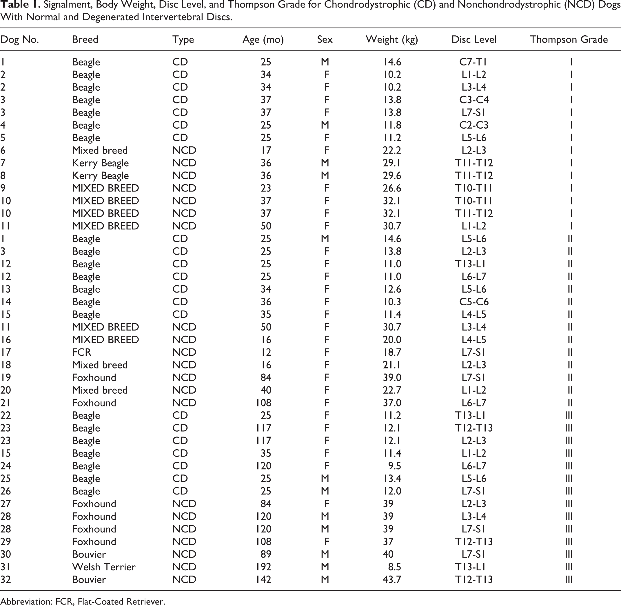
Abbreviation: FCR, Flat-Coated Retriever.
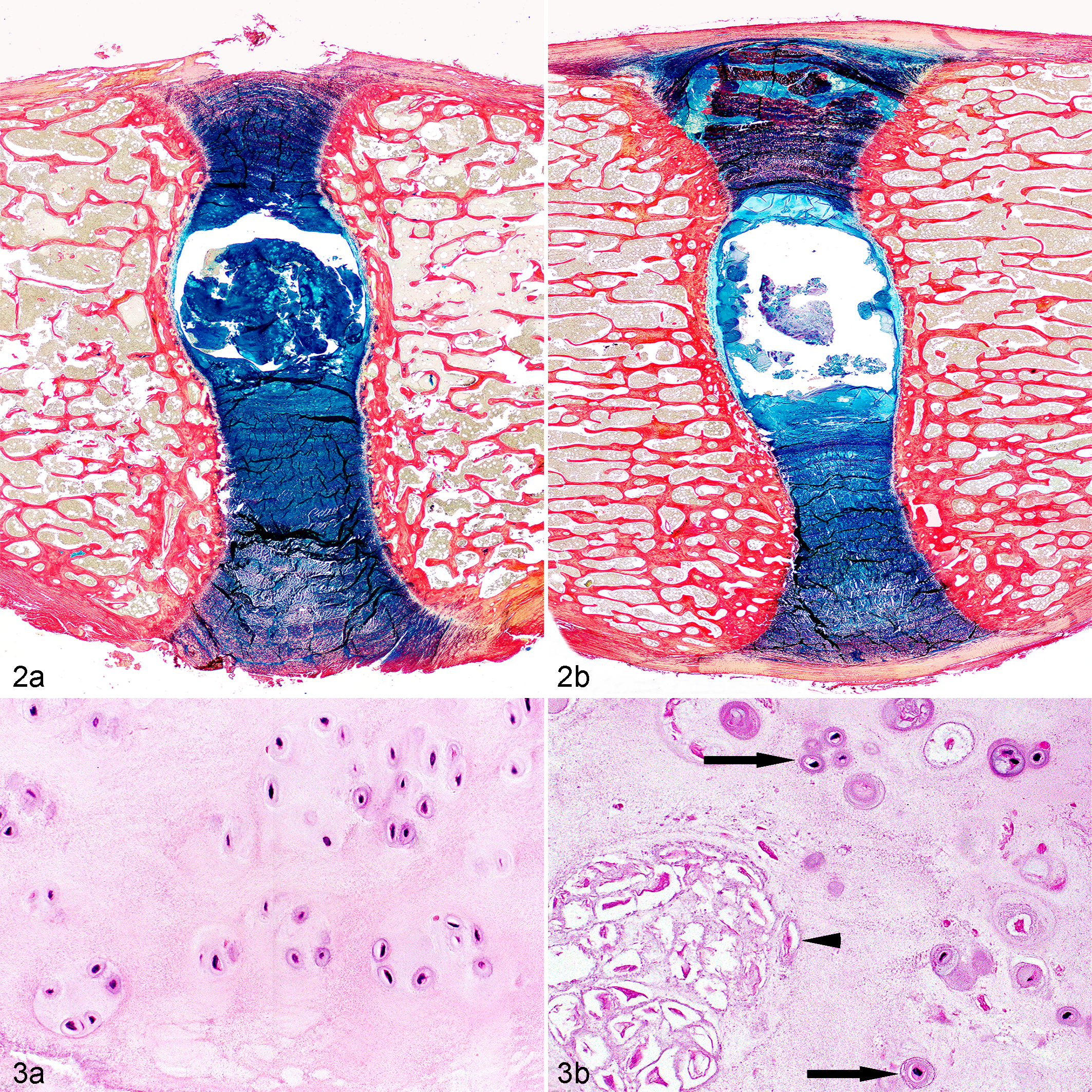
Each IVD was individually fixed in 10% neutral-buffered formalin and decalcified in EDTA (EDTA was used to preserve tissue morphology and quality). After decalcification, IVDs were embedded in paraffin, and tissue sections of 4 to 6 μm were stained with hematoxylin and eosin (HE) and Alcian blue/Picrosirius red. HE stain was used to evaluate the morphological changes in the IVDs and determine the type and estimated quantities of the different cell types. Alcian blue/Picrosirius red stain was used to evaluate the changes in the composition of the extracellular matrix, where Alcian blue stains proteoglycans and Picrosirius red stains collagen, with a higher affinity for collagen type I.
Histological Investigation
Histopathological changes in IVDs of Thompson grade I to grade III from NCD and CD dogs were investigated using a canine-specific histopathological scheme (Table 2). 2 Briefly, this scheme is a modification of a grading scheme for human IVDs 5 that was previously established by recording histopathological changes in 35 canine IVDs, including Thompson grades I to V IVDs of both CD and NCD dogs. Subsequently, the histopathological changes recorded were compared to the changes included in the human grading scheme, resulting in modification of the grading scheme designed by Boos et al. 5 As a result, the canine grading scheme included parameters like morphology of the AF, chondrocyte metaplasia of the AF, tears and cleft formation, chondrocyte proliferation of the NP, presence of notochordal cells in the NP, matrix staining of the NP with Alcian blue/Picrosirius red, end plate morphology, new bone formation, and subchondral bone sclerosis. Because fibroblast or fibrocytes were not noted in the NP of the IVDs that were examined for the modification of the human histopathological grading scheme, the presence of these cells was not included in the published canine scheme. 2 To answer the research question whether fibrous degeneration, histologically characterized by fibrous metaplasia, of the NP occurs in dogs, the additional parameter “presence of fibrocyte-like cells in the NP” was specifically evaluated for each IVD examined. The histological investigation was performed by 3 observers in consensus (T.H., N.B., and G.G.). All histological slides were evaluated in blinded fashion.
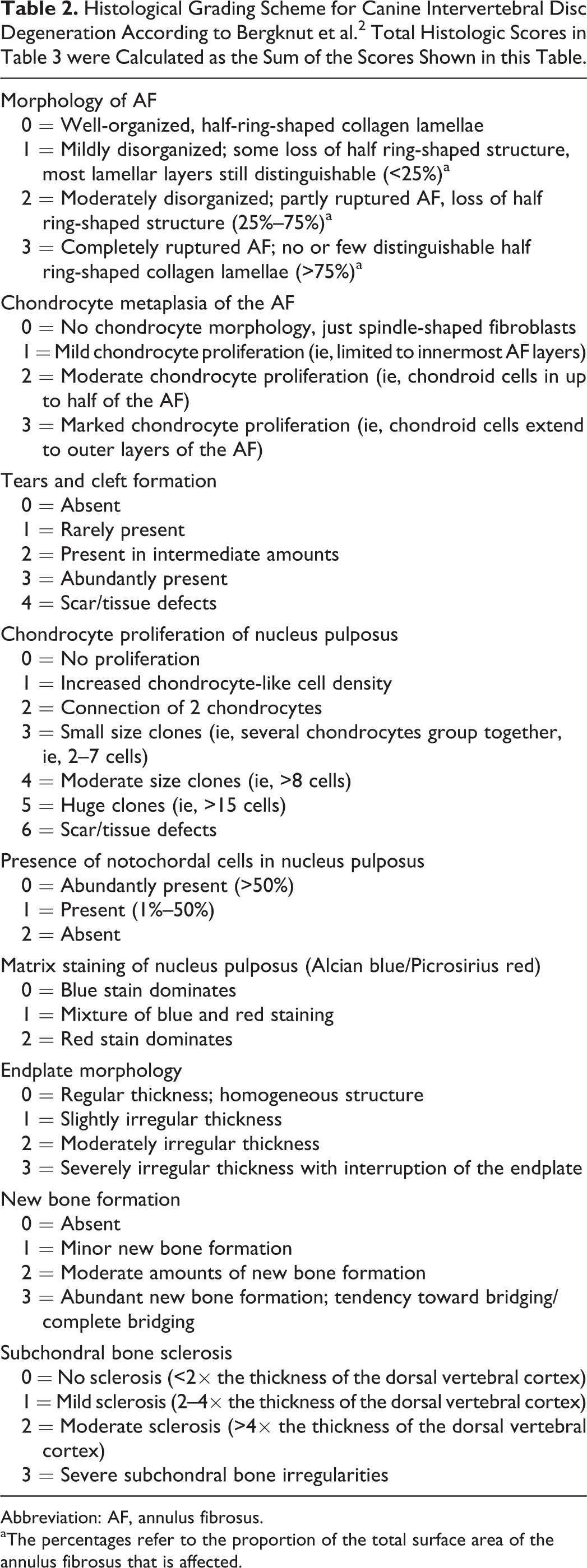
Abbreviation: AF, annulus fibrosus.
aThe percentages refer to the proportion of the total surface area of the annulus fibrosus that is affected.
Statistical Analysis
Total histological scores, based on the grading scheme, were calculated for all evaluated discs. Statistical analyses were performed using R Statistical Software. 27 To evaluate whether significant differences existed for mean total histological scores between CD and NCD dogs separately for Thompson grades I, II, and III, a general linear model was applied. Because some of the IVDs evaluated were collected from the same dogs, data sets for each possible combination of independent observations were created (1 observation per dog, hence 32 observations per data set; 768 data sets in total). Then, the same general linear model was applied with the total histological score as outcome variable. Thompson score (I, II, and III), breed group (CD and NCD), and the interaction between these factors were tested on each constructed data set. Parameter estimates were calculated and stored for each data set. Combining the results of all generated results, the median and the 95% percentile intervals (comparable to confidence intervals [CIs]) for each parameter were calculated to evaluate whether the mean total histological scores were different between CD and NCD dogs, separately for Thompson grades I, II, and III.
A statistical comparison between NCD and CD dogs for the individual histological parameters for the separate Thompson grades was not performed due to the small sample size, and descriptive statistics were used to describe these data. Median scores and ranges were calculated per individual parameter for both NCD and CD dogs for the 3 individual Thompson scores. Differences were highlighted in case a difference in median score larger than 2 points was found.
Results
Animals
Forty-two intervertebral discs from 32 different dogs were included in this study, of which 18 dogs were NCD and 14 were CD (Table 1). The respective ages (median) of the CD and NCD dogs were 34 and 25 months for the Thompson grade I discs, 28 and 73 months for the Thompson grade II discs, and 73 and 120 months for the Thompson grade III discs.
Total Histological Scores
Total histological scores, based on all 9 histological parameters, were significantly higher in CD dogs compared with NCD dogs for Thompson grade I IVDs (ie, a higher degree of degeneration in CD dogs; median difference 4.13; 95% CI, 2.95-5.07) (Fig. 1, Supplemental Table S2). For Thompson grade II IVDs, no significant differences in total histological scores were found between the 2 breed types (median difference = –0.08; 95% CI, –0.67 to 0.89). For Thompson grade III IVDs, total histological scores were significantly higher in NCD dogs compared with CD dogs (median difference = 2.53; 95% CI, 2.03-3.00).
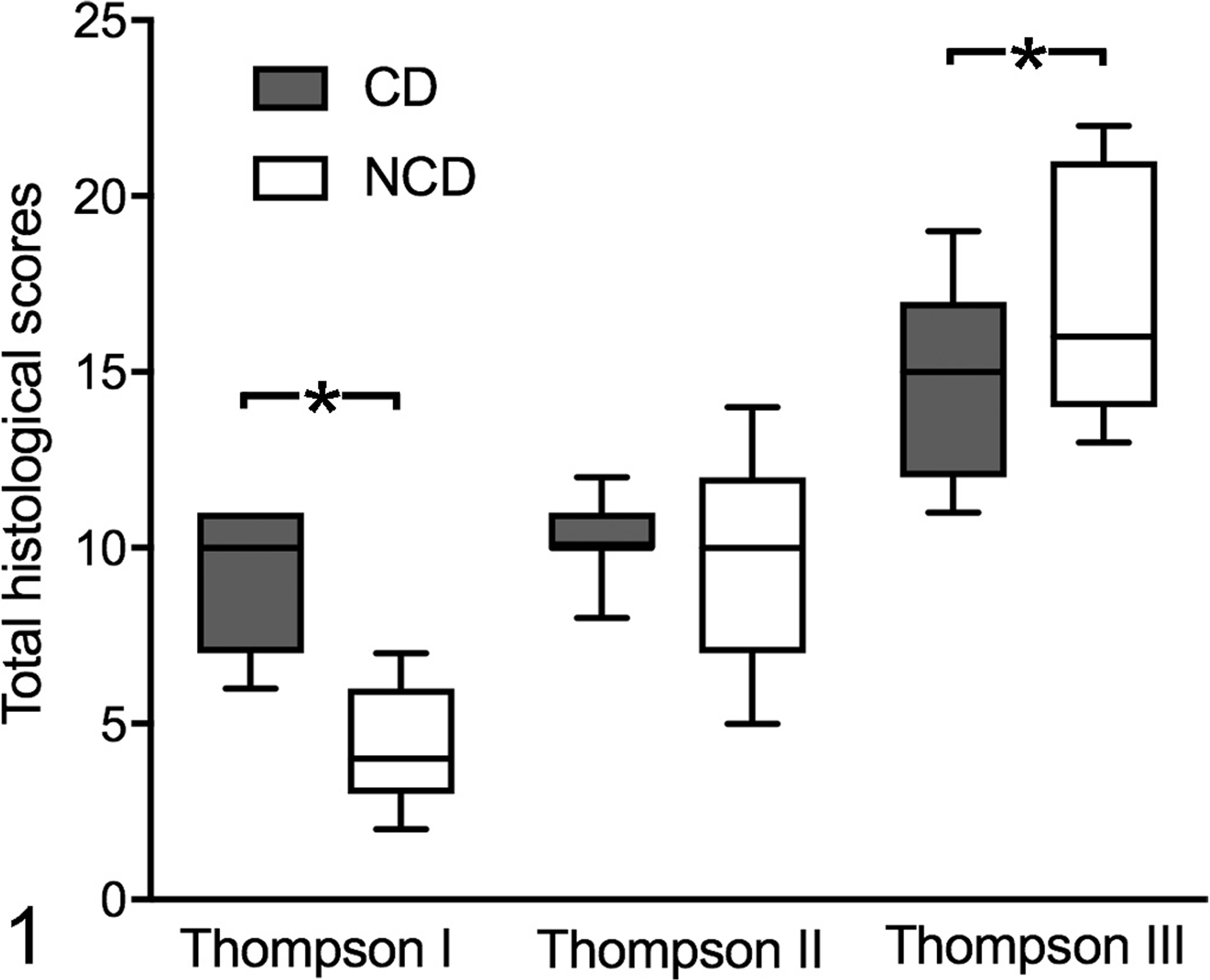
Total histological scores for intervertebral discs from chondrodystrophic (CD) and nonchondrodystrophic (NCD) dogs for Thompson grades I, II, and III. The boxplots display minimum, first quartile, median, third quartile, and maximum scores. Asterisks indicate a significant difference between NCD and CD dogs for a particular Thompson grade.
Individual Histological Parameters
No fibrocyte-like cells were identified in the NP in any of the NCD and CD dog samples (individual animal data: Supplemental Table S2). In all samples, the cell types found in the NP were either (viable or apoptotic) notochordal cells or chondrocyte-like cells (Figs. 2, 3). For the individual histological parameters, the majority of the parameters were similar between the NCD and CD IVDs (Table 3). The main difference between the CD and NCD dogs was found for the parameters chondrocyte proliferation of NP and presence of notochordal cells in NP in Thompson grade I. CD dogs showed a median score that was 2 points higher than NCD dogs for both parameters, indicating that in the macroscopically nondegenerated IVD, the NP of CD dogs contains mainly chondrocyte-like cells and less notochordal cells. The histological scores of the remaining individual histological parameters showed no differences larger than a median score of 1 between CD and NCD dogs for any of the 3 Thompson grades investigated in this study. However, for Thompson grade III, NCD dogs consistently showed a higher grade for the parameter chondrocyte metaplasia of the annulus fibrosus and showed a wider range for the parameter new bone formation. Furthermore, differences with regard to the fundamental histopathological processes involved in IVD degeneration were not identified between the different spinal levels examined.
Histologic Scores (Median and Range) for Intervertebral Discs of Chondrodystrophic and Nonchondrodystrophic Dogs for Thompson Grades I, II, and III.a
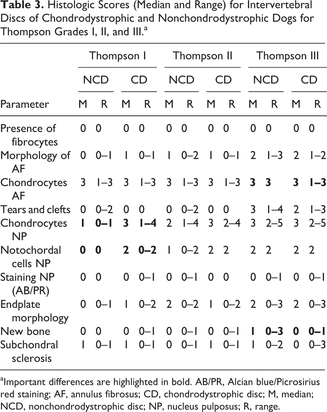
aImportant differences are highlighted in bold. AB/PR, Alcian blue/Picrosirius red staining; AF, annulus fibrosus; CD, chondrodystrophic disc; M, median; NCD, nonchondrodystrophic disc; NP, nucleus pulposus; R, range.
Discussion
The 1952 thesis by Hansen, 12 “A Pathologic-Anatomical Study on Disc Degeneration in Dog,” can be regarded as the histopathologic foundation of IVD degeneration in dogs. Hansen’s description of the distinction between CD and NCD dogs regarding different aspects of IVD degeneration has been accepted in the veterinary community for the past 60 years. Hansen highlighted important differences between NCD and CD dogs with regard to the spinal distribution, age of onset, frequency, and macroscopic and histopathological characteristics of IVD degeneration. Hansen used the famous key terms chondroid metamorphosis and fibroid metamorphosis not only to describe the histopathological events of the degenerating IVD but to summarize the complete degenerative process in CD and NCD dogs, respectively.
In many regards, the concept of this subdivision is correct and emphasizes the different character of IVD degeneration and disease in NCD and CD dogs. However, as a result of this thesis, it has been assumed by various authors that the fibroid metamorphosis occurring in NCDs involves the replacement of notochordal cells by fibrocyte-like cells, namely, a so-called fibrous degeneration, and thus it has been assumed that NCD and CD dogs suffer from fundamentally different types of degeneration. 7,14,20 However, the findings of the present study show that IVD degeneration in both CD and NCD dogs involves replacement of notochordal cells of the NP by chondrocyte-like cells, namely, chondroid metaplasia. No fibrocyte-like cells were found in the NP in any of the investigated IVDs. From a comparative perspective, this observation is in line with the histopathological features of IVD degeneration seen in humans, another species that also suffers from clinical IVD disease. 3 In addition, NCD and CD dogs were very similar regarding the changes observed within the annulus fibrosus, endplates, and adjacent subchondral bone. These results therefore show that NCD and CD dogs both suffer from chondroid metaplasia and are fundamentally similar regarding the histopathological aspects of IVD degeneration. Although the sample population of this study was limited to 18 NCD and 14 CD dogs, prohibiting statistical comparison of the individual histological parameters, the findings correspond with other recent studies on the histopathological appearance of surgically collected IVD tissue, which showed that the histologic differences between the 2 breed types were minimal. 19,23,25
However, the degenerative process in CD dogs occurs at a more accelerated rate than in NCD dogs. IVDs graded as Thompson I, II, and III were studied to identify differences between NCD and CD dog breeds, focusing on the initiating processes involved in IVD degeneration. Interestingly, CD dogs showed a significantly higher total histological score for IVDs that appear macroscopically healthy (Thompson grade I). The histological parameters explaining this difference were chondrocyte proliferation of NP and presence of notochordal cells in NP; specifically, more chondrocyte-like cells and less notochordal cells were seen in IVDs of CD dogs. These results correspond well with Hansen’s work and other previous publications that support the concept that the degeneration process starts in the NP and progresses in a more accelerated fashion in CD dogs compared with NCD dogs. 6,8,11 –13,15,18 In addition, for Thompson grade III IVDs, NCD showed a significantly higher total histological score, which may be explained by more excessive new bone formation and higher grades of chondrocyte proliferation within the AF. End stage degeneration of the NP (Thompson grade VI–V) may involve scarring defects with the formation of granulation tissue containing fibrocytes, 2 but these findings are expected to be generally similar in both CD and NCD breeds. The formation of granulation tissue (and hence the potential introduction of fibrocytes into the NP) needs to be seen as a secondary consequence to the initial degenerative process and does not reflect the initial morphological changes in IVD degeneration.
As mentioned previously, Hansen 12 used the terms fibroid and chondroid metamorphosis to describe a sequence of degenerative events in NCD and CD breeds, respectively. However, this potentially confusing subdivision is likely to have resulted in a significant body of literature citing that 2 fundamentally different forms of degeneration exist. 7,14,20 The statement that 2 kinds of canine IVD degeneration exist seems to be incorrect on several grounds. First, the terms fibrous and chondroid degeneration are never mentioned in Hansen’s thesis; therefore, we feel that the use of this terminology is an incorrect and imprecise reference to Hansen’s work. Second, the actual wording fibrocyte-like cell or cells with a certain similarity of fibrocytes within the NP is only found in 1 sentence of the entire thesis (Fig. 4). When carefully reading the associated passages to this picture, Hansen actually describes these degenerative processes as apoptosis of notochordal cells. Therefore, it is likely that the fibrocyte-like cells are apoptotic notochordal cells. This finding corresponds with other studies describing the cellular dynamics of the NCD NP. 17,25 Third, several passages of Hansen’s work indicate that fibroid metamorphosis in NCD dogs is an actual process of “maturation and collagenization” and not degeneration: “The fibroid metamorphosis of the NP has been conceived in the above as a continuously progressing maturation and not as the result of a degeneration, though it has been pointed out that degenerative processes are a usual component in the picture during this development.” 12 These degenerative changes are later described by Hansen as “cellular changes sometimes with pyknosis, sometimes with karyolysis, and by the changes in the collagenized intercellular substance,” once again referring to apoptotic notochordal cells of the NP (Fig. 4) and a change in composition of the associated intercellular matrix. Since Hansen only clearly described a process of early degeneration in NCD dogs, it is probable that in his studies, Hansen did not study IVDs in advanced stages of degeneration (Thompson III–V), in which chondroid metaplasia is the main cellular change. Lastly, several passages of Hansen’s thesis clearly indicate that he acknowledged a high degree of similarity between the histopathological processes of IVD degeneration in NCD and CD dogs, which is further supported by this study.
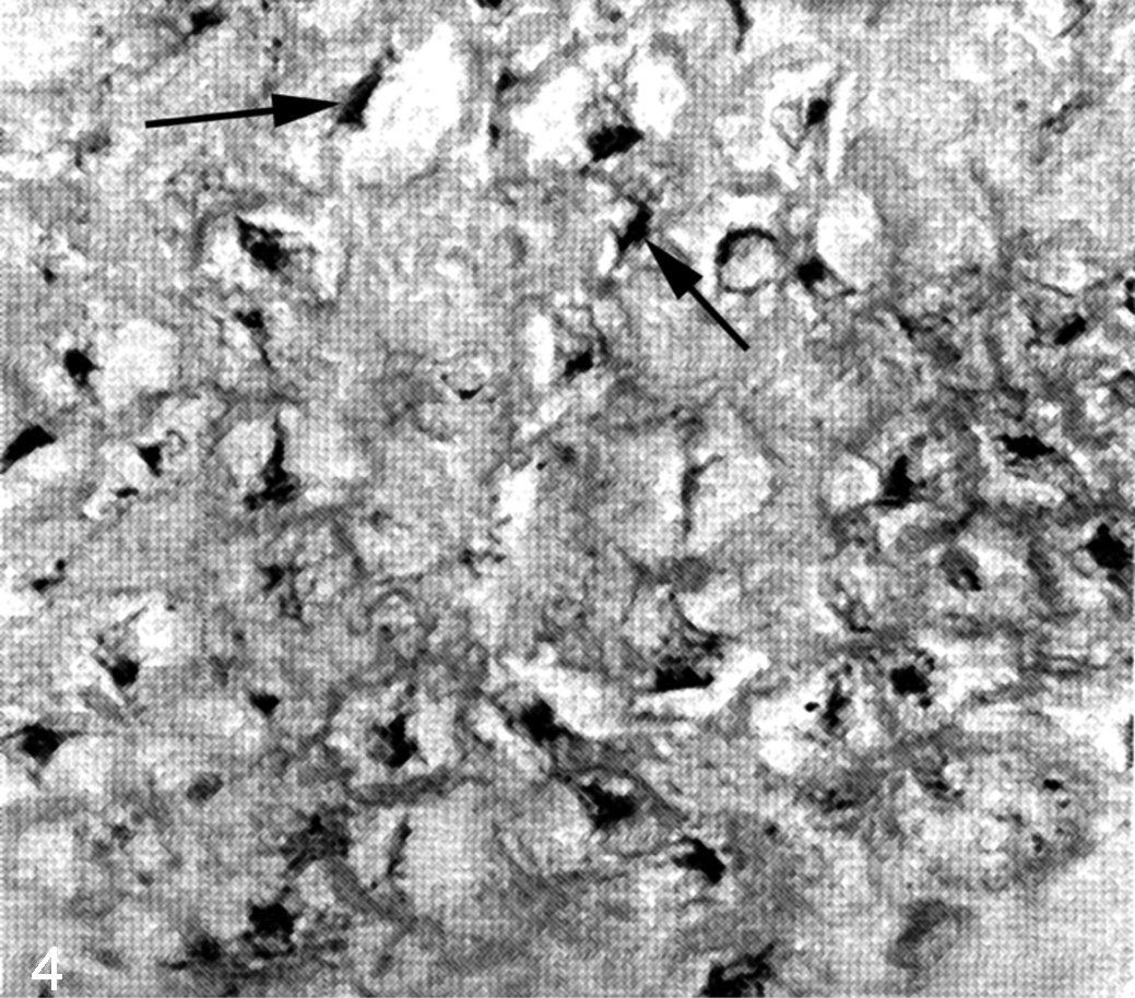
Degeneration of the nucleus pulposus, intervertebral disc, nonchondrodystrophic dog. Original picture from the 1952 thesis by Hansen 12 showing so-called fibrocyte-like cells (arrows) of the nucleus pulposus. Original legend: “Airdale terrier, 10 years old. N.p. (nucleus pulposus) from disc 9, showing still vital cells with a certain similarity of fibrocytes and an intercellular substance, rich in collagen fibers. Van Gieson 400x. 12 Figure used with permission from Acta Orthopaedica and The Veterinary Journal.
In conclusion, both NCD and CD dogs show chondroid metaplasia of the IVD; fibroblasts and fibrocyte-like cells were not seen as fundamental components of NP degeneration in both NCD and CD dogs. The distinction between fibrous degeneration and chondroid degeneration, terms often used in veterinary literature, is therefore a false concept. To avoid further confusion, we propose to describe the principal histological change characterizing the degenerative processes of the IVD in both NCD and CD dogs as chondroid metaplasia and abandon the use of the terminology fibrous and chondroid metaplasia/metamorphosis.
Footnotes
Declaration of Conflicting Interests
The author(s) declared no potential conflicts of interest with respect to the research, authorship, and/or publication of this article.
Funding
The author(s) received no financial support for the research, authorship, and/or publication of this article.
Supplementary material for this article is available online.
References
Supplementary Material
Please find the following supplemental material available below.
For Open Access articles published under a Creative Commons License, all supplemental material carries the same license as the article it is associated with.
For non-Open Access articles published, all supplemental material carries a non-exclusive license, and permission requests for re-use of supplemental material or any part of supplemental material shall be sent directly to the copyright owner as specified in the copyright notice associated with the article.