
Abstract
In humans and dogs, toxic epidermal necrolysis (TEN) is a life-threatening dermatosis characterized by sudden epidermal death resulting in extensive skin detachment. There is little information on the pathogenesis of keratinocyte cell death in canine TEN. We studied the occurrence of apoptosis in skin lesions of dogs with TEN to determine if apoptosis contributes to the pathogenesis of this disease. Immunostaining with antibodies to activated caspase-3 and the terminal deoxynucleotidyl-transferase (TdT)–mediated deoxyuridine triphosphate (dUTP) nick-end labeling technique revealed positive apoptotic keratinocytes in basal and suprabasal epidermal compartments in 17 biopsy specimens collected from 3 dogs with TEN and 16 from 3 dogs with erythema multiforme (EM). There was no significant difference in the number of positively stained epidermal cells between TEN and EM. These results suggest that apoptosis of epidermal keratinocytes and lymphocytic satellitosis represent one of the early steps in the pathogenesis of canine TEN, as in the human disease counterpart.
In humans and dogs, toxic epidermal necrolysis (TEN) is a life-threatening dermatosis characterized by sudden epidermal death resulting in extensive skin detachment, most often due to idiosyncratic drug reactions. 2,3 The keratinocyte cell death in skin lesions of human TEN exhibits features of apoptosis after examination with terminal deoxynucleotidyl-transferase (TdT)–mediated deoxyuridine triphosphate (dUTP) nick-end labeling (TUNEL) staining and electron microscopy. 12 Investigations on the nature of cell death in canine TEN cases are limited to a single study that failed to reveal lymphocytic satellitosis and apoptotic epidermal cells using the TUNEL method in areas of full-thickness necrosis or in the adjacent living epidermis. 10 These results suggested that necrosis, rather than apoptosis, was the mechanism underlying keratinocyte death in canine TEN. 10
In this study, our goal was to evaluate the occurrence of epidermal apoptosis in dogs with TEN to determine if apoptosis could contribute to its pathogenesis. We also wished to compare these findings to those present in dogs with erythema multiforme (EM), a disease associated with prominent lymphocyte-mediated keratinocyte apoptosis. 1 We used formalin-fixed, paraffin-embedded skin tissue collected from 3 dogs with TEN (17 skin samples) and 3 others with minor or major variants of EM (16 skin samples). The case selection criteria for TEN and EM subsets were based on clinical classification and supportive histopathological findings, as reported previously. 2,6 For negative controls, we used six 8-mm punches of skin tissues (abdomen, footpad) collected postmortem from 3 healthy adult dogs euthanized at a local shelter.
Two different methods for apoptosis detection were used on parallel formalin-fixed, paraffin-embedded skin sections: early stages of apoptosis were investigated by immunostaining for activated caspase-3, while end stages of apoptosis were examined using the TUNEL assay. Both techniques were identical to those we reported previously. 11 The detection of satellitosis with T-lymphocytes (T-lymphocytes closely apposed to early apoptotic cells) in TEN and EM sections was revealed using a double immunostaining technique with CD3 specific for T-lymphocytes and active caspase-3, as previously described. 5 Positive and negative immunofluorescence controls for caspase-3, TUNEL, and CD3 were performed using normal canine lymph node or omission of the primary antibodies, respectively. The quantitation of apoptotic keratinocytes stained for activated caspase-3 and TUNEL was conducted by manual count of the number of positively staining cells in the lesional interfollicular epidermis (divided in a basal and a suprabasal compartment) for the entire length of the histological section using adjacent 40× microscopic fields. Only keratinocytes containing a moderately intense and distinct cytoplasmic (activated caspase-3) or nuclear (TUNEL) immunostaining were counted. For the lymphocytic satellitosis evaluation, the presence of CD3-positive lymphocytes immediately adjacent to activated caspase-3 keratinocytes was evaluated, also in the entire interfollicular epidermis of the tissue sections. All results were expressed as number of positively staining cells per 40× high-power field (HPF). Descriptive and comparative statistics were performed using Prism 6 (GraphPad Software, San Diego, CA). The nonparametric Wilcoxon matched-pairs signed rank test was used to test the differences between immunostaining techniques (active caspase-3 vs TUNEL assay). The differences in the frequency of positive staining cells between diseases were assessed using the nonparametric unpaired Mann-Whitney U test. The level of significance was set at P < .05.
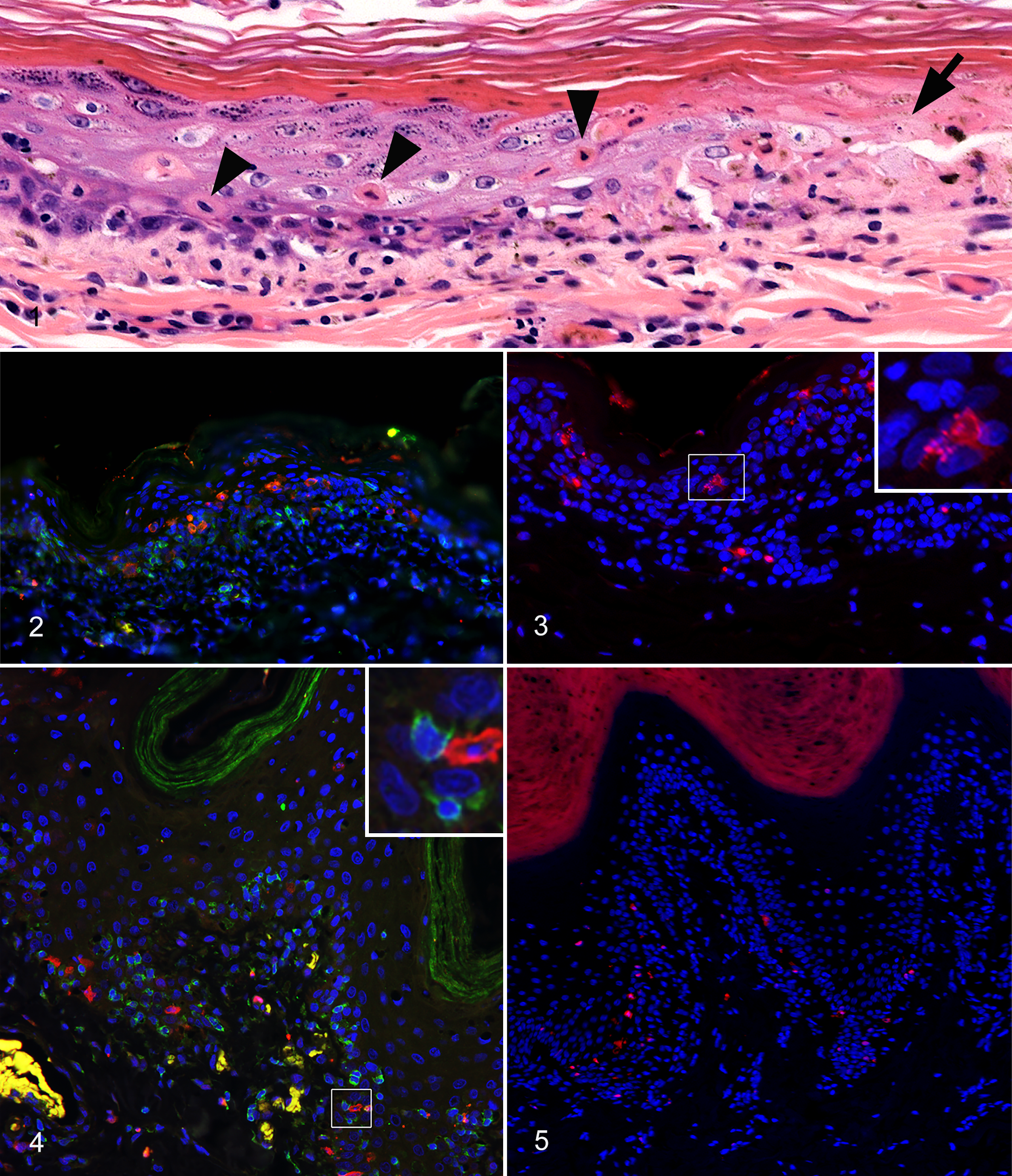
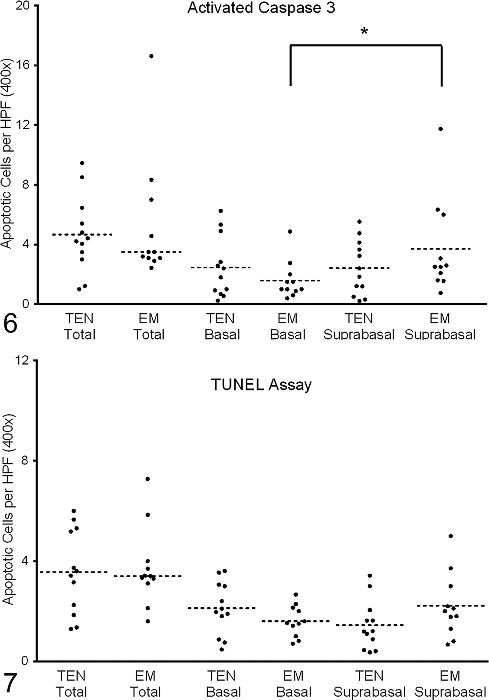
Healthy control abdomen and footpad epidermis did not contain any positive visible cell staining for either activated caspase-3 or TUNEL (Suppl. Figs. S1–S4). The cell death development in sections of skin from dogs with TEN was indicated by the presence of lymphocytic interface dermatitis with predominately deep epidermal apoptosis progressing to apoptosis at multiple epidermal levels at the border of full-thickness epidermal coagulation necrosis (Fig. 1). Activated caspase-3 (cytoplasmic) and TUNEL (intranuclear) positive apoptotic keratinocytes were observed in basal and suprabasal epidermal compartments of all canine TEN and EM skin sections (Figs. 2–5). In several sections of both groups, small TUNEL-positive apoptotic fragments of different sizes were in close proximity to TUNEL-positive apoptotic keratinocytes with pyknotic nuclei (Fig. 3). There was no significant difference in the number of positively stained epidermal cells between the TEN and EM biopsies, either when using the same immunostaining technique (Figs. 6 and 7; activated caspase-3: P = .72; TUNEL: P = .94) or when comparing caspase-3–stained sections to TUNEL assay (TEN group: P = .42; EM group: P = .63). The EM samples contained a higher number of activated caspase-3–positive apoptotic keratinocytes in the suprabasal compared with the basal compartment (P = .01), but this was not the case for TEN sections (P = .70). Disseminated foci of lymphocytic satellitosis, consisting of CD3-positive lymphocytes surrounding an activated caspase-3–positive keratinocyte, were present in basal and suprabasal epidermis of both TEN and EM biopsy samples (Fig. 5). There was no difference in the frequency of lymphocytic satellitosis between TEN and EM sections (P = .30). No positive staining was observed in negative controls, whereas positive staining was present in lymph node control sections.
The author of a recent review article, which compared the etiology, pathogenesis, and diagnostic and therapeutic aspects of EM and TEN, proposed that locally extensive to confluent waves of apoptosis with little lymphocyte-mediated cell attack represent the hallmark of epidermal TEN cell death in both humans and dogs. 17 Unfortunately, existing reports into the pathogenesis of keratinocyte cell death in canine TEN were not addressed, 8 –10 leaving uncertainty about the pathogenesis of disease, the understanding of which is critical for mechanism-based therapy to provide an optimal outcome to canine patients affected with this often fatal disease. The first investigation of cell death in various animal skin diseases, which included 4 dogs with TEN, showed no TUNEL-positive cells in the epidermis; the author concluded that massive necrosis rather than apoptosis was responsible for the epidermal cell death of animal TEN. 10 In contrast, 2 later case reports of 1 dog each affected with drug-induced TEN showed numerous activated caspase-3 and TUNEL-positive apoptotic epidermal keratinocytes, thereby challenging the initial hypothesis of massive necrosis in favor of a possible role of apoptosis in canine TEN development. 8,9 The results of our study extend these recent observations and provide additional support for epidermal keratinocyte apoptosis to represent one of the initial steps in canine TEN pathogenesis, as proposed 20 years ago for human TEN. 12
As recommended by the Nomenclature Committee on Cell Death, 4 2 different methods for detection of apoptotic cell death were used in this study. The detection of activated caspase-3, which leads to apoptosis-associated nuclear changes, is considered an important tool for identifying apoptotic cells in tissue sections before morphological changes typical of apoptosis are even visible. 16 In contrast, the TUNEL method uses the ability of the enzyme TdT to incorporate labeled dUTP onto the free 3′-hydroxyl termini of fragmented genomic DNA. 16 Small dispersed TUNEL-positive fragments associated with apoptotic keratinocytes in TEN and EM sections were identified in this study. These most likely represent apoptotic bodies containing well-preserved condensed nuclear fragments with DNA. In our study, we also detected lymphocytic satellitosis, which suggested lymphocyte-mediated killing of individual keratinocytes by apoptosis. This observation is consistent with the proposed pathogenesis of drug- or drug/peptide-specific T-lymphocyte and/or natural killer–cell initiation of human TEN. 15 Our findings contrast, however, with the previously reported lack of visible satellitosis in dogs with TEN. 10 The results of our study confirm that the histopathological features traditionally considered diagnostic for canine EM 1 —namely, apoptosis at multiple epidermal levels with lymphocytic satellitosis—can also be seen to a variable degree in dogs with TEN. This observation emphasizes the need for clinical information, rather than relying on microscopic findings, to differentiate these conditions in dogs.
A recent histopathological characterization of hematoxylin and eosin–stained sections of canine TEN identified a possible combination of morphological keratinocyte cell death patterns, which were suggestive of both early apoptosis with later rapid widespread coagulative necrosis. 2 As it is difficult to distinguish apoptosis from necrosis using light microscopy, further investigation of apoptotic markers was deemed necessary to better characterize the type of extensive keratinocyte death that occurs during canine TEN. Based on our earlier publication on the histologic appearance of canine TEN 2 and the lack of any significant difference in the total number of apoptotic keratinocytes in dogs affected with either TEN or EM, the rapid progression to full-thickness keratinocyte death and extensive epidermal detachment seen in canine TEN lesions cannot be explained only by confluent apoptosis as proposed recently by Yager. 17 Indeed, recent investigations propose that an alternative death pathway, named “programmed necrosis or necroptosis,” contributes to the pathogenesis of human TEN. 7,13 The programmed necrotic pathway involves the formation of a “necrosome” or a “ripoptosome” complex containing receptor-interacting protein (RIP) kinases 3 (RIPK3) and 1 (RIPK1) with downstream recruitment and phosphorylation of mixed-lineage kinase domain-like protein (MLKL). 7 This complex homo-oligomerizes and forms pores that disrupt membrane integrity, thereby resulting in morphological changes resembling necrosis. 7 Necroptosis is activated in response to death receptor ligands, such as tumor necrosis factor–α (TNF-α) 7 and annexin A1. 13 Importantly, the inhibition of necroptosis completely prevented TEN-like lesions in a mouse model, 13 thereby highlighting that RIPK3 could be a potential therapeutic target for the treatment of TEN, as RIPK3 inhibitors, a novel class of kinase inhibitors, are currently under preclinical development. 7 Finally, to investigate the presence of apoptotic keratinocytes in healthy canine epidermis, an additional control group of healthy dogs was included in this study. In contrast to a classic veterinary dermatology textbook that suggests that 1 or 2 apoptotic epidermal keratinocytes are normally found in 6-mm punch biopsy specimens of healthy canine skin, 14 we did not detect any apoptotic keratinocytes in the epidermis of normal skin sections in our study.
To summarize, the present study identifies apoptosis as an early mechanism of keratinocyte cell death in canine TEN skin lesions. As there was no increase in the total number of apoptotic keratinocytes in TEN compared with EM sections, the mechanism of abrupt and rapid TEN lesion progression to full-thickness keratinocyte death cannot be fully explained by an expansion of the apoptotic cell death pathway. Programmed cell necrosis or necroptosis has emerged as an important pathway in the pathogenesis of human TEN. Detailed studies using transmission electron microscopy and further understanding of canine necroptosis death cascade are necessary to elucidate whether canine TEN is a true mechanistic counterpart of human TEN.
Footnotes
Declaration of Conflicting Interests
The author(s) declared no potential conflicts of interest with respect to the research, authorship, and/or publication of this article.
Funding
The author(s) disclosed receipt of the following financial support for the research, authorship, and/or publication of this article: This study and publication were self-funded.
References
Supplementary Material
Please find the following supplemental material available below.
For Open Access articles published under a Creative Commons License, all supplemental material carries the same license as the article it is associated with.
For non-Open Access articles published, all supplemental material carries a non-exclusive license, and permission requests for re-use of supplemental material or any part of supplemental material shall be sent directly to the copyright owner as specified in the copyright notice associated with the article.