
Abstract
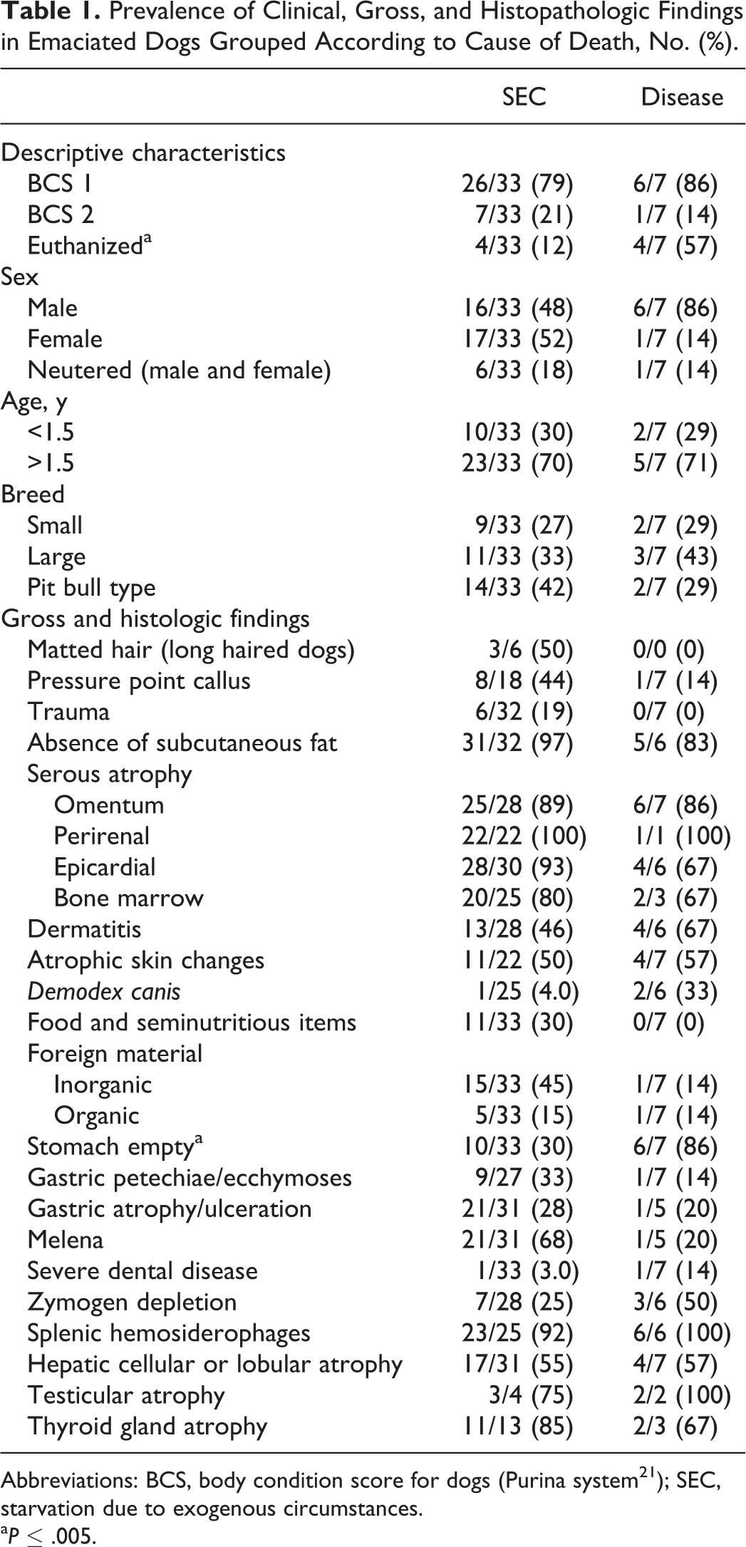
The authors reviewed the case circumstances, population characteristics, gross, and histopathologic findings in 40 cases of emaciated dogs with a suspected diagnosis of starvation. The dogs’ estimated age ranged from 3 months to geriatric. Nineteen breeds were represented, including small-breed (n = 11), large-breed (n = 13), and pit bull–type (n = 16) dogs. The median body condition score was 1 out of 9 (Purina scale). Various diseases were identified as the cause of death in 7 dogs, while the cause of death in the other 33 dogs was starvation due to exogenous causes (SEC). Circumstances associated exclusively with SEC included being found in a vacated residence and death during temperature extremes or severe weather. Dogs with SEC did not differ significantly from diseased dogs in body condition score, sex, neuter status, or breed category (small, large, or pit bull type). Gross findings associated exclusively with SEC included severe hair matting and traumatic injuries. Diseased dogs had an empty stomach significantly more often than SEC dogs, which frequently had food and/or foreign material in the stomach. In 5 of the 7 cases where disease was the cause of death, disease involved the gastrointestinal tract. Gross and histopathologic changes commonly found in SEC and diseased dogs included the following: gross loss of muscle mass and absence of subcuticular fat; serous atrophy of omental, perirenal, epicardial, and bone marrow fat; atrophy of the liver, skin, thyroid gland, and testicle; gastric mucosal petechiae and ecchymoses; melena; and splenic hemosiderophages.
Keywords
Diagnostic laboratories are increasingly presented with cases of suspected neglect, in which an animal may have been deprived of adequate food, water, shelter, or care. 27 In many cases of suspected neglect, the animal is emaciated, meaning that it has a severe and diffuse loss of fat and skeletal muscle. When an emaciated animal is submitted for necropsy, typically the cause of emaciation is sought, and a negligent or willful deprivation of food is suspected.
Emaciation is the corporeal result of 2 different chronic pathophysiologic mechanisms: cachexia and starvation. Cachexia is multifactorial, primarily cytokine-mediated wasting that occurs in conjunction with endogenous disease, such as cancer, organ failure, and some infections. Cachexia is characterized by voluntary reduction in caloric intake (anorexia), increased protein catabolism, and a disproportionate loss of lean body mass compared to adipose, and it is refractory to nutritional support. 11,14,29,31
In contrast, starvation is characterized by an involuntary reduction in caloric intake, decreased basal metabolic rate, and relative sparing of lean body mass until fat stores are depleted, and it is correctable with nutritional support. 12,41 Starvation typically occurs due to exogenous circumstances—adverse conditions that are not discoverable through an examination of the body. Examples of starvation due to exogenous circumstances (SEC) include inability to access food provided (eg, snowdrifts, hostile herd mates), inappropriate diets, and inadequate quality or quantity of food. Some endogenous diseases can also result in starvation culminating in emaciation (eg, exocrine pancreatic insufficiency). Some disease-associated clinical signs (eg, regurgitation, vomiting) and symptoms (eg, nausea, changes in taste and smell) may occur in conjunction with either mechanism—cachexia or starvation. Necropsy should reveal endogenous causes of emaciation, whereas the reasons behind SEC are revealed only through an inquiry into an animal’s environment and diet.
Despite the fact that emaciation is one of the most common reasons for law enforcement officers to seize dogs, 36 reports of gross and histopathologic findings in emaciated dogs are relatively uncommon and have focused on dogs with SEC. 30,39 Our purpose was to document and summarize the gross and histopathologic findings in emaciated dogs suspected of starving and to characterize differences, if any, in the circumstances, descriptive characteristics (age, breed, etc), gross findings, and histopathologic findings between dogs with endogenous disease and those with SEC.
Material and Methods
Case Selection
Between January 1, 2007, and December 31, 2013, a retrospective study of emaciated dogs or tissues from emaciated dogs submitted by or on behalf of a humane agency with the suspicion of starvation was conducted at the New York State Animal Health Diagnostic Center at the Cornell University College of Veterinary Medicine (CU-AHDC). Dogs were excluded if the body was not emaciated (body condition score [BCS] >2) or if the degree of autolysis, decomposition, and/or artifacts was severe enough to preclude meaningful histologic examination. Forty-one cases were identified, of which 40 were included in the study. In 1 case, a deceased dog found in an abandoned residence was eliminated because necropsy revealed thin (Purina score, 3 of 9) but not emaciated body condition; as there was no other evidence of disease, it was concluded that dehydration led to the dog’s death, not emaciation.
Case Review
At the end of the study period, all submission information, necropsy photographs, necropsy reports, and histologic sections were reviewed by 1 author (J.A.G.) or 2 (J.A.G., S.P.M). Review of all tissue sections was conducted prior to review of other case materials such that the reviewing pathologist was blinded to the necropsy findings. The underlying cause of death (COD) was determined by 1 author (J.A.G.) in consultation with the others and based on the sum of law enforcement findings, medical history (if any), necropsy, histologic examination, and ancillary laboratory tests. A COD of SEC was rendered when there was no gross or histopathologic evidence of disease that could account for emaciation. Any discrepancies between the initial gross or histologic interpretations and those at the time review (J.A.G.) were resolved by a third evaluation (S.P.M.) to arrive at a consensus diagnosis.
For each case, the submitter’s geographic location and the dog’s approximate age, breed, sex, neuter status, and BCS were determined. For descriptive purposes, age was classified as follows: young puppies (0–3 months), older puppies (3–6 months), young adult dogs (6 months–1.5 years), adult dogs (1.5–6 years), and geriatric dogs (≥6 years). Age was classified as young adult (<1.5 years) or older adult (>1.5 years) for statistical purposes, as insufficient numbers of dogs were present for comparisons using more narrow age ranges. Dogs were categorized into breeds based on appearance: small breeds, large breeds, and pit bull–type breeds, including the Staffordshire Bull Terrier, American Staffordshire Terrier, and American pit bull terrier.
The BCS was determined using the Purina system 21 and, with 1 exception, was determined by 1 author (J.A.G.) on the basis of either personal inspection (2 cases) or photos of the body immediately prior to necropsy (39 cases). In the case where photos were unavailable, the BCS assigned by the veterinarian who conducted the necropsy was used. A BCS of 1 was differentiated from a BCS of 2 based on the Purina BCS guideline criteria of “minimal loss of muscle” for a BCS of 2 and “obvious loss of muscle mass” for a BCS of 1. 21
Postmortem condition of the bodies varied from euthanasia with a brief postmortem interval resulting in minimal autolysis and artifacts to moderate to severe autolysis and decomposition with an unknown postmortem interval. Freeze artifact was present in many cases, due to either the body being found in subfreezing temperatures or intentional postmortem freezing in semicontrolled environment (freezer). One case had mild scavenging, limited to the extremities. The condition of the body at the scene and postmortem handling of the body was not always disclosed.
Statistical Analysis
All data analyzed were categorical and thus summarized with percentages. Comparisons between diseased and SEC dogs were made through Fisher exact test, since at least 1 cell in all comparisons had an expected number <5. Dogs with missing data for particular variables were omitted from analysis involving those variables. Statistical tests were performed with Statistix 9 (Analytical Software, Tallahassee, Florida). P ≤ .05 was considered significant.
Results
Forty-one cases of emaciated dogs submitted by or on behalf of a humane agency with the suspicion of starvation were identified in the study period. Forty cases were included in the study. In 7 cases, the necropsy was conducted at the CU-AHDC. For 33 cases, tissue samples collected at necropsy, accompanied by photos of the necropsy (not available for 1 case), were submitted to the CU-AHDC. Necropsies were performed by a variety of veterinarians, including referring veterinarians (1 case), veterinarians whose practice is limited to forensic medicine and pathology (23 cases), or a board-certified veterinary pathologist (16 cases). Thirty-three cases were from New York City, 6 cases from central New York State, and 1 from Florida. Of 40 dogs, 33 (82%) died of SEC and 7 (18%) died of disease.
Circumstances
Two circumstances occurred exclusively in association with dogs that died of SEC: death inside a vacated residence (5 cases) and death during extreme weather (8 cases). Extreme weather included below-freezing temperatures (5 cases), hot weather (2 cases), and hurricanes (1 case). In 1 case of hot weather, the body was found in an abandoned apartment, and in 1 case during cold weather, the location of the body was not specified. In the case of the dog found deceased outside shortly after a hurricane, 2 other dogs were found alive in the same yard, and the deceased dog showed no postmortem evidence of drowning.
Descriptive Characteristics
Descriptive characteristics are summarized in Supplemental Table 1. The median BCS was 1. There were 0 puppies <3 months of age, 1 older puppy, 11 young adult, 19 adult, and 10 geriatric dogs. Of the 7 dogs with disease, 2 had endogenous causes of starvation (megaesophagus and masticatory muscle fibrosis).
Eight dogs were euthanized, 30 were found dead, and 2 died within 3 days of being surrendered or confiscated. These 2 cases of spontaneous death in custody were diseased dogs. Euthanasia in custody occurred either within hours of assessment or after weeks of care (Supplemental Table 1). Of the 8 dogs that were euthanized, 5 were promptly euthanized in extremis (eg, recumbent); 2 were euthanized due to lack of response to weeks of intensive care; and 1 was promptly euthanized due to human-directed aggression. In 4 of the 8 euthanized dogs, the underlying COD was SEC.
SEC and diseased dogs did not differ significantly in BCS, sex, neuter status, age category, or breed category (Table 1). Dogs with disease were significantly more likely to have been euthanized than those with SEC, which most often were found deceased by law enforcement officers or third parties or were presented deceased to a veterinarian or animal shelter (P = .008). Pit bull–type dogs composed a majority (40%) of the population, with fewer large-breed (32.5%) and small-breed dogs (27.5%).
Prevalence of Clinical, Gross, and Histopathologic Findings in Emaciated Dogs Grouped According to Cause of Death, No. (%).
Abbreviations: BCS, body condition score for dogs (Purina system 21 ); SEC, starvation due to exogenous circumstances.
a P ≤ .005.
Gross and Histopathologic Findings
In 5 of the 7 cases where disease was the COD, disease involved the gastrointestinal (GI) tract (Supplemental Table 1). Significantly more diseased dogs had an empty stomach compared to SEC dogs (Table 2; P = .02). All dogs that had food in their GI tract were SEC dogs: 11 of 33 SEC dogs had digesta in the GI, and 7 of those 11 had food in the stomach specifically. The presence of food in the GI was not significantly associated with the COD or euthanasia, however. Of the 7 dogs with food in the stomach at necropsy, 4 were alive at the time of surrender or confiscation. Of these 4 dogs, 1 was fed bones and meat by the owner; 1 was fed by the shelter staff; and in the remaining 2 cases, food was obtained from unknown sources.
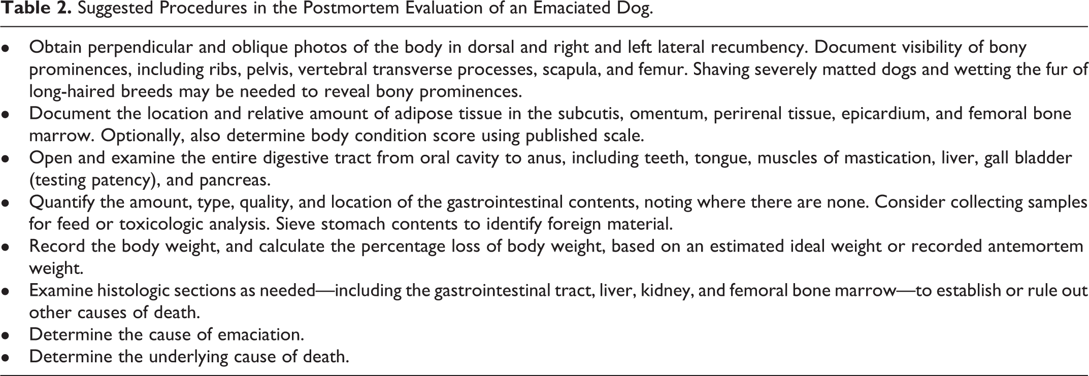
Suggested Procedures in the Postmortem Evaluation of an Emaciated Dog.
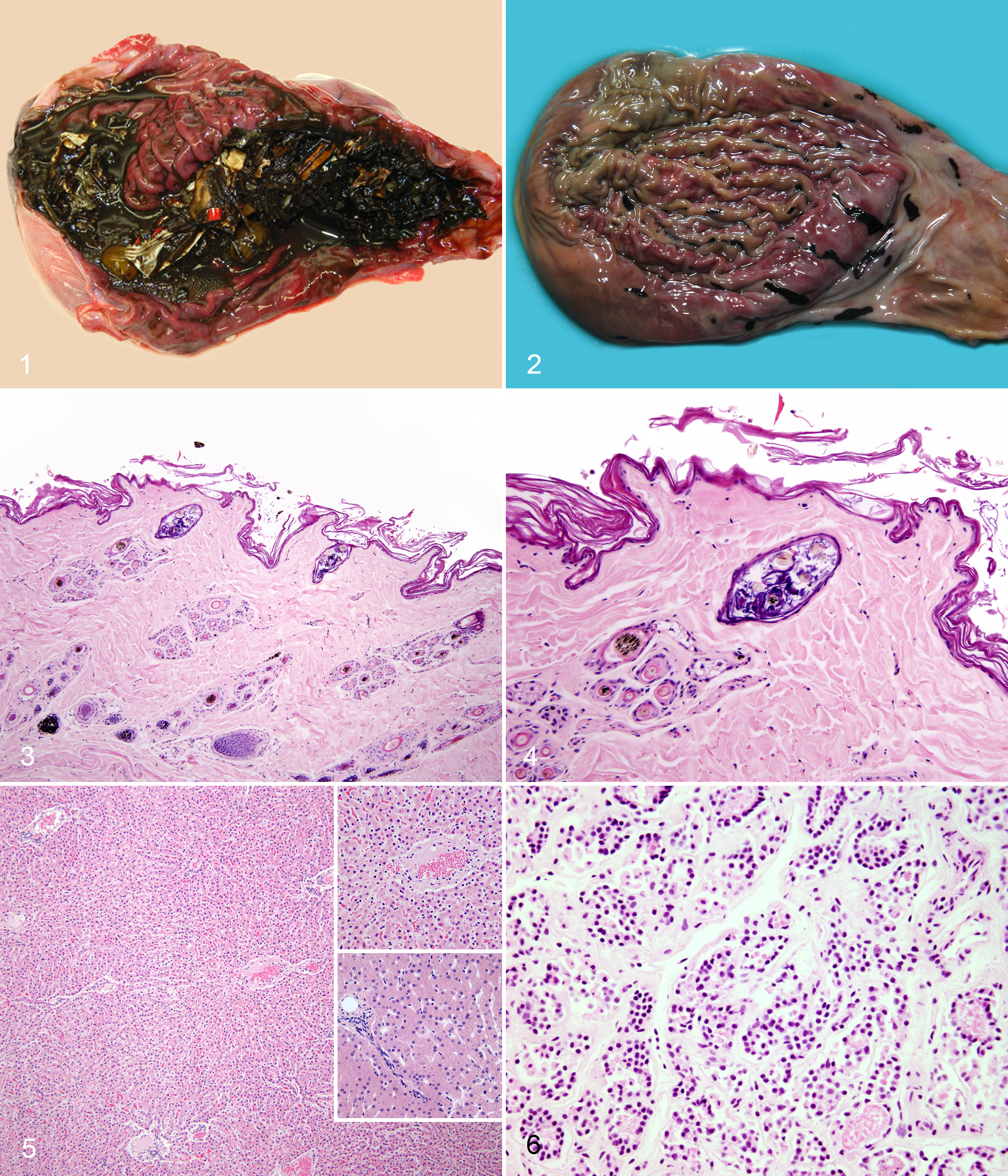
The stomach of SEC dogs frequently contained food (eg, kibble, meat, rice), marginally nutritious items (eg, bones, packets of ketchup), inorganic foreign material (eg, rocks, bits of plastic, razor blades), organic matter (eg, plant matter, hair), and combinations of these materials (Table 1). Melena (Fig. 1), gastric ulcers, and gastric mucosal petechiae and ecchymoses (Fig. 2) were frequently noted alone and in combination in both SEC and diseased dogs. Out of 36 dogs for which data were available, 11 (31%) had 1 or more of these lesions. Although melena was often found in association with ulceration, ulceration was not always present, and melena was not significantly correlated with it. Two of the 5 cases associated with extremely cold temperatures had gastric petechiae/ecchymoses, out of a total of 11 dogs with this lesion. There was no significant correlation between dogs exposed to cold weather and gastric petechiae/ecchymoses.
Starvation of exogenous causes, dogs.
Mild to moderate GI parasitism was seen at necropsy in 1 diseased dog (Ancyclostoma sp) and 1 dog that died of SEC (Dipylidium caninum). Toxocara spp were detected in tissue sections in 1 other dog that died of SEC. Severe dental disease—in the form of abrasion, tooth loss, and fractured teeth—was noted in 2 dogs, one of which was euthanized with a stomach full of food and the other of which died with an empty stomach.
Gross absence of fat and/or histopathologic evidence of serous atrophy of fat of the subcutis, omentum, perirenal area, epicardium, and bone marrow was noted in a majority of cases, with comparable frequency in both SEC and diseased dogs (Table 1). There was no significant difference between SEC and diseased dogs in depletion of fat stores in any location.
Gross and/or histopathologic evidence of dermatitis was noted occasionally in both SEC and diseased dogs. Demodex canis was frequently noted in diseased dogs and rarely in SEC dogs, although the difference was not significant. Atrophic changes in the skin—specifically, follicular miniaturization with telogen dominance, sebaceous gland miniaturization, and epidermal atrophy with mild to moderate orthokeratotic hyperkeratosis—were typically seen concurrently and with comparable frequency in both SEC and diseased dogs (Figs. 3, 4).
Splenic hemosiderophages were noted in the majority for which a histologic section of the spleen was available, and they occurred with nearly equal frequency in SEC and diseased dogs (Table 1). Hepatocyte and/or hepatic lobule atrophy (Fig. 5), thyroid gland atrophy (Fig. 6), and testicular atrophy were noted often in both SEC and diseased dogs. Pancreatic zymogen granule depletion was no more frequent in diseased dogs versus SEC dogs or in dogs with an empty stomach versus dogs with food in the stomach.
Discussion
We investigated the circumstances, descriptive characteristics, and gross and histopathologic findings of 40 dogs that were emaciated and suspected to have been starving. Diseased dogs were significantly more likely than SEC dogs to have an empty stomach. We found no other statistically significant differences in circumstances, descriptive characteristics, or gross or histopathologic findings of dogs that died of SEC versus disease.
Dogs with disease were significantly more likely to have been euthanized compared to dogs that died of SEC. We suspect that this occurred because of case selection bias. When dogs are confiscated alive and emaciated, dogs with clinical signs of disease or dogs that fail to gain weight are more likely to be euthanized. In contrast, dogs that gain weight with refeeding alone and have no clinical signs of disease are not usually submitted for necropsy, even if they are ultimately euthanized for nonmedical reasons.
Among dogs in which the weather was suspected by the submitters to have played a role in death, most dogs died outside during very cold weather. This may be a reflection of the comparatively mild summers in the area from which our study dogs originated, or it may stem from physiology—it is likely easier for emaciated dogs to keep cool in the heat than to keep warm in the cold. Further studies from different geographic regions might further elucidate the relationship among emaciation, death, and exposure.
Diseased dogs were significantly more likely to have an empty stomach than dogs that died of SEC. Most diseased dogs were probably unwilling to ingest food, as loss of appetite is a hallmark of cachexia. Two diseased dogs were unable to ingest (masticatory muscle fibrosis) or digest (megaesophagus) food. In contrast, the presence of material in the stomach of SEC dogs demonstrates both a willingness and an ability to ingest material—any material—including food, marginally nutritious items (eg, garbage), and various nonnutritious organic and inorganic materials. Thus, the presence of material in the stomach is an important and informative necropsy finding. Gastric inorganic foreign material has been documented in dogs that died of SEC, 30,39 and our findings further support these observations.
Food was found in the GI tract of 11 of the 33 SEC dogs. The fact that digesta was found in the stomach in 7 of these 11 cases suggests that the dogs were fed within a few hours of death, given the average gastric emptying time in dogs. 2,37 At first glance, it might seem counterintuitive that a starving dog would have food in its stomach; however, dogs may be given food by 1 or more people around the time of confiscation (J.A.G., R.R., personal experience). First, owners often feed their animals when law enforcement officers arrive on the scene, in an effort to demonstrate compliance with the law. Second, law enforcement officers and shelter staff will often feed emaciated dogs treats or meals at the scene, upon transport or intake at a shelter, or just prior to euthanasia, for various reasons (eg, enticement into or out of kennels). It may be warranted to state plainly in the necropsy report that the presence of food in the stomach in an emaciated animal is not proof that a diet of sufficient quality or quantity was being provided on a regular basis.
An emaciated animal that dies or is found deceased with a GI tract containing food, especially in normal to large amounts, raises the suspicion of refeeding syndrome. Refeeding syndrome is an assortment of subclinical to fatal metabolic disturbances that can occur in the first few days to weeks after implementation of nutrition in the chronically undernourished. 33 The phenomenon is well characterized in people and typically involves abnormalities in phosphorus, magnesium, other electrolytes, or thiamine, resulting in fatal cardiovascular, neuromuscular, or respiratory compromise. 38 Refeeding syndrome occurs in undernourished horses 42 and has been documented in 1 cat 3 and in dogs under artifactual experimental conditions, 20 but no clinical cases of refeeding syndrome in dogs have been documented. Ascribing COD to refeeding syndrome in a dog should thus be done with extreme caution, especially in postmortem cases with a lack of clinical data.
Although gastric ulcerations, petechiae/ecchymoses, and melena have been noted in SEC dogs 28,30 and although mice, rats, and pigs are known to develop gastric ulcers during starvation, 32,19,35 the pathogenesis and significance of these lesions are unclear. Decreased cell turnover, decreased trophic factors, stress, and ingestion of foreign material may compromise GI epithelial barrier function, leading to ulcerations and melena. In addition, gastric petechiae/ecchymoses may be seen in people who have died of hypothermia, but we found no significant correlation between dogs exposed to cold weather and gastric petechiae/ecchymoses.
The combination of histopathologic atrophic changes in the skin common to both SEC and diseased dogs is unique and distinct from those seen in association with endocrinopathies. Although epidermal and follicular atrophy, telogenized hairs, and hyperkeratosis are features seen in hyperglucocorticoidism, hypothyroidism, and other endocrinopathies, other diagnostic features of these endocrinopathies were absent—such as follicular hyperkeratosis and mineralization of the external root sheath (hyperglucocorticoidism) or epidermal and follicular infundibular hyperplasia (hypothyroidism).
We presume that most of the observed skin changes in these cases ultimately result from chronic, severe caloric and protein deficits, mediated in part by concomitant endocrine changes. Hair is 95% protein, and hair growth and keratinization can account for up to 30% of an animal’s daily protein requirement. 15 Sebum is lipid rich and thus also profoundly affected by diet. In fasting people, sebum composition changes in a few days and explains the dull hair and flaky skin noted grossly in starved individuals. 5,8,34 Starvation results in endocrine changes, 7,12 and while the endocrine changes that occur in dogs experiencing prolonged negative energy balance are unknown, we assume that they have some effects on the skin. A confounding effect in this retrospective study was the lack of standardization and frequent lack of information regarding of the anatomic site from which samples of skin were obtained.
We regularly observed atrophic changes in the liver, testicles, skin, and thyroid gland, as well as large numbers of splenic hemosiderophages, in both SEC and diseased dogs. The finding of atrophy in some organs (liver, testicles) was somewhat unsurprising considering the fact that these lesions have also been noted to accompany negative energy balance and emaciation in a variety of species. 26 Splenic hemosiderophages have been noted in starved reindeer, although their pathogenesis and significance are unknown. 18 While pancreatic zymogen granule depletion— a change noted in starved rats 19 — was noted in almost a third of the dogs, the presence or absence of this finding should not be considered diagnostic, as postmortem studies have shown that the pancreas of dogs autolyzes unpredictably and unevenly, even in experimental conditions. 10
Given these and previous findings, we suggest certain procedures for the postmortem evaluation of an emaciated animal (Table 2). The difference between a dog’s ideal body weight (BW) and weight at the time of necropsy is of keen interest to the courts. Consulting with a clinical veterinarian or breeder may help establish an approximate ideal weight. Ideally, individual-specific information, such as a photograph or medical record, is used to establish prior BCS or weight.
The duration and completeness of starvation are also of interest to the courts. These questions are impossible to answer on the basis of postmortem diagnostics alone, in which only BCS and final BW are known. Furthermore, they are—if not impossible—exceedingly difficult to answer with even a modest degree of medical confidence even when clinical data (eg, recorded weight) and husbandry information are available. Answers to these questions depend on starting BW, body fat percentage, caloric intake, available water, exercise, ambient temperature and calories needed to thermoregulate, and many other often unknowable factors, including individual-specific differences in physiology and metabolism.
Unfortunately, no information was available for any of our cases regarding the length of time that the dogs starved or how completely they starved (zero caloric intake versus some caloric intake). However, information gleaned from the literature can aid the pathologist in drafting informative comments for the court. Clearly, survival time for complete starvation is heavily dependent on initial BCS / percentage body fat. In people, death can occur when 35% to 50% of ideal BW is lost, 25 and some published data on emaciated dogs suggests a similar range. 36 Various poorly controlled and often terminal studies from the early 20th century showed that dogs with zero caloric intake and free access to water may be moribund or die in as few as 15 days (47% loss of BW) or survive for 117 days or longer (62% loss of BW). 23 Later better-controlled nonterminal experiments showed that dogs given only water but no food for 2 weeks lost between 11% and 15% of initial BW. 22 A similar study in which dogs were given only water for 3 weeks showed an average 18% loss of initial BW by nonobese dogs and 24% loss by obese dogs. 6
The effects of dehydration are more difficult to assess. Postmortem evaluation of dehydration rests in part on qualitative changes such as dry and tacky mucous membranes, reduced skin turgor, wrinkled skin, and sunken eyes, the last 3 factors of which are strongly influenced by the relative abundance of local fat deposits. Also, decomposition, autolysis, and postmortem dehydration of the body may significantly alter these parameters. There are reports in the literature of 6 dogs deprived of both food and water, with 1 death at 11 days (33% BW loss), 4 dogs that survived 14 to 15 days (3 dogs lost between 22% and 25% BW; 1 was moribund with a 33% BW loss), and 1 dog that was still alive at 20 days (35% BW loss). 9,17 For the 33 cases of SEC in our study, we believe that the primary COD was starvation, not dehydration. Based on basic physiology and some experimental evidence, 1 death due to dehydration occurs before the extreme loss of fat (BCS 1 and 2) and muscle (BCS 1) that defines emaciation. However, we cannot be certain that terminal dehydration played no role in death once an emaciated state was reached.
In people, comparing morphometric measurements (eg, femur length) and organ weights to a reference population are a cornerstone of the diagnosis of starvation, especially in infants and children, whose growth is stunted from chronic undernutrition. 25 With the possible exception of Beagles, 4,24 adoption of these practices for dogs is problematic, given the limited amount of published information available on canine morphometrics, growth, and organ weights 13 and the profound effect that breed, sex, and age have on these parameters. 16,40 In our population, death due to emaciation in dogs <6 months of age was uncommon (1 in 33 dogs).
We maintain that the necropsy of an emaciated animal should document the extent and degree of emaciation, rule in or out disease that could cause emaciation, and determine the COD. It is worth keeping in mind that a necropsy cannot prove neglect—this is a legal determination, not a medical one. Regardless of the cause of emaciation, it is often the failure to seek medical advice that is considered neglectful. Finally, careful use and definition of terms throughout the report are key, especially when laypeople may interpret “starvation” to mean the intentional withholding of food—something only an investigation, not a necropsy, can prove.
This study was limited by its retrospective nature—specifically, the lack of standardization in reporting gross findings and the tissues collected for histopathologic examination. The low number of diseased dogs also limited the power of statistical analyses, and only 5 of the 7 diseased dogs were likely cachectic. It would be interesting to compare the gross and histopathologic findings in cachectic dogs with SEC dogs, especially in light of the fact that gross and histopathologic changes accompanying cachexia have not been described in dogs, despite the fact that cachexia is a clinically significant syndrome responsible for much morbidity and mortality. 14 Prospective studies of blood chemistry and body composition changes in emaciated dogs receiving nutritional support could shed light on the changes, if any, which occur during refeeding. Finally, studies similar to this are needed for other species, as dogs are not the only animals subject to starvation.
In summary, emaciated dogs suspected of being starved have gross fat and muscle wasting and often have serous atrophy of omental, perirenal, epicardial, and bone marrow fat. A variety of usually adult to geriatric small- and large-breed dogs—most frequently, pit bull–type breeds—may present for emaciation. In the majority of cases, the COD is SEC, with a minority due to endogenous disease, usually of the GI tract. Being found in a vacated residence, death during temperature extremes or severe weather, severe hair matting, and traumatic injuries were each associated exclusively with SEC. Diseased dogs had an empty stomach significantly more often than SEC dogs, which frequently had food and/or foreign material in the stomach. Both groups often had gastric petechiae/ecchymoses and melena. Histopathologic lesions commonly found in all emaciated dogs included atrophy of the liver, skin, thyroid gland, and testicle, as well as splenic hemosiderophages.
Footnotes
Acknowledgement
We thank Dr Jeanine Peters, DACVP, DACVD, for her evaluation of the histopathologic changes in the skin.
Declaration of Conflicting Interests
The author(s) declared no potential conflicts of interest with respect to the research, authorship, and/or publication of this article.
Funding
The author(s) received no financial support for the research, authorship, and/or publication of this article.
References
Supplementary Material
Please find the following supplemental material available below.
For Open Access articles published under a Creative Commons License, all supplemental material carries the same license as the article it is associated with.
For non-Open Access articles published, all supplemental material carries a non-exclusive license, and permission requests for re-use of supplemental material or any part of supplemental material shall be sent directly to the copyright owner as specified in the copyright notice associated with the article.