
Abstract
Sodium fluoroacetate is an organofluorine compound toxic to mammals, insects, and birds, currently registered for use only in livestock protection collars as a predacide in some North American states, with restricted use in California. A flock of 445 lambs and ewes in California were moved into a native pasture on a municipal refuse disposal site. Within 24 hours, 14 ewes were found dead, and the remaining sheep were moved off the site. Both ewes and lambs exhibited disoriented running, followed by apparent blindness, weakness, ataxia, coma, and death. Over the next 4 days, 63 ewes and 80 lambs died with a peak at 3 days after grazing the suspect pasture (157/445, 35% mortality). Two dead 4-month-old lambs and 2 ewes were submitted to the California Animal Health and Food Safety laboratory for necropsy. Grossly, there were bilateral diffuse pulmonary congestion and edema, hydrothorax and hydropericardium with fibrin clots, and multifocally extensive areas of epicardial petechiae, ecchymoses, and pallor. In 1 ewe, there was regional caudodorsal pulmonary hemorrhage and intraluminal tracheal clotted blood. Microscopically in all cases, there was multifocal acute myocardial degeneration and necrosis with nonsuppurative pleocellular myocarditis. Sodium fluoroacetate was detected in kidney from a lamb and a ewe at 27.5 and 12.5 parts per billion, respectively. All sheep were selenium deficient, and concurrent copper deficiency was diagnosed in 3. The pathological and toxicological findings were consistent with 1080 poisoning, possibly exacerbated by micronutrient deficiency. This outbreak raised an alert about the use of restricted products with potential lethal effect in animals in California.
Sodium fluoroacetate (compound 1080) is an organofluorine compound toxic to mammals (including humans), insects, and birds introduced in the United States as a rodenticide in 1946. 8 Although the compound is used extensively in some parts of the world, 6 in the United States, it is currently registered for use only in livestock protection collars as a predacide mainly for coyote control in sheep and goat flocks in states that have registrations and US Environmental Protection Agency (EPA)–approved certification and training programs; it is not approved for use in California. 3 The oral route of exposure is most toxic for mammals. 3 The toxicity of sodium fluoroacetate stems from its chemical similarity to acetate. Fluoroacetate combines with coenzyme A (CoA) to form fluoroacetyl-CoA, which substitutes acetyl-CoA in the tricarboxylic acid (Krebs) cycle, halting the cycle and thereby impairing oxidative metabolism and energy (adenosine triphosphate [ATP]) production. 8 In addition, fluoride liberated from fluoroacetate is a calcium chelator, and there are data to support hypocalcemia also as a mechanism of fluoroacetate toxicity. 8 Clinical signs in experimental acute 1080 intoxication in sheep (single oral dose) included excessive salivation, lethargy, tachycardia, tachypnea, dyspnea, tremors and muscle spasms, coma, and terminal tonic convulsions, with death occurring between 22 and 96 hours after exposure. 4 Death is attributed to ventricular fibrillation and/or respiratory failure. The postmortem changes are characterized by myocardial necrosis and inflammation, as well as pulmonary edema.4,6 The present work describes the epidemiological, pathological, and toxicological findings in a field case with unique exposure to 1080 in sheep in California and the laboratory investigations conducted to achieve the diagnosis.
Materials and Methods
History
The outbreak started on March 13, 2011, when a flock of 296 lambs and 149 headlong ewes that were strip grazing a municipal refuse disposal site (landfill) in northeast San Joaquin County, California, were moved into a native pasture containing red clover (Trifolium pratense), Johnson grass (Sorghum halepense), Lupinus spp, and Amsinckia spp. As soon as the outbreak was reported to the referring veterinarian (N. E. E.), 4 dead animals (two 4-month-old lambs and 2 ewes) were submitted to the California Animal Health and Food Safety (CAHFS) laboratory in Davis for necropsy and diagnostic workup on March 14 and 16, respectively, along with plants collected from the pasture.
Pathology
All 4 sheep had thorough postmortem examinations, and samples of heart, liver, spleen, kidney, lung, trachea, adrenal gland, thyroid gland, thymus, lymph node, skeletal muscle (thigh), diaphragm, tongue, esophagus, rumen, abomasum, pancreas, small intestine, cecum, colon, and brain (cerebral cortex, hippocampus, brainstem, and cerebellum) were immersion-fixed in 10% buffered formalin (pH 7.4) for 24 hours, embedded in paraffin, and processed by standard histological methods for the production of 5-μm-thick sections that were stained with hematoxylin and eosin (HE) for light microscopic examination.
Toxicology
Extensive laboratory ancillary testing was conducted. Heavy metal screens (HMS) were performed on fresh liver in all 4 cases. Samples were digested with nitric acid and subsequently analyzed for lead, manganese, cadmium, copper, iron, zinc, molybdenum, arsenic, and mercury by an inductively coupled argon plasma emission spectrometer (FISONS, Accuris Model; Thermo Optek Corp., Franklin, MA). For selenium analysis, liver tissue was digested in an oxidizing mixture of nitric, sulfuric, and perchloric acids, followed by reduction with 5 M hydrochloric acid. Selenium concentration was determined by a hydride vapor generation inductively coupled plasma spectrometer (FISONS, Accuris Model; Thermo Optek Corp.). In both lambs, vitamin E concentrations were analyzed in fresh liver samples by high-performance liquid chromatography. Cyanide and nitrate/nitrite were evaluated in rumen contents and aqueous humor in both lambs using commercially available products (Cyantesmo [CTL Scientific Supply Corp., Deer Park, NY] and EM Quant Test Strips [EMD Millipore, Merck KGaA, Darmstadt, Germany]), respectively. Cardiac glycosides (oleandrin and strophanthidin) were investigated in 2 pooled samples of rumen content (both ewes and both lambs separately) by liquid chromatography/tandem mass spectrometry. Alkaloid screen for the detection of 4-hydroxyspartein-2-one, anabasine, anagyrine, atropine, coniine, deltaline, gramine, lupanine, nicotine, scopolamine, sparteine, and taxus was also conducted in rumen contents of 1 lamb and both ewes using gas chromatography/mass spectrometry (GC/MS). All the aforementioned toxicological testing was conducted according to CAHFS standard operating procedures (SOPs). In addition, frozen kidney from 1 lamb and 1 ewe was submitted to the North Dakota State University Veterinary Diagnostic Laboratory for determination of sodium fluoroacetate. Homogenized kidney samples were extracted with tungstic acid, partitioned into ethyl acetate and evaporated, derivatized with pentafluorobenzylbromide, and analyzed by GS/MS (Agilent 6890 GC/5973 MS; Agilent, Wilmington, DE) using specific ion monitoring. Recovery of fluoroacetate during the procedure was monitored using a 14C-fluoroacetate spike. Last, plants from the pasture were referred to the herbarium (UC Davis Center for Plant Diversity) for botanical identification.
Microbiology and Immunology
Lung and liver swabs from all 4 sheep, pericardial sac swabs from both lambs, and spleen swabs from both ewes were processed routinely for aerobic bacterial culture (sheep blood agar, incubation at 35°C–39°C in 5%–10% CO2 for 48 hours). A sample of feces from each animal was processed for the detection of Salmonella spp by polymerase chain reaction (PCR). Small intestinal contents from both lambs were also processed for bacterial anaerobic culture (pre-reduced anaerobically sterilized Brucella blood agar and egg yolk agar [Anaerobe Systems, Morgan Hill, CA] incubated anaerobically at 37°C for 48 hours). In addition, samples of intestinal contents (small intestine in all 4 animals and cecal contents in both lambs) were tested for Clostridium perfringens alpha toxin (CPA), beta toxin (CPB), epsilon toxin (ETX), and C. perfringens structural antigen using a commercially available capture-qualitative enzyme-linked immunosorbent assay (ELISA) kit (Bio-X Diagnostics SPRL, Jemelle, Belgium) following the instructions of the manufacturer. Spleen and serum samples from both ewes were analyzed for the detection of bluetongue virus (BTV, Orbivirus) genomic RNA and antibodies by quantitative real-time reverse transcriptase PCR and capture ELISA, respectively. All the aforementioned testing was performed according to CAHFS SOPs.
Results
History
Within a few hours of moving the flock into the new pasture, 2 ewes were found dead. The next morning, 12 more ewes were found dead, and the remaining live sheep were moved off the site. Both ewes and lambs exhibited a brief period of disoriented running, breaking through the electric fence followed by apparent blindness, weakness, ataxia, coma, and death. Over the next 4 days, 63 ewes and 80 lambs died with a peak at 3 days after grazing the suspect pasture. The mortality rate was 35% (157/445), and the direct economic loss was approximately $33 000.
Pathology
Grossly, in both lambs, there was diffuse severe bilateral pulmonary congestion and edema with large amounts of white froth within the lumen of the distal trachea, major bronchi, and intrapulmonary airways (Fig. 1). The pericardial sacs contained 50 to 100 ml of serous clear amber fluid that clotted when exposed to air, admixed with fibrin clots, and there were multifocally extensive areas of left ventricular epicardial pallor and moderate petechiation (Fig. 2). One lamb had diffuse thymic petechiation. The ewes had mild hydrothorax and pericardial effusion with fibrin clots, mild epicardial petechiae, and ecchymoses on the right ventricle extending to the junction with the interventricular septum. In 1 ewe, there was also diffuse epicardial hemorrhage in the left auricle, severe regionally extensive right caudodorsal pulmonary hemorrhage with multifocal lobar hemorrhage in the ventral caudal and middle lobes, and abundant intraluminal tracheal clotted blood (Fig. 3).
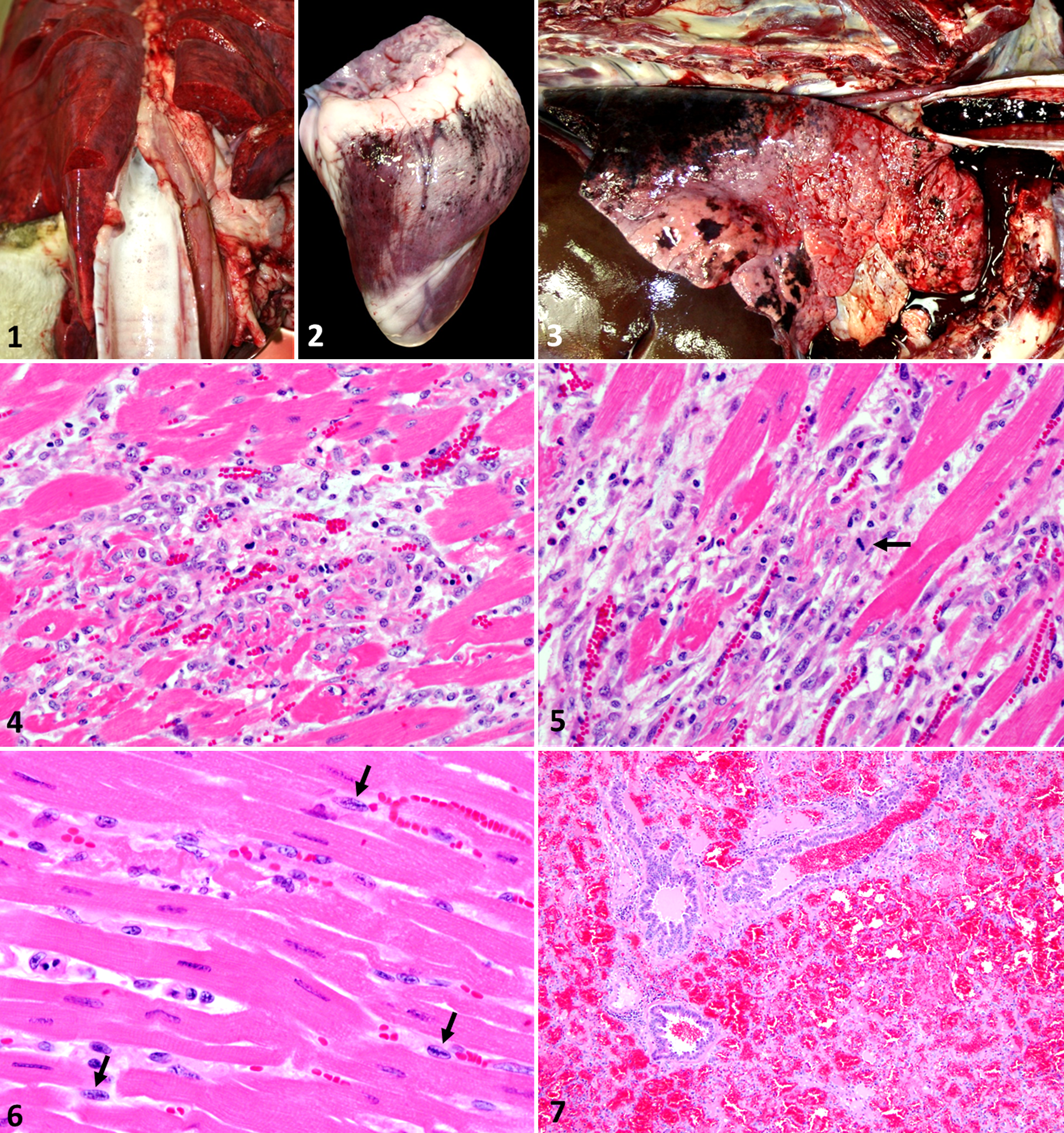
Microscopically, in all cases, the most significant changes were seen in the heart, with multifocal to coalescing areas of acute degeneration, necrosis, and inflammation in which the cardiomyocytes were segmentally hypereosinophilic with loss of transverse striations (degeneration) or had fragmented sarcoplasm and occasionally pyknotic nuclei (necrosis). Frequently, there was collapse of small fascicles with segmental loss of adjacent cardiomyocytes and replacement by hypertrophied and hyperplastic reactive spindled stromal cells that sometimes were undergoing mitosis and occasionally had an oval nucleus with chromatin condensed centrally in a linear undulated wavy pattern (resembling Anichkov cells). These areas were also frequently infiltrated by variable numbers of histiocytes and fewer lymphocytes and viable neutrophils (Figs. 4–6). There was also mild interstitial edema. In addition, in 1 ewe, there was multifocal lymphocytic interstitial myocarditis, although the inflammation was not directly associated with areas of myocardial degeneration or necrosis. The left ventricular myocardium, including the papillary muscles, was most commonly affected, but changes were scattered throughout the heart (auricles, interventricular septum, and right ventricular free wall). Other changes in all animals included diffuse pulmonary alveolar edema with scattered alveolar hemorrhages and multifocal hemorrhage in the thymic cortices in the lambs. In 1 ewe, there was severe diffuse acute alveolar and bronchiolar hemorrhage (Fig. 7) and minimal individual skeletal muscle myocyte degeneration with sarcoplasmic swelling and granularity, as well as formation of hypereosinophilic sarcoplasmic bands (contraction bands), which was not observed in the other 3 animals. No other significant microscopic lesions were observed in the other examined tissues.
Toxicology, Microbiology, and Immunology
The results of the different toxicology, microbiology, and immunology ancillary tests are presented in Table 1. Three of the 4 sheep were found to have copper deficiency, and all 4 were deficient in selenium. The liver level of vitamin E was low in 1 of 2 animals in which this analysis was done. Sodium fluoroacetate was detected in kidney in both animals tested for this compound (1 lamb and 1 ewe). Low concentrations of 1 or more of the following plant alkaloids were detected in the rumen contents: nicotine, lupanine, anagyrine, 4-hydroxyspartein-2-one, gramine, and/or several other unidentified lupine alkaloids. Cyanide, nitrate/nitrite, oleandrin, and strophanthidin testing was negative. Streptococcus bovis was isolated in rare numbers on aerobic culture from the lung of 1 lamb. C. perfringens toxin ELISA, bacterial anaerobic cultures, Salmonella spp, and BTV PCR were all negative. Antibodies against BTV were detected in 1 ewe. The plants from the pasture were identified as Lolium perenne (perennial ryegrass), Lolium multiflorum (annual ryegrass), Hordeum jubatum (foxtail barley), Medicago polymorpha (California burclover), Erodium sp (filaree), Geranium sp (geranium), and Amsinckia sp (fiddleneck).
Laboratory Test Results.
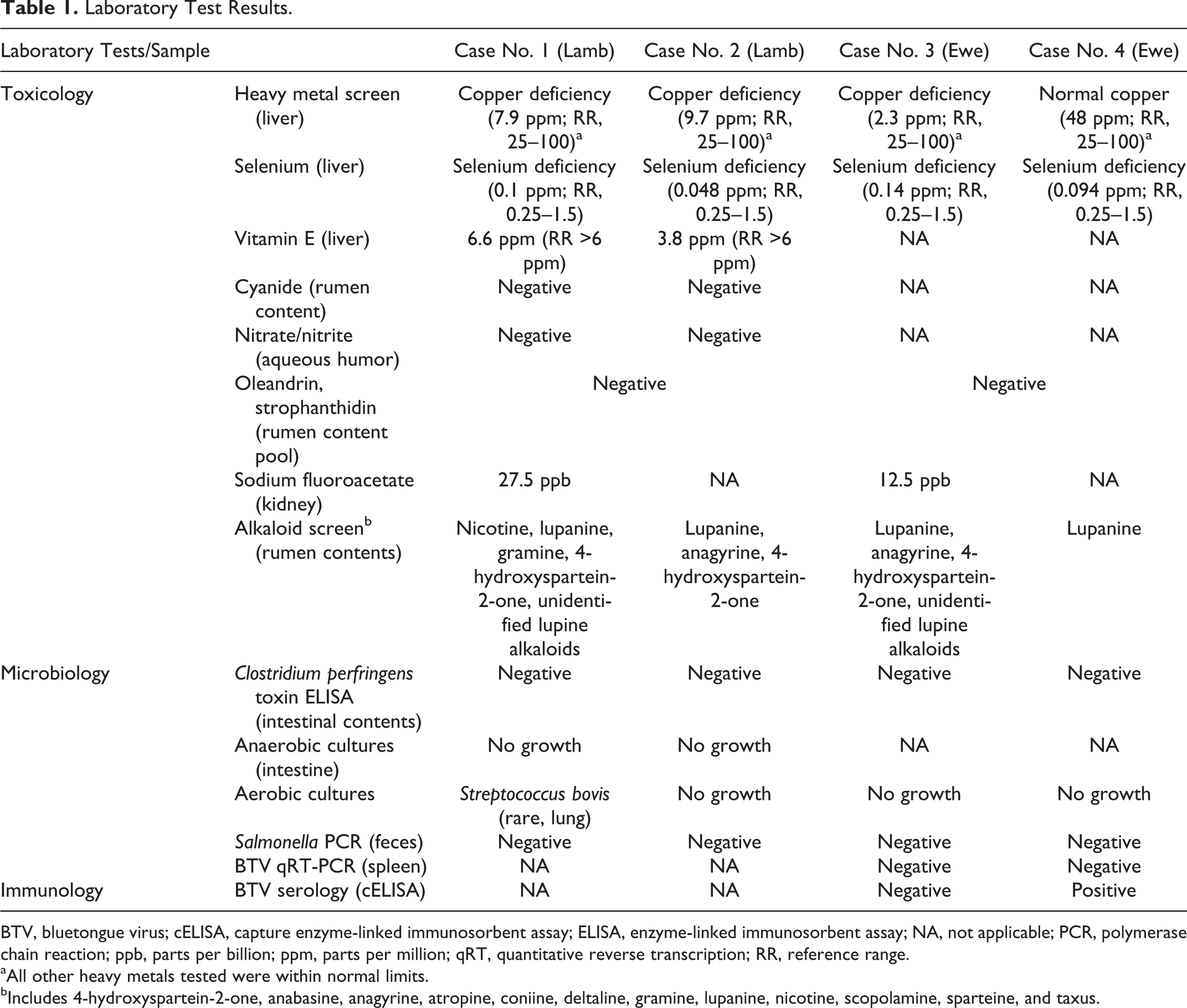
BTV, bluetongue virus; cELISA, capture enzyme-linked immunosorbent assay; ELISA, enzyme-linked immunosorbent assay; NA, not applicable; PCR, polymerase chain reaction; ppb, parts per billion; ppm, parts per million; qRT, quantitative reverse transcription; RR, reference range.
aAll other heavy metals tested were within normal limits.
bIncludes 4-hydroxyspartein-2-one, anabasine, anagyrine, atropine, coniine, deltaline, gramine, lupanine, nicotine, scopolamine, sparteine, and taxus.
Discussion
The history with sudden onset of clinical signs and mortality in a large number of animals after moving into a new pasture in this flock was highly suggestive of a toxic condition. Necropsy and histopathological findings revealed primary cardiac and pulmonary involvement. Several infectious and toxic conditions that can cause acute cardiorespiratory collapse were investigated and will be discussed here.
C. perfringens type D enterotoxemia has been associated with acute death, pulmonary edema, and hydrothorax and hydropericardium in sheep, frequently after sudden changes in the diet. 10 This condition was ruled out by C. perfringens major toxin testing (ELISA for CPA, CPB, and ETX) and anaerobic cultures from intestinal contents.
BTV infection can cause acute fatal disease in sheep characterized by severe pulmonary edema. 7 Acute BTV infection was ruled out by RT-PCR in both ewes, although one of the animals had serum antibodies against the agent, as demonstrated by ELISA. Since BTV is endemic in California, this most likely represents previous contact with a field strain of the virus or vaccination rather than a current infection. In addition, most cases of bluetongue disease in California occur in late summer and autumn, 7 and the mortality reported here occurred in winter.
The results of the bacterial aerobic and anaerobic cultures and the Salmonella sp PCR were unremarkable in all animals and samples. Small numbers of S. bovis were isolated from the lung of 1 lamb, which was considered an incidental finding. These results ruled out bacterial infections that can occasionally occur in outbreaks with sudden onset such as anthrax.
Reportedly, the pasture where the sheep were moved onto contained a proportion of Sorghum halepense (Johnson grass), although this plant was not submitted to the laboratory for botanical identification. S. halepense may contain cyanide, and species of Sorghum can also accumulate nitrates, and both toxic principles have been associated with livestock mortality. 1 Detection of cyanide and nitrate/nitrite was negative in both lambs.
Cardenolide cardiac glycoside-containing plants, such as Nerium oleander (common oleander) and Adonis aestivalis (pheasant’s eye), are common in California and have been associated with myocardial necrosis and sudden death in livestock. 6 Toxicosis by these plants was ruled out by oleandrin and strophanthidin testing in rumen contents, respectively.
One of the plants identified in the pasture in this case (Amsinckia spp [fiddleneck]) has been described as a pyrrolizidine alkaloid-containing plant, 9 but the epidemiological, clinical, and pathological findings in these sheep were not consistent with pyrrolizidine alkaloid intoxication. No other plants identified from submitted samples were considered to have toxicological effects consistent with the case history and postmortem findings. Several plant alkaloids were investigated in rumen contents in 3 animals. Low concentrations of nicotine, lupanine, anagyrine, 4-hydroxyspartein-2-one, gramine, and several other unidentified lupine alkaloids were detected in 1 or more of the rumen contents. Lupinus sp was reported in the pasture but was not submitted to the laboratory for botanical identification. Lupine alkaloids (eg, lupanine) and nicotine stimulate muscarinic and nicotinic receptors, causing acute neurologic signs. 1 However, the low concentrations detected in rumen contents, lack of history of significant pasture contamination with Lupinus spp or nicotine-containing plants, and other postmortem findings suggested that the presence of the alkaloids was not associated with the reported mortality. The other detected alkaloids were not considered to be of toxicological significance in this case.
Furthermore, the pasture was also reported to contain Trifolium pratense (red clover; although again this plant was not submitted for botanical identification), which has been associated with ruminal frothy-bloat and sudden death in ruminants after sudden changes in diet, under grazing conditions. Necropsy findings typical of ruminal bloating (esophageal “bloat line”) were not observed in any of the 4 animals in this study. 6
Due to negative results for a variety of toxins and the diagnostic challenge these cases represented, testing for sodium fluoroacetate in kidney samples from 2 sheep was performed and detected in both samples (27.5 and 12.5 parts per billion [ppb]). These results, in conjunction with the pathological findings in the cardiopulmonary system, were consistent with sodium fluoroacetate poisoning.4,6 Detection of 1080 in tissues suggests recent exposure as experimental treatment with a single oral dose did not detect 1080 in the serum of surviving sheep 3 days after dosing, and 1080 was not detected in skeletal muscle, kidney, and liver at day 14 (limit of detection = 1.5 ppb). 4
In the United States, sodium fluoroacetate is manufactured by only 1 chemical company in Alabama, and the compound remains in use but only as a predacide for livestock protection collars, and US EPA-approved certification must be obtained. 3 No livestock protection collars had been used in this sheep flock. Sodium fluoroacetate also occurs naturally in several plant species, and it has been extensively described in toxic plants in South Africa, Australia, and Brazil.2,6 However, it has not been found in plants in the United States. The source of compound 1080 in this case could not be determined, although according to the referring veterinarian (N.E.E.), treated grain illegally used to control burrowing mammals is a plausible scenario.
Acute selenium toxicosis has also been associated with cardiac hemorrhages and pulmonary edema in sheep. There was no recent history of selenium supplementation in the flock, and selenium toxicosis was further ruled out by selenium determination in liver. In fact, selenium levels were deficient in all 4 sheep, and vitamin E concentration was also low in one of the lambs but within normal limits in the other. Nutritional myopathy (white muscle disease, selenium and vitamin E deficiency) is frequently diagnosed at CAHFS as a cause of myocardial and skeletal muscle degeneration, necrosis, and mineralization in growing ruminants from California (authors’ personal observation, 2013), but this disease is usually chronic and does not occur in outbreaks. Furthermore, the microscopic lesions in the heart and skeletal muscles of these sheep were not typical of white muscle disease due to lack of myocardial mineralization, 6 and this condition rarely occurs in adult animals. However, since selenium and vitamin E are antioxidants, it is plausible that selenium/vitamin E deficiency may have contributed to or potentiated the 1080-associated myocardial damage in these sheep. Overexposure to various heavy metals included in the HMS was ruled out in all animals.
Interestingly, copper deficiency was demonstrated in 3 animals in this report. “Falling disease,” a syndrome of cattle in Australia and Florida, is characterized by sudden death and is believed to result from prolonged copper deficiency. Myocardial scars accompany acute lesions, suggesting repetitive episodes of myocardial injury, and these lesions resemble those of fluoroacetate poisoning in cattle. 6 The cardiac muscle of copper-deficient laboratory animals has a significant decreased concentration of ATP, since copper deficiency significantly decreases cytochrome c oxidase activity in the cardiac muscle, which makes it prone to oxidative damage. 5 The copper status in the sheep of this report may have also had a contributory effect on the pathogenesis of the myocardial lesions.
In conclusion, the detection of 1080 in 2 of these sheep demonstrated sodium fluoroacetate exposure, and based on the pathological changes and the epidemiological aspects, a diagnosis of 1080 intoxication was established. The deficient levels of selenium and copper may have played a contributory or predisposing role. This outbreak demonstrates the potential harm from the use of restricted products that can have lethal effects in wild and domestic animals, and potentially humans. This report demonstrates the possible risk associated with contractual targeted grazing of novel areas for vegetation control. Sodium fluoroacetate should be considered in the differential diagnoses of myocardial degeneration, necrosis, and inflammation in sheep, including those from states where this product is restricted.
Footnotes
Acknowledgements
We thank all CAHFS toxicology and pathology personnel and Beth Tackle at NDSU-VDL for their technical assistance.
Declaration of Conflicting Interest
The author(s) declared no potential conflicts of interest with respect to the research, authorship, and/or publication of this article.
Funding
The author(s) received no financial support for the research, authorship, and/or publication of this article.