
Abstract
Trema micrantha, a fast-growing tree distributed throughout the Americas, produces palatable leaves that have been associated with hepatic necrosis and acute death when consumed by livestock. This report describes fatal pulmonary disease of sheep triggered by consumption of Trema micrantha. Affected sheep had severe progressive dyspnea for a few days before death. Subcutaneous and mediastinal emphysema, reddened lungs, interalveolar septal thickening, and diffuse type II pneumocyte proliferation were the main pathological findings. After ingesting 77.5 and 102.5 g/kg (divided in 3 doses, at 30-day intervals) of T. micrantha leaves, 2 additional sheep developed the same condition. These findings indicate that T. micrantha toxicosis should be considered in the differential diagnosis of ovine respiratory disease.
Trema micrantha is a fast-growing pioneer tree widely distributed across tropical and subtropical zones in South, Central, and North America. This plant has been used in reforestation systems, particularly for the recovery of degraded soils. 9 T. micrantha leaves are highly palatable and readily consumed by herbivores. Ingestion of this plant has been associated with poisoning in goats 5,16 and horses. 2 Poisoning by T. micrantha is characterized predominantly by acute hepatic insufficiency due to centrolobular hepatic necrosis 16 and is often accompanied by neurologic signs 2 of hepatic encephalopathy. 6 In southern Brazil, sheep were traditionally grazed in large extensions of the native rangelands; recently, however, sheep have been intensively managed on small pastures where supplementation of the daily feed often includes alternative sources of forage. Toxicosis, including those caused by plants, is a leading cause of death in sheep in southern Brazil. 12 This report describes T. micrantha poisoning in sheep with emphasis on pneumopathy.
Materials and Methods
Natural Cases: Clinical History and Pathological Characteristics
Clinical information was obtained during visits to the farm. Necropsy was performed on 2 dead sheep. Representative samples of major organs were fixed in 10% neutral buffered formalin. Trimmed tissues were routinely processed, paraffin embedded, and sectioned at 5 μm. Staining methods included hematoxylin and eosin (HE), Masson’s trichrome (MT), and immunohistochemistry (Tables 1 and 2). Necropsy specimens from the lungs of 2 sheep that died from nontoxic, infectious, or neoplastic diseases served as the controls.
Immunohistochemistry Techniques.
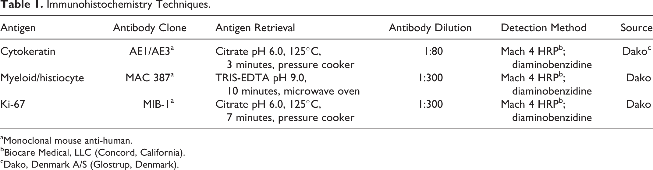
aMonoclonal mouse anti-human.
bBiocare Medical, LLC (Concord, California).
cDako, Denmark A/S (Glostrup, Denmark).
Immunohistochemistry Results in Lungs of 4 Sheep Exposed to Trema Micrantha.
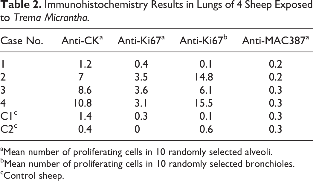
aMean number of proliferating cells in 10 randomly selected alveoli.
bMean number of proliferating cells in 10 randomly selected bronchioles.
cControl sheep.
Experimental T. Micrantha Toxicosis
To reproduce the respiratory disease experimentally, 2 Maedi-Visna-seronegative, 1-year-old Texel sheep (sheep Nos. 3 and 4) were fed T. micrantha leaves in 3 doses at 30-day intervals. Dose calculation was based on a trial in goats. 15 Sheep No. 3 consumed 20, 22.5, and 35 g/kg body weight at each interval, respectively; sheep No. 4 consumed 22.5, 30, and 50 g/kg body weight. Pathological examination was conducted in the experimental cases as for the natural cases. Immediately after euthanasia, lung samples from the sheep No. 3 were fixed in 2% glutaraldehyde + 2% paraformaldehyde and sodium cacodylate buffer solution, dehydrated in ethanol, and embedded in epoxy resin. Semithin sections were stained with methylene blue. Ultrathin sections from selected areas of resin blocks were contrasted with uranyl acetate and lead citrate. An electron microscope was used at 80 kV.
Results
Natural Cases: Clinical History and Pathological Characteristics
On a farm in southern Brazil, a flock of sheep grazed native pastures and was occasionally fed leaves from mulberry (Morus nigra) and grape-Japan (Hovenia dulcis). Three days after receiving the pruned branches from a T. micrantha tree, two 1-year-old mixed Texel sheep fell ill. Sheep No. 1 died 5 hours after the onset of clinical signs. Sheep No. 2 developed lethargy, ataxia, and severe, progressive dyspnea with death 11 days after consuming the plant. At necropsy, both sheep were in good nutritional status. Sheep No. 1 had a pale liver with enhanced lobular pattern and dark red lungs. Sheep No. 2 had cyanotic and congested mucous membranes, cervical subcutaneous and mediastinal emphysema, yellowish liver, red lungs with rib markings (Fig. 1), scanty tracheal and bronchial foam, and subepicardial hemorrhages.
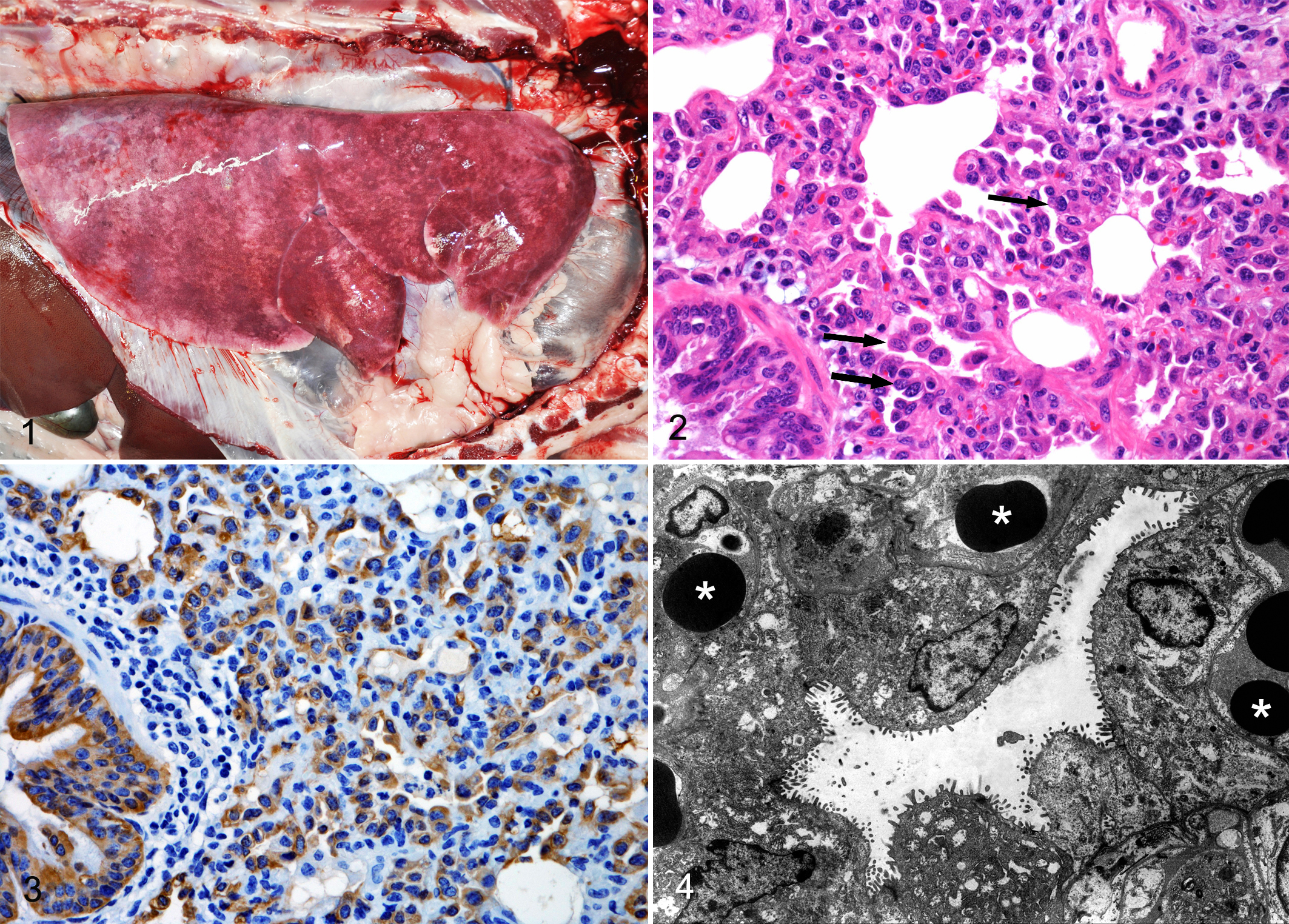
Histologically, sheep No. 1 had severe centrolobular hepatic necrosis and severe pulmonary edema and congestion. Sheep No. 2 had pulmonary congestion with thickening of interalveolar septa and diffuse type II pneumocyte proliferation. Syncytial cells, hyaline membranes, and occasional neutrophils within alveoli and bronchioles were also observed. Immunohistochemistry results are summarized in Table 2. Histologic findings in the liver included hepatocellular vacuolation and tawny pigmentation within macrophages (both mainly in lobular centers), expanded sinusoids and centrolobular veins, reduced sinusoidal eosinophilic globules, and hepatomegalocytes. Lesions in other tissues included diffuse myocardial congestion, eosinophilic globules in renal tubules and urinary spaces, and multifocal hemorrhages in the thymus.
Experimental T. Micrantha Toxicosis
The mean time of ingestion of each dose was 4 hours and 30 minutes. Nine days after the first dose, sheep No. 3 had anorexia, hyperthermia (40°C–41°C), tachypnea (60–160 breaths per minute), and severe dyspnea, with cyanotic mucosae and mild mucinous nasal discharge. However, the sheep spontaneously recovered 3 days after the initial dose. The sheep appeared asymptomatic after the second dose but responded severely 78 hours after the third dose, whereupon it was euthanatized in extremis. Clinical illness in sheep No. 4 was not recognized until 5 days after the third dose, when it also was euthanatized in extremis. Postmortem examination revealed gross and histologic (Figs. 2 and 3) lesions indistinguishable from those in sheep No. 2. Immunohistochemistry results are summarized in Table 2.
Upon evaluation of MT-stained sections, no increase in pulmonary interstitial fibrous tissue was identified in naturally or experimentally exposed sheep compared with controls. Ultrastructurally, type I pneumocytes had fragmented or absent cytoplasm but intact basal lamina. Type II pneumocytes covered extensive areas of the alveolar surface (Fig. 4).
Discussion
The epidemiological, clinical, and pathological findings in this study provide evidence for the toxicity of T. micrantha in sheep. Poisoning by T. micrantha has been reported in other herbivores; however, the reports were focused on the hepatotoxic effects. 2,5,16 Although ingestion of T. micrantha was associated with hepatic necrosis in sheep, the most striking finding in this study was pneumotoxicosis, similar to that in acute respiratory distress syndrome, which is characterized by acute onset, severe hypoxemia, and diffuse alveolar damage with proteinic edema and hyaline membrane formation. 3,8 In South Africa, ingestion of Crotalaria globifera and Crotalaria dura has been associated with pulmonary adenomatosis of sheep and horses. In Brazil, ingestion of Crotalaria sp has also been linked to hepatopathy and pneumopathy in pigs and horses. 14
The toxic components in T. micrantha are unknown. However, Trema tomentosa (Trema aspera), a Trema species endemic to Australia, contains trematoxin, 11 a glycoside that can induce hepatocellular necrosis in several animal species. 7,17 The pathogenesis of the pulmonary lesions induced by T. micrantha poisoning in sheep is likewise unknown but could be the result of damage to alveolar and bronchiolar epithelial cells and, consequently, the integrity of the blood-air barrier. In cattle, acute pulmonary edema and emphysema have been associated with the ingestion of plants rich in L-tryptophan, which results in the ruminal production of 3-methylindole. Subsequently, pulmonary mixed function oxidases can convert 3-methylindole to intermediate electrophilic products that induce pneumotoxicosis. 3,8
In sheep No. 2 and the experimental sheep (Nos. 3 and 4), the pulmonary lesions included diffuse alveolar damage, indicative of type I pneumocyte injury, increased vascular permeability, hyaline membrane formation, and type II pneumocyte proliferation. Type II pneumocytes can be indistinguishable from alveolar macrophages by light microscopy, 3 so immunohistochemistry for cytokeratins was used to document type II pneumocyte proliferation in the lungs of the naturally and experimentally exposed sheep. No difference in the number of macrophages (anti-MAC387 IHC) was observed when comparing naturally or experimentally exposed sheep to controls. Type II pneumocyte proliferation was also confirmed by immunohistochemistry for Ki-67 antigen. Moreover, proliferation of the bronchiolar epithelial cells was observed and confirmed by anti-Ki-67 immunohistochemistry. Electron microscopy also showed proliferation of type II pneumocytes and thickened blood-air barrier, which, although a normal mechanism of alveolar repair, impairs gas exchange.
Sheep No. 1, which died 5 hours after onset of clinical signs, had the typical hepatic necrosis of T. micrantha toxicosis, which has been reported in herbivores. 2,5,15,16 The hepatic or pulmonary manifestations in sheep could be due to factors such as variation in species susceptibility, individual susceptibility, and the amount of ingested plant material. The emphysema of the mediastinum and subcutis of the neck in these sheep could have resulted from increased intra-alveolar pressure generated by increased expiratory effort and altered interstitial permeability. Viral diseases, such as pulmonary adenomatosis and Maedi-Visna, cause chronic respiratory distress and progressive respiratory insufficiency in sheep. 4,10 Bacterial or viral pneumonia could also induce respiratory insufficiency, and altered leukograms could reflect such infection. 13 However, the hematologic values and the bacterial cultures from specimens of the experimental sheep were within laboratory reference range and devoid of growth, respectively. Severe ovine respiratory disease in bluetongue viral infection has been associated with dysphagia and esophageal myonecrosis, 1 two findings that were absent in the sheep of this study. In summary, the evidence indicates that T. micrantha toxicosis should be considered in the differential diagnosis of ovine respiratory disease.
Footnotes
Acknowledgement
We thank Dr Nelson I. Matzenbacher for the identification of T. micrantha.
Declaration of Conflicting Interests
The authors declared no potential conflicts of interest with respect to the research, authorship, and/or publication of this paper.
Funding
The author(s) disclosed receipt of the following financial support for the research, authorship, and/or publication of this article: This work was supported by the Conselho Nacional de Desenvolvimento Científico e Tecnológico (CNPq), Brazil (grant 478266/2012-0).