
Abstract
A 9-month-old female Yucatan pig was euthanized after acute onset of paraplegia. Gross and microscopic examination revealed dorsal dissection of the nucleus of the L2–L3 intervertebral disk through the annulus fibrosus, extrusion of nucleus pulposus material through the overlying dura mater and into the spinal cord, and associated acute spinal hemorrhage and necrosis. This is, to the authors’ knowledge, the first reported case of intervertebral disk disease in swine.
Intervertebral disk disease (IVDD) is common in people and dogs. 2 Degeneration of intervertebral disks leads to injury to the overlying spinal cord, from parenchymal contusion, compression, or penetration. IVDD is generally divided into 2 different processes: acute extrusion of degenerate nucleus pulposus material into the vertebral canal (Hansen type I) or chronic protrusion of the annulus fibrosus (Hansen type II). Among domestic animals, IVDD is most commonly observed in dogs but has also been reported in cats, 4 horses, 3 and ferrets. 8 Herein we describe a case of spontaneous IVDD in a pig, caused by the extrusion of nucleus pulposus material into the vertebral canal and the overlying spinal cord.
The subject was a 9-month-old female Yucatan pig weighing 29 kg at acquisition. The animal was part of a cohort of 30 healthy young adult animals used in a study evaluating safety and efficacy of stents implanted in coronary arteries via femoral artery catheterization. The animal received implantation of 3 coronary stents using standard methods and recovered uneventfully from surgery. It was administered acetylsalicylic acid (aspirin, 325 mg) and clopidogrel (Plavix, 75 mg) daily in feed, according to a routine poststenting protocol. Twenty-two days after surgery, the animal was heard emitting a sudden loud cry and upon immediate examination was found in a “dog-sitting” position, with pelvic limbs extended. The animal could not get up or stand on its pelvic limbs. Neurologic examination showed a mild hyperreflexia in both pelvic limbs. The animal was kept under observation until the next day but showed no signs of improvement. It was euthanized because of the poor prognosis for recovery or for its continued use in the study.
A full necropsy was performed. In the vertebral column, the intervertebral disk between the second and third lumbar vertebrae had a reddened and irregular surface within the vertebral canal, but no compressive dorsal protrusion was apparent. There was increased mobility between the 2 vertebral bodies compared with adjacent vertebrae. The spinal cord overlying the L2–L3 disk was softened, and the dura mater at that level was torn on its ventral aspect. The spinal cord and the vertebral column from L2 to L3 were immersion-fixed in neutral-buffered formalin. After formalin fixation, the cut surface of the spinal cord at the L2–L3 level contained a focal area of hemorrhagic malacia, measuring approximately 5 mm diameter by 10 mm long. Transverse sections of the spinal cord at this level were embedded in paraffin. The L2–L3 intervertebral disk with adjacent vertebral bodies was decalcified en bloc, then cut into sagittal sections and embedded in paraffin. Sections of the spinal cord and intervertebral disk were stained with hematoxylin and eosin, Masson’s trichrome, and Alcian blue (at pH 1.0).
Histologic examination of decalcified sections of the intervertebral disk and vertebrae at the L2–L3 junction showed irregularity of the dorsal margin of the disk, with an amorphous, poorly staining material dissecting between the collagenous bands of the dorsal annulus fibrosus close to the vertebral canal, with associated mild hemorrhage and neutrophilic infiltration (Figs. 1, 2). Alcian blue staining showed this material to be alcianophilic, as was the material present in the in situ nucleus pulposus. Underlying the dorsal annulus fibrosus, the attachment between one side of the disk and the adjacent vertebral endplate was torn, forming a clear tract that extended to the central area of the disk; this tract contained some of the same alcianophilic material, admixed with red blood cells.
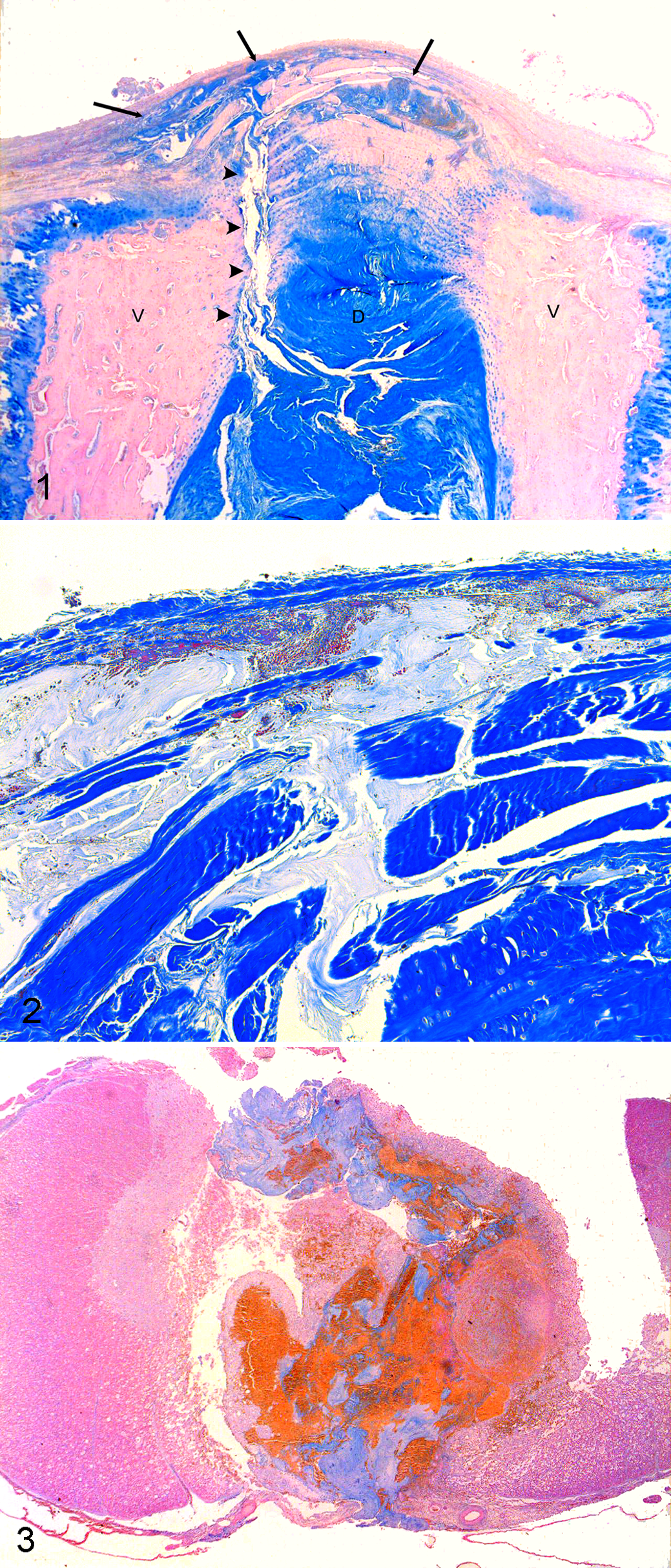
L2–L3 intervertebral disk (D) with adjacent vertebral endplates (V), sagittal section; pig. The disk material at the level of the nucleus pulposus is Alcian blue positive. Alcianophilic material has dissected between the collagenous bands of the dorsal annulus fibrosus, close to the vertebral canal (arrows). A tear is present at the attachment between one side of the disk and the adjacent vertebral
Examination of the spinal cord lesion at L2–L3 showed loss of structural integrity, with a large central area of acute hemorrhage admixed with necrotic neural tissue and a nonstaining, amorphous, poorly cellular material. Alcian blue staining revealed the material to be alcianophilic (Fig. 3), with a fine fibrillar structure, and similar in appearance to the material observed in the intervertebral disk nucleus pulposus and dissecting the annulus fibrosus. This material appeared to be extending from the vertebral canal into the spinal cord through a discontinuity of the ventral dura mater. Scattered throughout this material were small numbers of darker blue, elongate to polygonal structures resembling cells but without visible nuclear staining. The surrounding spinal cord white matter showed extensive edema and axonal degeneration.
The various changes observed (a tear within the dorsal intervertebral disk, alcianophilic material infiltrating between the dorsal layers of the annulus fibrosus, similar material extending through a tear in the overlying dura mater and present in large amounts within a malacic area of the spinal cord) were considered diagnostic of an acute extrusion of nucleus pulposus material from the disk into the spinal cord, causing acute paraplegia. Such an “explosive” extrusion of disk material with penetration of the dura and extension into the spinal cord is a rare manifestation of IVDD but has been described previously in dogs 5,7 and a cat. 6 To our knowledge, this is the first reported case of spinal cord injury due to IVDD in a pig. Although IVDD may appear to be an exceptional occurrence in pigs, this may be a function of the early age at which most pigs are sacrificed for food or research since IVDD is generally observed in mature dogs and cats. In dogs, it is recognized that IVDD due to acute extrusion of nucleus pulposus material (Hansen type I) is more frequently observed in chondrodystrophic breeds, such as the Dachshund, Pekingese, French Bulldog, and Beagle. 2 Anomalies of skeletal cartilage metabolism or development are therefore thought to be associated with premature disk degeneration and subsequent IVDD in these breeds. It is interesting to note that the pig affected in this report was of the Yucatan breed, which is significantly smaller than the usual pig breeds used for food and shares some phenotypic similarities with some of the chondrodystrophic dog breeds (shorter limbs and shorter snout than standard pig breeds). However, it is not currently known whether these characteristics of Yucatan pigs are due to anomalies of cartilage metabolism. Spontaneous canine IVDD has been proposed as a research model for human IVDD. 1 The present case suggests that if Yucatan pigs do indeed have a predisposition to develop type I IVDD, they might also constitute a useful research model for the human disease.