
Abstract
Aprosencephaly is a rare condition in veterinary and human medicine characterized by the complete absence of telencephalon and diencephalon. Some cases are accompanied by a facial dysmorphism designated as otocephaly. A stillborn lamb had splanchnocranial anomalies that were classified by computed tomography, magnetic resonance imaging, and pathologic examination as aprosencephaly and otocephaly. The brain included parts of the cerebellum and brainstem but no telencephalon, diencephalon, or mesencephalon. The cerebellum had a structurally normal cortex with expression of neuronal nuclear antigen in the inner and doublecortin in the outer granular cell layers, as well as an irregularly situated nucleus dentatus. Aprosencephaly with otocephaly has been described in mice with heterozygous mutations in the Otx2 gene; however, no causative polymorphisms were detected in the Otx2 gene region of this lamb.
Keywords
Aprosencephaly is a rare defect in forebrain induction 12 that is characterized by the absence of prosencephalic structures, including the telencephalon and diencephalon. The term otocephaly has been applied to cases of dysgnathia in which the upper and lower jaws, tongue, and frontonasal bones are absent or hypoplastic, and the pinnae are often fused in the midbasal part of the head. 8 Mutations in genes of the Otx family, which encode highly conserved transcription factors required for forebrain morphogenesis, are potential causes of alterations in the specification, regionalization, and terminal differentiation of anterior neuronal patterning. 4,5 Otx2 protein is expressed during embryogenesis within the rostral neuroectoderm destined to form anterior parts of the brain. Heterozygous mutations in the Otx2 gene cause severe brain and craniofacial abnormalities reminiscent of otocephalic phenotypes in several mouse models. 5,19 The purpose of this report is to present the computed tomographic and magnetic resonance imaging, pathologic, and genetic analyses in a case of aprosencephaly in a lamb.
History
A Texel lamb was stillborn after normal duration of gestation in March 2009; its 2 siblings were born alive without macroscopic or behavioral abnormalities. The multiparous ewe was from a flock of 300 sheep with regular anthelmintic treatment, vaccination against Clostridium perfringens and Fusobacterium necrophorum, and natural insemination with annual introduction of different rams to decrease inbreeding. There was no history of infectious disease or medical treatment of the ewe during pregnancy. No information concerning other congenital anomalies in the flock was available.
Postmortem Examination
The lamb had severe craniofacial alterations. Its head was small and lacked eyes, upper or lower jaws, and associated structures of the muzzle. The pinnae extended laterally from a narrow pharyngeal aperture that opened into the esophagus and trachea (Fig. 1).
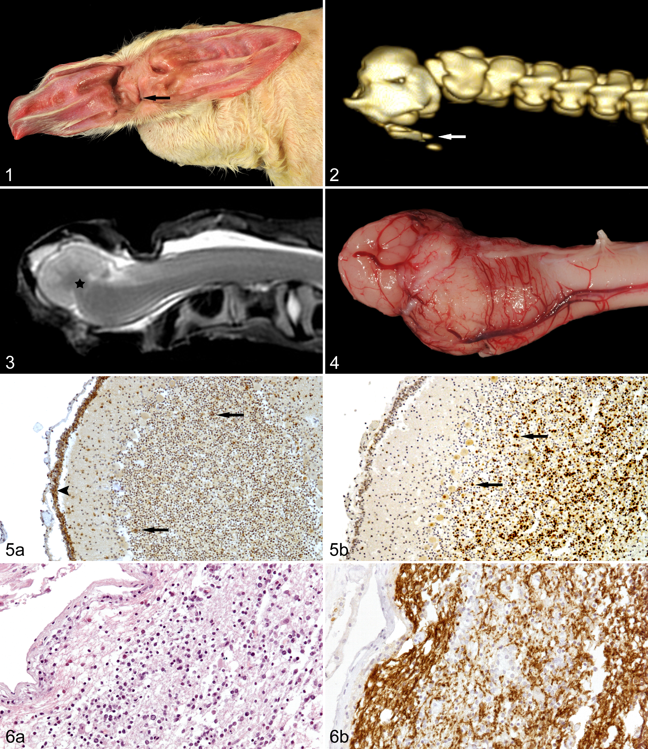
Head, lamb. Otocephalic phenotype with central pharyngeal aperture (arrow).
With computed tomography (CT), skeletal abnormalities were restricted to the skull. The neurocranium included parts of the parietal, temporal, and occipital bones, with occipital condyles and foramen magnum. The splanchnocranium was dysplastic and without recognizable frontonasal bones, orbits, osseus portion of the ears, maxillae, or mandibles. Hyoid bones were rudimentary (Fig. 2). With magnetic resonance imaging (MRI), the arbor vitae of the cerebellum filled the small cranial vault and was continued caudally by the medulla oblongata and spinal cord. The pons was not evident (Fig. 3).
At necropsy, the brain was rudimentary, less than 3 cm long, and without visible telencephalic, diencephalic, or mesencephalic structures. The cerebellum and medulla oblongata were evident, but the pons was not discernible (Fig. 4). No anomalies besides those of the head and brain were observed.
Histopathology
Brain, spinal cord, and tissues from visceral organs (lung, heart, liver, spleen, kidney, mesenteric lymph nodes, intestine) were fixed in 10% neutral buffered formalin, processed routinely, and embedded in paraffin. Sections were stained with hematoxylin and eosin (HE). Luxol fast blue-cresyl violet staining was used to demonstrate myelin and Nissl substance. Immunohistochemistry was performed with primary antibodies against neuronal nuclear antigen (NeuN), synaptophysin, doublecortin (DCX), glial fibrillary acidic protein (GFAP), and 2′,3′-cyclic nucleotide 3′-phosphodiesterase (CNPase) (Table 1). Tissues that express these markers were used as positive controls. Consecutive sections incubated with a nonreactive mouse monoclonal antibody against chicken lymphocytes or polyclonal goat or rabbit serum served as negative controls.
Antibodies and Detection Systems Used for Immunohistochemistry
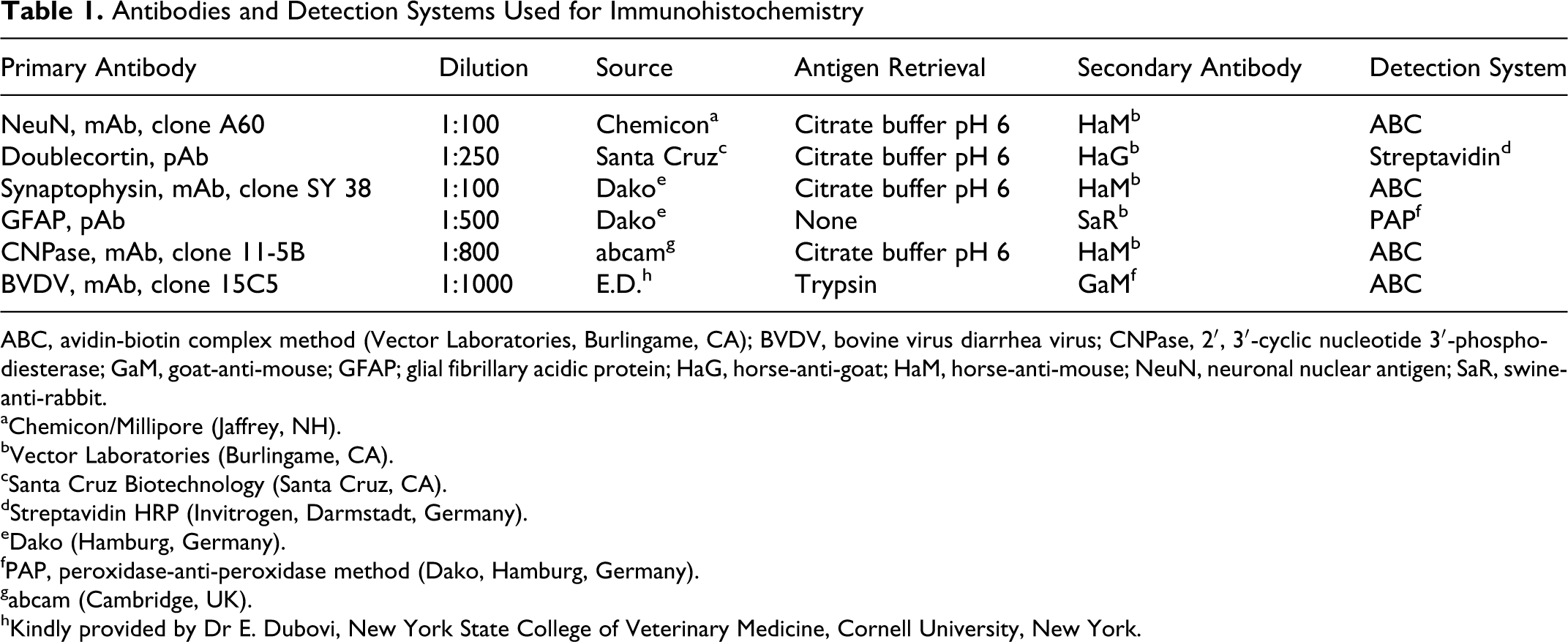
ABC, avidin-biotin complex method (Vector Laboratories, Burlingame, CA); BVDV, bovine virus diarrhea virus; CNPase, 2′, 3′-cyclic nucleotide 3′-phosphodiesterase; GaM, goat-anti-mouse; GFAP; glial fibrillary acidic protein; HaG, horse-anti-goat; HaM, horse-anti-mouse; NeuN, neuronal nuclear antigen; SaR, swine-anti-rabbit.
aChemicon/Millipore (Jaffrey, NH).
bVector Laboratories (Burlingame, CA).
cSanta Cruz Biotechnology (Santa Cruz, CA).
dStreptavidin HRP (Invitrogen, Darmstadt, Germany).
eDako (Hamburg, Germany).
fPAP, peroxidase-anti-peroxidase method (Dako, Hamburg, Germany).
gabcam (Cambridge, UK).
hKindly provided by Dr E. Dubovi, New York State College of Veterinary Medicine, Cornell University, New York.
Histologically, the diagnosis of aprosencephaly was confirmed by the complete absence of both the telencephalon and diencephalon. In addition, no mesencephalic structures could be identified. In coronal sections of the brain remnants, cerebellar tissue had a normal architecture. Neurons of the external granular cell layer and individual cells in inner cortical layers were immunoreactive for DCX, a marker of differentiating and migrating neurons (Fig. 5a), whereas immunoreactivity for NeuN, a marker for mature neurons, was restricted to the inner granular cells as expected (Fig. 5b). 10,25 The cerebellar white matter was mixed with irregularly arranged fragments of deep cerebellar nuclei surrounded by neuronal processes and glial cells. Neurons in the nuclei expressed synaptophysin on the cytoplasmic membrane, interpreted as synaptic vesicle formation, but were not labeled by NeuN, consistent with the nonreactivity of neurons of the cerebellar dentate nuclei. 25,26 The brainstem sections contained a misshapen fourth ventricle with multifocal subpial accumulations of loosely arranged cells (Fig. 6a) that were immunohistochemically positive for CNPase (Fig. 6b) or GFAP, consistent with a disorganized focus of oligodendroglial cells and astrocytes. A few neurons in the spinal cord had swollen perikarya with loss of Nissl substance staining by cresyl violet, consistent with chromatolysis, probably as a consequence of failure to connect to intrathalamic neurons.
Otx2 Analysis
Genomic DNA from the lamb and 2 healthy control sheep was isolated from skeletal muscle using the Gentra Puregene Tissue Kit (Qiagen, Hilden, Germany) according to the manufacturer’s protocol. Otx2 nucleotide sequence of Bos taurus (NCBI Acc. No. NM_001193201) was blasted against whole-genome shotgun sequences of Ovis aries. 1 Four clones containing parts of exons 1 to 3 were identified (NCBI Acc. No. ACIV011001973.1, ACIV011001974.1, ACIV011001976.1, ACIV011001977.1). Polymerase chain reaction (PCR) was performed with specific primers for the 3 exons of the Otx2 gene, generating amplicons of 228, 254, and 930 base pairs. PCR products were visualized by electrophoresis in a 2% agarose gel, treated with ExoSapit for purification according to the manufacturer’s protocol (GE Healthcare, Munich, Germany) and commercially sequenced by GATC (Konstanz, Germany). Subsequently, the nucleotide sequences were blasted against homologous sequences of B. taurus and the 2 control sheep. Further sequencing of introns and promoter region was performed to search for polymorphisms. Nucleotide sequences of the lamb and the 2 control sheep were identical. Single-nucleotide substitutions between O. aries and B. taurus within the coding sequence were neutral mutations without impact on the amino acid sequence. (Primer sequences are available upon request.)
Virology
Border disease virus (BDV) antigen was not detected by immunohistochemistry using an antibody against an epitope of the 48-kDa glycoprotein of the bovine virus diarrhea virus (BVDV, Table 1) with known cross-reactivity with BDV (M. Hewicker-Trautwein, personal communication). 14 Genomic RNA of bluetongue virus (BTV) was not detected by quantitative real-time RT-PCR. For RT-PCR, total RNA was isolated from formalin-fixed, paraffin-embedded (FFPE) spleen using the QuickExtract FFPE RNA Extraction Kit (Epicentre Biotechnologies, Madison, WI) according to the manufacturer’s instructions.
Discussion
Congenital defects of the ovine nervous system are rare. In a 3-year necropsy study, 67 of 4417 lambs were affected, a prevalence of 1.5%. 7 Of these 67 cases, 19.4% were solitary central nervous system defects not associated with defects of other organ systems. Hydrocephalus (15 cases) and spina bifida (9 cases) were most common. Three lambs had aprosopia, 3 had anencephaly, and 2 had microencephaly. In case reports, lambs with similar phenotypes have been diagnosed grossly with synotia, otocephalus aprosopus, or acephaly. 22,23,28
Although mentioned in textbooks, aprosencephaly is rare, with about 22 published human cases. 27 The condition was first described by Iivanainen et al 17 and Garcia and Duncan 11 in 1977. The exact incidence is unknown due to inconsistent use of nomenclature and lack of histologic examination. In true aprosencephaly, as present in this case, telencephalic structures (cerebral cortex, hippocampus, and striatum) as well as diencephalon (globus pallidus, thalamus, hypothalamus, hypophysis) and the eyes are absent. In contrast to anencephaly, which is regularly associated with cranioschisis and, therefore, is a neural tube closure defect, aprosencephalic individuals have an intact cranial vault, indicating that the developmental errors occurred after neurulation. 18 Imaging techniques, such as CT, were helpful in ruling out a neural tube closure defect in this case. Nevertheless, the term anencephaly is a misnomer. Because only the telencephalon or prosencephalon is absent in almost all reported cases, they should be classified instead as cerebral aplasia or prosencephalic hypoplasia. 6,20 Furthermore, aprosencephaly differs from holoprosencephaly, the result of impaired prosencephalic cleavage. Holoprosencephaly is often accompanied by various facial dysmorphisms, including hypoplasia or absence of jaws and tongue. Those cases with ear anomalies are consequently designated as an otocephalic phenotype with synonyms such as synotia or agnathia. 8 In addition, there may be extracranial anomalies of the reproductive tract or limbs, 21 which were not found in the lamb of this case.
Cases of pseudo-aprosencephaly have been described with features of primitive prosencephalic anlage that remains vesicular without further differentiation of a holospheric brain mantle. These cases should be classified in the complex of holoprosencephaly in its most primitive form. 27 Because it can be difficult to recognize a pseudo-aprosencephaly when the forebrain vesicle is collapsed upon opening the cranial vault, pre-necropsy MRI is helpful to distinguish between tissue and fluid filling the cranial vault. In contrast to pseudo-aprosencephaly, the present case had no remnants of prosencephalic vesicle wall and thus was classified as true aprosencephaly.
In human medicine, severe anomalies of the forebrain together with facial and radial limb defects are often syndromic and have been reported, among others, in Steinfeld syndrome (Online Mendelian Inheritance in Man [OMIM] ID 184705) and Meckel-Gruber syndrome (OMIM ID 603194). Aprosencephaly syndrome or Garcia-Lurie syndrome (OMIM ID 207770), which is also reported as XK aprosencephaly, is suspected to be inherited in an autosomal recessive manner. Studies in animal models suggested a critical role of the homeobox gene Otx2, which is a vertebrate homologue to the Drosophila orthodenticle gene (otd) with essential functions in the development of the forebrain and midbrain. Mouse Otx2 null mutants are embryonic-lethal and lack the rostral neuroectoderm destined to become the forebrain, midbrain, and rostral hindbrain. In addition, they have an abnormal body axis. 5 Heterozygous Otx2 mutants have loss of forebrain and midbrain as well as eye anlagen and display an otocephalic phenotype. 8,15,18,19 The Otx2 gene in humans has been analyzed only in a few cases of aprosencephaly but without causal association, whereas several cases of ocular malformations were associated with Otx2 heterozygous loss-of-function mutations. 9,24 Genetic analysis of the complete ovine Otx2 gene region in the present case (NCBI Acc. No. JN581044) provided no indication for an involvement of this gene in the pathogenesis of the malformation, although the spectrum of lesions matched exactly the Otx2 expression profile.
Aprosencephaly may arise either as a primary malformation, secondary to an encephaloclastic process, or a combination of the two. 18,29 The absence of inflammation, hemorrhage, degeneration of neuroglial tissue, calcification, or other signs of tissue destruction in this case is suggestive of a primary malformation due to genetic or environmental inhibition of forebrain induction.
In sheep, infections with BDV, BTV, and Akabane virus have been associated with congenital anomalies of the nervous system. 2,13,16 Akabane virus mainly infects cattle and is exotic to Europe. In this lamb, neither BDV nor BTV could be detected by immunohistochemistry or RT-PCR, respectively. A virus infection with clearance during gestation cannot be excluded, but the birth of 2 normal siblings argues against this possibility. Toxic plants (eg, Veratrum californicum) or prolonged hypoxia during gestation have also been linked to nervous system defects. 3 However, no evidence of exposure to environmental teratogens was found in this case.
In conclusion, this lamb was stillborn with true aprosencephaly and otocephaly, probably due to arrest of prosencephalic development after neurulation. Otx2 gene mutations were not found so another mutation or metabolic/hypoxemic disturbances in a triplet pregnancy could have been etiologic factors in the pathogenesis of this rare malformation.
Footnotes
Acknowledgements
The authors thank Prof Dr M. Hewicker-Trautwein, Institut für Pathologie, Stiftung Tierärztliche Hochschule Hannover, Germany, for performing the immunohistochemistry for border disease virus and for careful reading of the manuscript; Dr E. Dubovi, New York State College of Veterinary Medicine, Cornell University, New York, for kindly providing the BVDV-specific antibody; and Dr H.-P. Hamann and C. Sauerwald from the Landesbetrieb Hessisches Landeslabor in Gießen, Germany, for performing PCR for bluetongue virus.
Declaration of Conflicting Interests
The authors declared no potential conflicts of interest with respect to the research, authorship, and/or publication of this article.
Funding
The authors received no financial support for the research, authorship, and/or publication of this article.