
Abstract
Cynomolgus macaques were exposed to the Angola strain of Lake Victoria Marburg virus (MARV) by aerosol to examine disease course and lethality. Macaques became febrile 4 to 7 days postexposure; the peak febrile response was delayed 1 to 2 days in animals that received a lower dose; viremia coincided with the onset of fever. All 6 macaques succumbed to the infection, with the 3 macaques in the low-dose group becoming moribund on day 9, a day later than the macaques in the high-dose group. Gross pathologic lesions included maculopapular cutaneous rash; pulmonary congestion and edema; pericardial effusion; enlarged, congested, and/or hemorrhagic lymphoid tissues; enlarged friable fatty liver; and pyloric and duodenal congestion and/or hemorrhage. Fibrinous interstitial pneumonia was the most consistent pulmonary change. Lymphocytolysis and lymphoid depletion, as confirmed by TUNEL (terminal deoxynucleotidyl transferase-mediated deoxyuridine triphosphate nick end labeling), were observed in the mediastinal lymph nodes and spleen. MARV antigen was detected in the lungs, mediastinal lymph nodes, spleen, and liver of all animals examined. In infected macaques, nuclear expression of interleukin-33 was lost in pulmonary arteriolar and mediastinal lymph node high endothelial venule endothelial cells; interleukin-33-positive fibroblastic reticular cells in the mediastinal lymph node were consistently negative for MARV antigen. These macaques exhibited a number of features similar to those of human filovirus infections; as such, this model of aerosolized MARV-Angola might be useful in developing medical countermeasures under the Animal Rule.
Marburg viruses (MARVs)—small negative-stranded RNA viruses of the Filoviridae family—cause some of the most devastating viral hemorrhagic fevers in humans. To date, Ebola virus (EBOV) is the only other known virus belonging to the Filoviridae family. There are currently five species of EBOV—Zaire, Sudan, Ivory Coast, Reston, and Bundibugyo—with each species having one or more strains. There is only one species of MARV (Lake Victoria), but at least nine genetically distinct isolates have been identified, 7,83 including the Ci67 77 and Popp strains, 14 different isolates derived from the initial, simultaneous outbreaks in Germany and Yugoslavia, respectively, in 1967; the prototype Musoke strain, from the 1980 cases in Kenya; 79 the Ravn strain, from the 1987 outbreak in Kenya; 46 and the most recent strain, from a 2004–2005 outbreak in Angola. 16
MARV was first isolated in 1967 from laboratory workers in Marburg, Germany, who were exposed to tissues from infected African green monkeys. Of the 32 patients who were confirmed as being infected, 7 succumbed to the infection. Secondary transmission by direct contact with infected patients or tissues occurred but was limited. 51,56,76 The 2004–2005 MARV-Angola outbreak was among the most virulent outbreaks of viral hemorrhagic fever yet recorded, 16 in which an estimated 227 out of 252 patients succumbed to infection. The MARV-Angola virus therefore appears similar to the typically more virulent Ebola viruses—in particular, EBOV-Zaire, which is often fatal in a majority of human cases. 17 Why the disease caused by MARV-Angola is more severe than other MARV strains is not clear. However, an initial study with MARV-Angola in rhesus macaques found that time to death was shorter than that previously observed for parenteral (intramuscular) inoculation with other strains of MARV. 33
Epidemiological analysis of MARV and EBOV outbreaks indicated that inhalation is not a prominent means of natural transmission between humans. 5,25,51,68,73,78,87 However, transmission of EBOV has been observed from infected nonhuman primates (NHPs) to naive animals housed in the same room. 42 In an outbreak of the Reston strain of EBOV in the Hazleton facility in Reston, Virginia, aerosol transmission between NHPs might have occurred. 22,44,58,59 Reports from the late 1980s and 1990s demonstrated that guinea pigs and NHPs could be lethally infected by aerosol transmission with either MARV or EBOV. 8,13,31,43,45,54,55,65,70 In the early 1990s, a defector from the former Soviet Union revealed that the Soviets had an active offensive biological weapon program that included development of MARV and EBOV as biological weapons for aerosol dispersal. 1 For these reasons, MARV and EBOV are listed as category A select agents. 52
Licensure of vaccines and therapeutics for MARV (and other biological threat agents) where clinical efficacy cannot be ethically or logistically determined is feasible through only the Food and Drug Administration’s Animal Rule, 29 under which pivotal efficacy is demonstrated in animal models rather than clinical cohorts. To meet the requirements of the animal rule, animal models must be well characterized; the pathology of the disease must be well understood; the disease in the animal must closely mimic the human condition; and the route of exposure must be the same for the animal model as that proposed for human exposure (most likely, aerosol exposure in the case of a biological weapon attack). In the case of MARV and EBOV, only NHPs have been shown to be susceptible to infection with wild-type virus; guinea pig and mouse models exist but require adaptation of the virus.
Human infections by strains of EBOV and MARV begin to clinically manifest after an incubation period that can range from 2 to 21 days. There is typically an early nonspecific flulike illness, as characterized by fever, headache, nausea, vomiting, and watery diarrhea and followed by a maculopapular rash on the face, trunk, and extremities. An early lymphopenia is characteristic of human patients. The liver is a key target organ of EBOV and MARV, with elevated liver enzymes occurring in many patients. Coagulopathy is another common feature of these infections, and as many as 30 to 50% of patients exhibit overt hemorrhage. Death usually occurs 8 to 16 days after infection and is often attributed to multiorgan failure with disseminated intravascular coagulation and cardiovascular collapse. These and other clinical features of human infections have been described in a number of reports. 9,15,17,57 A delicate temporally associated balance between the development of a protective immune response and a deleterious inflammatory response appears critical to determining the outcome of infection. For example, patients who recover from EBOV-Zaire infection develop an early immune response involving specific humoral factors and a tightly regulated cytotoxic T-lymphocyte response. 53 However, fatal outcome has been associated with suboptimum humoral immunity, unregulated T-cell activation, lymphocyte apoptosis, and uncontrolled mononuclear phagocyte activation resulting in elevated cytokine levels that contribute to the multisystemic dysfunction seen terminally. 4,86 Autopsy studies of EBOV and MARV cases are limited 24,26,67,93 in large part because of the difficulty of performing autopsies in isolated areas where these cases occur and because of the biosafety concerns of performing such autopsies. Nonetheless, human cases demonstrate lesions in both the liver and the lymphoid tissues, with specific virus targeting of mononuclear phagocytes, fibroblasts, hepatocytes, and endothelial cells. Hepatocellular necrosis is the dominant liver finding, and lymphocyte destruction is seen in the spleen and lymph nodes. Endothelial damage is also reported. 92
Lethal experimental infections by EBOV and MARV have been induced in mice, guinea pigs, and several NHP species, including African green monkeys, rhesus and cynomolgus macaques, squirrel monkeys, and baboons. These models have been described in several primary and review articles. 6,9,12,19,23,28,34,35,71,72,88 –90 The features of disease in these models depend on a variety of factors, including virus strain, dose and route of administration, and the age, species, and strain of animal involved. Whereas NHPs are often susceptible to lethal infection by wild-type strains of EBOV and MARV, immunocompetent adult mice and guinea pigs are not; they are susceptible to some attenuated strains, however. All three animal models typically demonstrate debilitating disease characterized by weight loss, fever (measured in NHPs and guinea pigs), viremia, and measurable virus titers in a variety of other tissues. There is also early targeting of mononuclear phagocytes, followed by infection of fibroblasts, fibroblastic reticular cells (FRCs), hepatocytes, adrenal cortical cells, endothelial cells, and some other cell types. The liver and lymphoid tissues especially incur significant damage attributed to virus infection. Hepatocellular damage and other changes in the liver are generally considered the result of direct virus infection, whereas lymphocyte destruction is reportedly a bystander effect. Not surprisingly, lymphopenia and increased hepatic enzymes are important clinicopathological features of animal models, as are neutrophilia and thrombocytopenia. The NHP models of EBOV and MARV more closely resemble other reported features of the human diseases than do the mouse and guinea pig models. In particular, NHPs more consistently develop hemorrhagic lesions and severe coagulopathy late in the course of infection. NHPs infected with EBOV demonstrate elevated expression of a variety of cytokines, 34,69 which is comparable to human cases. These cytokines are believed to contribute to coagulopathy and hemorrhage, as are other factors, such as virus-induced damage to endothelial cells and FRCs. In addition, NHPs exhibit altered expression of a number of soluble factors related to immune function and apoptosis. Another key aspect of the NHP model is that dendritic cells (DCs) have been shown to be a key early target of infection by EBOV. 34 Human DCs have also been shown to be susceptible to infection by EBOV and MARV, though only in vitro. 11 DC dysfunction, whether the direct effect of virus infection or the result of indirect factors, is considered an important aspect of the altered immune status characteristic of filovirus infection. 61 In many ways, the animal models of Filovirus infections have been characterized to a much greater extent than that of the human diseases themselves. Accordingly, it is difficult to determine the suitability of the various animal models in some regards because the applicable features in the human cases have not been fully investigated.
MARV infection in NHPs closely resembles what has been reported for the human disease, including the development of the characteristic rash and fibrin deposition and disseminated intravascular coagulation. 27,33,39 To our knowledge, only one study has described the pathology (albeit limited) associated with MARV-Angola infection in NHPs, and these animals were experimentally challenged via the intramuscular route. 20 Aerosol infection with the Popp strain of MARV has been shown to be lethal to guinea pigs and NHPs. 54,55 The clinical signs and disease course after aerosol exposure for MARV-Popp appear to resemble those of parenteral exposure; however, data are limited. The transmissibility and lethality of MARV-Angola in humans, evident in the 2004–2005 outbreak, have caused concern that, like MARV-Popp, it might be used as a biological weapon. Animal models are needed to evaluate vaccines and therapeutics for protection against aerosol exposure to MARV-Angola.
We report here for the first time the clinical, virology, and pathology findings in cynomolgus macaques exposed to aerosols containing MARV-Angola, with emphasis on the pathologic changes in the lung and hematopoietic tissues. In addition to conducting thorough postmortem and routine histologic examinations, we performed histochemical and immunohistochemical staining for fibrin in the lung, spleen, liver, and kidney; TUNEL (terminal deoxynucleotidyl transferase-mediated deoxyuridine triphosphate nick end labeling) in the mediastinal lymph node and spleen for identification of apoptotic cells; and immunohistochemical and double immunofluorescence staining in the lung, spleen, and mediastinal lymph node for MARV antigen and different cell markers to include FRCs (interleukin-33 [IL-33], nuclear factor from high endothelial venules [HEVs]), endothelial cells (IL-33 and factor VIII–related antigen / von Willebrand factor [vWF]), a DC marker (DC-SIGN/CD209), and a macrophage marker (MAC387).
Materials and Methods
Animals
This study used 6 healthy adult cynomolgus macaques (Macaca fascicularis) of both sexes (4 males, 2 females) from the NHP colony at the US Army Medical Research Institute of Infectious Diseases (USAMRIID). Macaques were surgically implanted with telemetry devices before entry into a biosafety level 4 laboratory. Macaques were put into biosafety level 4 a total of 10 days before study initiation for acclimation and to collect baseline telemetry data. Research was conducted in compliance with the Animal Welfare Act and other federal statutes and regulations relating to animals and experiments involving animals, and it adhered to the principles stated in the 1996 edition of the National Research Council’s Guide for the Care and Use of Laboratory Animals. The facility where this research was conducted is fully accredited by the Association for Assessment and Accreditation of Laboratory Animal Care International.
Surgical Implantation of Telemetry Devices
A radiotelemetry device (TL11M2-D70-PCT implant, Data Sciences International, St Paul, MN) to monitor body temperature, heart rate, blood pressure, and activity was surgically implanted into macaques at least 24 days before exposure, as previously described. 66
Virus
The MARV-Angola strain in this study was originally isolated from a patient during the 2005 outbreak in Angola and was passed twice in Vero cells. 33 For aerosol exposures, virus was diluted to an appropriate concentration in Hank’s buffered saline solution containing 1% fetal bovine serum.
Aerosol Exposure
Six cynomolgus macaques were separated into 2 groups of 3 and individually exposed to small-particle aerosols containing MARV-Angola. Immediately before aerosol exposures, macaques were anesthetized by intramuscular injection of tiletamine/zolazepam (6 mg/kg), and a whole-body plethysmograph was performed (Buxco Research Systems, Wilmington, NC) to determine the animal’s respiratory capacity. The macaque was then inserted into a class III biological safety cabinet inside a biosafety level 4 suite and exposed in a head-only aerosol chamber for 10 minutes to an aerosol created by a 3-jet Collison nebulizer (BGI, Inc, Waltham, MA) controlled by an automated bioaerosol exposure system. 40 Hank’s buffered saline solution containing 1% fetal calf serum and 0.001% antifoam A was used as the collection medium in the all-glass impinger (AGI, Ace Glass, Vineland, NJ). Virus concentrations in starting solutions and AGI were determined by plaque assay. The first group received a low dose (2 to 14 plaque-forming units [pfu]) whereas the second group received a higher dose (99 to 705 pfu) (Table 1 ). Determination of presented dose was calculated by respiratory minute volume from the plethysmograph. Presented dose was calculated by multiplying the total volume of inhaled experimental atmosphere (total volume = respiratory minute volume × length of exposure) by the aerosol concentration (presented dose = concentration × total volume).
Challenge Dose, Outcome, and Viremia in Macaques Exposed to Aerosolized Marburg Virus–Angola
a Virus plaque-forming units inhaled.
b Plasma viremia, plaque-forming units per milliliter.
c Number of days positive for virus.
Postexposure Cage-Side Observations
Macaques were observed daily for 3 days preexposure and at least twice daily for 9 days after aerosol exposure. Animals were scored for signs indicating infection according to 5 criteria:
Appearance: 0, normal; 1, reduced grooming; 2, dull/rough coat; 3, piloerection/hunched
Changes in clinical signs: 0, normal; 1, slight changes in temperature/pulse; 2, fever / pulse increase; 3, hypothermia / pulse increase / prostration
Natural behavior: 0, normal; 1, minor changes; 2, little peer interaction, less mobile; 3, no peer interaction, vocalization, or self-mutilation
Provoked behavior: 0, normal; 1, subdued when not stimulated; 2, subdued when stimulated; 3, unresponsive/weak, precomatose
Dyspnea: 0, normal; 1, mild; 2, moderate; 3, severe
Observers were blinded with regard to inoculation dose. The clinical score for each animal was recorded as the sum of the 5 criteria. Animals that were either comatose or moribund (defined as a combined score of ≥ 13) were promptly euthanized by a barbiturate overdose.
Virology and Clinical Laboratory Determinations
Beginning 3 days before exposure and continuing until day 9 postexposure, macaques were anesthetized daily with tiletamine/zolazepam (3 mg/kg, intramuscular), and blood samples from the femoral vein were collected to assess complete blood counts and viremia. Viral titers in plasma were measured by standard plaque assay methodologies in Vero cells. Blood cell counts were determined with a Coulter ACT-Diff instrument (Beckman Coulter, Inc, Brea, CA) and a manual differential count. Cytokine and chemokine levels in plasma were measured with a Bio-Plex Human Cytokine 17-Plex Panel on a BioPlex instrument (Bio-Rad Life Science Group, Hercules, CA).
Telemetry Data Analysis
Body temperature, heart rate, and blood pressure were recorded every 15 minutes by the DataQuest ART 4.0 system (Data Sciences International). Monitoring began 14 days preexposure to develop a baseline to fit an autoregressive integrated moving average model. Forecasted values for the postexposure period were based on the baseline and extrapolated forward with NCSS 2004 software (Kaysville, UT). Residual changes were determined by subtracting the predicted value from the actual value recorded for each point. For body temperature, residual changes greater than three standard deviations were used to compute fever duration (number of hours of significant temperature elevation), fever hours (sum of the significant temperature elevations), and average fever elevation (fever hours divided by fever duration in hours)
Necropsy
Necropsies were performed on each animal at biosafety level 4. Tissues were collected from organ systems previously characterized as target organs for filoviral infections, with emphasis on the lungs and mediastinal organs: lymph nodes (mandibular, axillary, inguinal, tracheobronchial, and mesenteric), mediastinum (thymus, aorta) with mediastinal lymph node, lung, tongue, tonsil, larynx, trachea, thyroid gland, trachea, esophagus, spleen, heart, kidney, liver, adrenal gland, testes/ovary, and brain. The kidney from 1 NHP was not processed. Tissues were fixed for a minimum of 21 days in neutral buffered formalin. Representative samples of unfixed tissues were submitted for virus isolation: lung, axillary lymph node, inguinal lymph node, mesenteric lymph node, testes/ovary, liver, spleen, kidney, adrenal gland, and brain.
Histology
The tissues from each animal were trimmed and processed according to standard protocol. Histology sections were cut at 5 to 6 μm on a rotary microtome, mounted on glass slides, and stained with hematoxylin and eosin. Replicate sections of lung, mediastinal lymph node, and spleen were mounted on positively charged glass slides and immunohistochemically stained for detection of viral antigen or by a fluorescence-based method. Sections of the lung, liver, spleen, and kidney from each monkey that were stained with phosphotungstic acid–hematoxylin (PTAH) were evaluated to subjectively determine the degree of polymerized (aged) fibrin deposited in the tissues.
Immunohistochemistry: MARV
Serial sections of lung, mediastinal lymph node, thymus, esophagus, aorta, spleen, and liver were sectioned and stained with a monoclonal antibody (USAMRIID immunohistochemical No. 1286) for a yet-to-be-defined MARV structural protein. An immunoperoxidase kit was used according to the manufacturer’s recommendations (EnVision System, Dako Inc, Carpinteria, CA). Normal liver from an adult male cynomolgus macaque served as the negative tissue control; known MARV-infected liver served as the positive tissue control, also from an adult male cynomolgus macaque. Commercial nonimmune mouse immunoglobulin G was used as the negative serum control. Unstained tissue sections were deparaffinized, rehydrated, subjected to methanol–hydrogen peroxide block, and rinsed, and antigen retrieval was performed by incubation with Tris–EDTA buffer at 9.0 pH for 30 minutes at 97°C. A serum-free protein block (Zymed, San Francisco, CA) plus 5% normal goat serum was applied for 30 minutes. The monoclonal antibody was diluted 1:4,000 and incubated at room temperature for 30 minutes. The secondary antibody was a horseradish peroxidase–labeled anti-mouse antibody (EnVision kit), incubated for 30 minutes at room temperature. All sections were exposed to DAB (3,3′-diaminobenzidine) permanent chromogen for approximately 5 minutes, rinsed, counterstained with hematoxylin, dehydrated, and cover-slipped with Permount.
Fibrin
Serial sections of lung, liver, spleen, and kidney were sectioned and immunostained for fibrin II using a mouse monoclonal antibody (fibrin II Bβ, 15-42, Accurate Chemical, Westbury, NY) and the immunoperoxidase method described previously. Normal lung tissue served as the negative tissue control; the positive control tissue was lung from a monkey euthanized because of a known virally induced fibrinous pneumonia. The monoclonal antibody was then applied to the tissue at a dilution of 1:100 and incubated at room temperature for 2 hours.
Immunofluorescence Method: IL-33, DC-SIGN, or MAC 387 and MARV Immunofluorescence Assay
Five-micrometer-thick paraffin-embedded tissue sections were deparaffinized in Xyless and rehydrated in ethanol. Slides were then incubated for 30 minutes at room temperature in a 0.3% hydrogen peroxide–methanol solution to quench endogenous peroxidase activity. Heat-induced epitope retrieval was then performed for 30 minutes in a Tris–EDTA buffer (10mM Tris base, 1mM EDTA solution, 0.05% Tween 20, pH 9.0) in a vegetable steamer. After a 20-minute cooldown period, sections were washed in phosphate buffered saline (PBS), and a protein-blocking solution containing 5% normal goat serum was applied for 30 minutes at room temperature to inhibit nonspecific binding of subsequent antibodies. Then one of the following monoclonal antibodies was applied for 30 minutes at room temperature and washed three times with PBS: IL-33 (diluted 1:300; Abcam, Cambridge, UK), DC-SIGN (diluted 1:18,000; R&D Systems, St Paul, MN), or MAC387 (diluted 1:24,000; Serotec, Raleigh, NC). Next, an anti-mouse immunoglobulin conjugated to horseradish peroxidase (CSA II system, Dako) was applied and incubated for 10 minutes at room temperature. Slides were once again rinsed in PBS; a fluorescyl-tyramide solution (Dako) was applied; and slides were incubated for 10 minutes in the dark. Slides were rinsed in PBS; then, a monoclonal anti-MARV antibody (diluted 1:1000; USAMRIID) was applied and incubated for 1 hour at room temperature. After rinsing in PBS, an anti-mouse Alexa 594 and an anti-fluorescein / Oregon Green Alexa 488 antibody (both diluted 1:200; Invitrogen, Carlsbad, CA) were applied for 30 minutes. Slides were then rinsed in PBS, then stained with DAPI (4′,6-diamidino-2-phenylindole) and coverslipped.
vWF and MARV Immunofluorescence Assay
Briefly, sections were deparaffinized and rehydrated in graded alcohols; endogenous peroxide was quenched; and slides were pretreated with a Tris–EDTA buffer as above. A biotin-blocking procedure was added and performed as suggested by the manufacturer (Vector Labs, Burlingame, CA); then the following primary antibodies were incubated for 1 hour at room temperature: a polyclonal factor VIII antibody (diluted 1:800; Dako) and a monoclonal antibody to MARV (diluted 1:1,000; USAMRIID). Sections were rinsed with PBS, and a biotinylated goat anti-rabbit immunoglobulin G (diluted 1:200; Vector Labs) was used for 30 minutes at room temperature. After rinsing with PBS, sections were then incubated with a Streptavidin Alexa 488 or an anti-mouse Alexa 594 antibody (diluted 1:200; Invitrogen) for 30 minutes. Slides were rinsed, then stained with DAPI and coverslipped.
TUNEL Staining
In situ end labeling with the ApopTag detection kit (S7100 kit; Chemicon, Temecula, CA) was used to detect cells undergoing apoptosis in the mediastinal lymph node and spleen. Sections were stained in accordance with kit instructions and the following modifications: A 13-minute room-temperature protein digestion in prediluted proteinase K (Dako) was used. Slides were incubated in equilibration buffer for 5 minutes, then in working strength terminal deoxynucleotidyl transferase enzyme at 37°C for 60 minutes, as followed by a stop/wash step and an anti-digoxigenin peroxidase step, which were identical to kit instructions. Color development was with DAB chromogen; then, slides were counterstained with hematoxylin.
Results
Lethality of Aerosolized MARV-Angola Virus
The first outward indication that macaques were ill was seen between days 5 and 6; anorexia and depression were the chief signs noted (data not shown). For all 6 animals, a cutaneous petechial rash on the face, across the chest, and in the axillary and inguinal regions, consistent with Filovirus infection in macaques, was noted on days 6 and 7—typically, 1 day after onset of the febrile response. Viremia was first detected between days 4 and 6 after exposure, and it peaked between 3.8 × 10⁷ and 1.1 × 10⁸ pfu/ml of blood (Table 1, Fig. 1). The macaque that received the highest dose (705 pfu) was moribund on day 7 and subsequently euthanized; the remaining 2 animals in the high-dose group were moribund on day 8 and also subsequently euthanized. All 3 macaques in the low-dose group became moribund on day 9 and were promptly euthanized.
Febrile response and viremia after inhalation of Marburg virus–Angola. Graphs show viremia (dotted lines with filled triangles) overlaid with predicted and actual body temperature (gray lines and black lines, respectively) for individual macaques exposed to low and high doses of aerosolized Marburg virus (left and right graphs, respectively). Telemetry data were lost for all animals for a period of 13 hours between days 4 and 5 for all macaques and for a substantially longer period for monkey No. 5.
Febrile Response
Analysis of the telemetry data collected during the study showed that all 6 macaques developed a significant fever postexposure (Fig. 1). A 13-hour gap in the telemetry data between days 4 and 5 for all the macaques and a substantially longer gap for 1 macaque (case No. 6, Fig. 1) made it impossible to determine the exact duration of the febrile response. However, the fever response was clearly between 2 and 4 days duration, and the first signs of fever were detected at the same time that virus was first detected in blood. Analysis of heart rate, as detected by telemetry, indicated perturbations coinciding with onset of fever and viremia consistent with sinus tachycardia (data not shown).
To analyze clinical pathology changes in leukocyte populations, complete blood counts were conducted daily (Fig. 2). Significant depletion of white blood cells was not seen in the low- or high-dose macaques; in the low-dose macaques, the trend was toward a gradual increase until late in the infection, when white blood cell counts sharply increased. Lymphopenia was seen but only in the high-dose macaques and only between days 3 and 7; there was a sharp increase in the number of lymphocytes of nearly all macaques at the time that they were determined to be moribund. In contrast, granulocyte counts were relatively stable in the high-dose macaques, but in the low-dose macaques, there was a significant increase in granulocytes postexposure, peaking by day 9 at levels 2 to 4 times higher than those at preexposure. Platelet counts declined throughout the course of the disease (Fig. 3 ). A sharp rise in platelet distribution width late in the course of infection (Fig. 3) suggested that platelet loss was even greater than that reflected in the absolute platelet counts, because an increase in platelet distribution width is consistent with a premature release of immature platelets from the bone marrow.
Leukocyte changes after inhalation of Marburg virus–Angola. Graphs show postexposure counts for white blood cells (WBC), lymphocytes, and granulocytes for individual macaques exposed to low and high doses of the virus (left and right graphs, respectively).
Changes in platelets after inhalation of Marburg virus–Angola. Graphs show postexposure counts for platelets and platelet distribution width (PDW) for individual macaques exposed to low and high doses of the virus (left and right graphs, respectively).
To look for signs of an inflammatory response or an activation of the host’s immune system, plasma from macaques exposed to MARV-Angola was analyzed with a Bio-plex analyzer for inflammatory cytokines and chemokines. Figure 4 shows the results of 3 chemokines (CCL2, CCL4, CXCL8) and 1 cytokine (IL-6). In general, all 3 chemokines increased over the course of the infection, but only increases in CXCL8 were seen before onset of fever; increases in CCL2 and CCL4 occurred on the same day as fever onset or slightly after. An increase in IL-6 was seen in sera from 5 of the 6 macaques, but the increase came after initiation of the febrile response in 3 of those 5. Other cytokines and chemokines (granulocyte colony-stimulating factor, granulocyte–macrophage colony-stimulating factor, interferon γ, IL-1β, IL-2, IL-4, IL-5, IL-7, IL-10, IL-12, IL-13, IL-17, tumor necrosis factor α) analyzed by the Bio-plex showed little if any change postexposure (data not shown).
Changes in plasma levels of cytokines and chemokines after inhalation of Marburg virus–Angola. Graphs show postexposure levels for CCL2 (solid lines), CCL4 (dotted lines), CXCL8 (dashed lines), and interleukin-6 (dot/dash lines) overlaid with actual body temperature (gray lines) for individual macaques exposed to low and high doses of the aerosolized virus (left and right graphs, respectively). Telemetry data were lost for all animals for a period of 13 hours between days 4 and 5 for all macaques and for a substantially longer period for monkey No. 5.
Gross Necropsy Findings
Thoracopulmonary changes were observed in 4 of 6 animals and consisted of mildly to moderately enlarged lung lobes with variable congestion and edema. Fibrinous pleural tags, loosely adhered between lung lobes and the adjacent thoracic walls, were rare findings and only seen in case No. 3 (Fig. 6). Additionally, 4 to 5 ml of serosanguineous pericardial fluid was noted in 2 of 3 of the high-dose animals. Other gross lesions typically seen and described in MARV-infected NHPs 32,33,72 included mild to moderate cutaneous petechiation (skin rash) on the face (particularly around the eyes and chin), within the axillary and inguinal areas (bilaterally), across the chest, and along the medial aspects of both arms (Fig. 5). Lesions also included enlarged, congested, and/or hemorrhagic lymph nodes; enlarged and congested spleen; enlarged friable tannish-yellow liver (Fig. 7); and pyloric and duodenal congestion.
Histopathologic and Immunohistochemical Findings
Lung
Notable pulmonary changes, without definable airway or vascular distribution, were observed in 5 of 6 macaques. The pulmonary changes in 3 animals (case Nos. 2, 3, 6) were multifocal to diffuse and consisted of variable amounts of intra-alveolar hemorrhage, fibrin, and proteinaceous fluid admixed with few to many alveolar macrophages (Fig. 8A). The alveolar septae in the 2 remaining macaques, case Nos. 4 and 5, were variably disrupted by scattered interstitial single-cell necrosis or mildly expanded by fibrin and edema, both consistent with acute to subacute interstitial pneumonia. Multifocally, hemorrhage, fibrin, and proteinaceous fluid were present in alveolar lumina. An additional finding in both NHPs was focal peribronchial/peribronchiolar inflammation consisting of mostly mononuclear cells and fewer neutrophils admixed with small amounts of necrotic debris. In case No. 4, the inflammation extended into the bronchial subepithelium with disruption and loss of the overlying bronchial epithelium. Perivascular edema and pulmonary congestion with increased numbers of circulating neutrophils and mononuclear cells were observed in all 5 MARV-infected animals. Intravascular fibrin thrombi occasionally occluded pulmonary vasculature. Significant pulmonary intra-alveolar, interstitial, and/or intravascular fibrin deposition was confirmed by PTAH histochemical staining for aged fibrin (Fig. 8B) and immunohistochemistry (IHC) for detection of fibrin II.
Intense MARV immunoreactivity was observed in all 6 animals, with the most consistent finding being strong free and cell-associated alveolar septal immunostaining (Fig. 9). Scattered MARV-positive alveolar macrophages were also present (Fig. 9). In 2 of 6 animals, MARV antigen localized to the endothelium of larger pulmonary arteries. In the lung of a noninfected cynomolgus macaque, strong IL-33 nuclear expression was demonstrated in the endothelial cells of small- to medium-caliber pulmonary arterioles (Fig. 10) but, rarely, capillaries. In contrast, rare arteriolar endothelial cells in the MARV-infected macaques continued to exhibit IL-33 nuclear immunoreactivity, and double staining determined that these endothelial cells were immunonegative for MARV antigen (Fig. 11). Likewise, the MARV immunostaining in the alveolar septae was often closely associated with the vWF-positive capillary endothelial cells, but the two antigens did not appear to colocalize, as depicted by the absence of yellowish-gold color change when panels were merged (Fig. 12). DC-SIGN marker for DCs was diffusely negative in all lung tissues examined from the MARV-Angola-challenged animals.
Mediastinal lymph node, spleen, and tonsil
Extensive lymphoid depletion and lymphocytolysis were consistently observed, particularly in the tracheobronchial and mediastinal lymph nodes (6 of 6), spleen (6 of 6), and tonsils (4 of 6). The most severe lymphocytolysis, as confirmed by numerous TUNEL-positive lymphocytes and apoptotic bodies, occurred in the follicular centers and paracortex and in the periarterial lymphatic sheath (PALS) and red pulp parenchyma of the lymph nodes and spleen, respectively (Fig. 13). Often, there was partial to complete obliteration and loss of nodal architecture and cellular debris, hemorrhage, and neutrophils variably filled the remaining subcapsular and medullary sinuses. Fibrinoid vascular necrosis of postcapillary venules (HEVs) was observed in severely affected areas, and prominent perivenular fibroblast-like cells were associated with affected HEVs (Fig. 14A). A frequent finding in the spleen was perilymphoid (perifollicular) red pulp hemorrhage and congestion, as well as a noticeable loss of the extramedullary hematopoietic elements. Additionally, the red pulp was often mildly expanded by a seroproteinaceous and fibrinous material, apoptotic and cellular debris, and variable neutrophilic inflammation (Fig. 20). Despite the fibrinous appearance of the red pulp, fibrin immunostaining was variable, but polymerized fibrin was rarely observed with PTAH. Increased numbers of circulating mononuclear and polymorphonuclear cells tightly filled the red pulp sinuses.
The strongest MARV immunostaining was associated with HEVs, followed by a punctate to vaguely linear staining pattern between lymphoid cells adjacent to the HEVs that continued multifocally throughout the cortex (Fig. 14B). Lymphocytes were consistently negative for viral antigen. Although vWF recognized HEV endothelium by immunofluorescence, viral antigen appeared primarily perivenular and rarely, if at all, colocalized with the HEV endothelium (Fig. 18). As identified by MAC387 and DC-SIGN immunolabeling, increased numbers of granulocytes and macrophages were recognized but, rarely, DCs (respectively). Viral colocalization with these cells was not a feature. The few remaining DCs—which numbered fewer than 5, on average, in the entire section of lymph node—had lost their characteristic dendritiform appearance. We also wanted to determine the extent of viral colocalization, if any, to FRCs and HEV endothelial cells using IL-33. In the mediastinal lymph node of a noninfected cynomolgus macaque, we observed strong IL-33 nuclear staining (interpreted as FRCs) primarily in the subcapsular sinus, in follicular marginal zones, and throughout the paracortex but variable nuclear staining of HEV endothelial cells (Figs. 15, 16). In the MARV-Angola-infected animals, IL-33 nuclear staining continued to be expressed in the marginal zones and paracortex but never in the HEV endothelium (Fig. 17). Additionally, double labeling of MARV antigen with IL-33-labeled FRCs was not observed (Fig. 19).
In the spleen of the MARV-infected animals, an increase in circulating and parenchymal MAC387-positive cells but few DC-SIGN-labeled cells were identified. Like the mediastinal lymph nodes, viral antigen did not appear to localize to either cell type. However, strong MARV antigen staining was present in the red pulp of all 6 animals, particularly along the basement membrane of venous sinuses and in the adjacent supporting reticulin framework (Fig. 21). Although vWF did localize to the luminal surface of venous sinuses, MARV antigen was often associated with, but did not appear to colocalize to, the vWF-positive venous sinus endothelium. Case No. 3 showed significant fibrillar to linear MARV antigen staining at the periphery of lymphocytolytic T-cell areas of PALS (Fig. 22). Rare IL-33 nuclear staining without MARV colocalization was seen in the endothelial cells of central arterioles and in marginal zones. Splenic lymphocytes were consistently negative for viral antigen.
Variable lymphocytolysis, lymphoid depletion, lymphoblast proliferation, hemorrhage, edema, sinus histiocytosis, and congestion were frequently present in the mesenteric, axillary, and inguinal lymph nodes in all 6 animals, although less severe than in the mediastinal and tracheobronchial lymph nodes. We did not evaluate the mesenteric, axillary, or inguinal lymph nodes for MARV immunoreactivity in this study. Thymus. The thymus from 4 of 6 animals was submitted for histologic examination. All 4 animals had a mild to moderate increase in the number of tingible body macrophages, with variable congestion and multifocal mild hemorrhage.
Liver
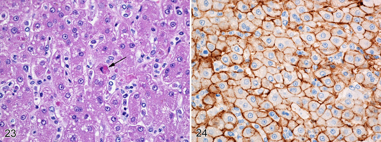
Although MARV antigen staining was intense and diffuse in all 6 macaques, the histologic lesions were relatively mild in comparison. Diffuse mild to moderate hepatocellular cytoplasmic vacuolation (interpreted as fatty/lipoidal degeneration) was a consistent finding with sinusoidal congestion and increased numbers of circulating mononuclear and polymorphonuclear cells (Fig. 23). There were occasional nondiscrete collections of inflammatory cells that disrupted hepatic cord architecture and surrounded individual necrotic hepatocytes. Eosinophilic pleomorphic intracytoplasmic viral inclusions were readily observed in degenerate and necrotic hepatocytes in all 6 animals. Low numbers of mostly mononuclear cells infiltrated the portal areas in 4 of 6 animals. IHC examination in all 6 animals revealed diffuse strong MARV antigen staining associated with the sinusoidal lining and hepatocellular membrane surfaces (Fig. 24). Immunolabeling of the cytoplasm of hepatocytes was not observed. With the exception of rare intrasinusoidal granular fibrin deposits in the livers of 2 animals (case Nos. 2 and 3) and fibrin strands observed with PTAH in a hepatic artery of 1 animal (case No. 2), hepatic fibrin deposition was unremarkable.
Kidneys
Histologic examination was performed on the kidneys of 5 of 6 animals. Two animals (case Nos. 2 and 3) exhibited acute degeneration of the proximal convoluted tubules with fibrin thrombi that often occluded medullary vessels. Interestingly, fibrin was observed by IHC in the kidneys of all animals in this study. Fibrin immunoreactivity, most commonly detected at the corticomedullary junction and within medullary vessels, was also occasionally seen in cortical vessels and within glomerular capillaries. Likewise, individual fibrin strands, clumps of fibrin, and/or fibrin thrombi were confirmed by PTAH staining but were not as extensive as those seen by IHC. The most intense PTAH staining was seen in case No. 2. Although the kidneys were not evaluated for MARV immunoreactivity nor were renal function tests performed, the acute renal tubular degeneration with fibrin deposition was most likely due to MARV-Angola infection. We have observed similar renal histologic changes in other MARV-infected cynomolgus macaques, in addition to strong MARV antigen staining in the proximal convoluted tubular epithelium, as well as elevations in serum blood urea nitrogen and creatinine (D.A.A., personal observation).
Adrenal gland
Mild to moderate adrenal cortical congestion with increased numbers of circulating neutrophils was noted in multiple animals. In 3 of 6 animals, variably sized foci of mostly degenerate adrenocortical cells with fewer individual necrotic cells and apoptotic-like debris were observed in primarily the zona fasciculata. As with the kidneys, immunostaining for MARV antigen was not performed.
Gastrointestinal tract
The esophagus was the only section of gastrointestinal tract submitted for histologic and immunohistochemical examination. Transmurally, nests of immunolabeled squamous epithelial cells were observed in 3 of 6 animals.
Discussion
This study showed that cynomolgus macaques are susceptible to infection by aerosolized MARV-Angola and develop lethal disease characterized by a number of features typical of human infections by MARV and EBOV and their applicable animal models. Prominent among the clinical findings in these macaques was the development of fever in conjunction with the presence of viremia, followed by the onset of anorexia and depression and, later, the development of a typical skin rash. All macaques ultimately became moribund before euthanasia. Key gross lesions included hepatomegaly, pyloric congestion, and the presence of enlarged congested spleens and lymph nodes. Histologically, lymphocyte destruction, loss of hematopoietic elements, hepatocellular and adrenocortical degeneration, and fibrin thrombi, primarily in the lung and renal medullary vessels, were noteworthy lesions. Clearly, the main cellular targets of aerosolized MARV were mononuclear phagocytes—particularly, alveolar macrophages—and hepatocytes. FRCs and some endothelial cells were involved, but the extent of direct infection was less clear. In these terminally infected cases, infected DC-SIGN-labeled DCs were not identified in the mediastinal lymph nodes or spleens. In addition, gross and histological changes were prominent in the lungs. Common findings included the presence of viral antigen in the alveolar septa, as well as the filling of alveolar spaces or expansion of alveolar walls by edema, fibrin, and erythrocytes. Similar histologic and immunohistochemical findings have been seen in the lungs of macaques parenterally infected with other strains of MARV. In general, the pulmonary changes in those animals appeared less severe when compared to those of the MARV-Angola-infected animals (D.A.A., personal observation).
A key finding of this study is that even at relatively low doses (2 to 14 pfu), aerosolized MARV-Angola produced lethal disease in cynomolgus macaques and resulted in a shorter time to death than what has been reported for parenteral (intramuscular) inoculation with substantially higher doses of other MARV strains (1,000 pfu) . 20,21,41 At higher doses, the time to death after infection with MARV-Angola was shorter still and in the same range as parenteral infection with EBOV-Zaire for cynomolgus macaques. Despite the loss of telemetry data between days 4 and 5, the febrile response appears to be roughly 3 to 4 days, with fever onset occurring on the same day as virus detection in the blood (between days 4 and 6). The peak of the fever response was 1 to 2°C higher than that seen with other MARVs or EBOVs (D.S.R., manuscript in preparation). The first major clinical sign of illness was lethargy and anorexia at day 5, followed by a characteristic cutaneous petechial rash observed at day 6, a clinical finding described in macaques infected parenterally with EBOV.
The most consistent pulmonary histologic changes occurred in the interstitium, often resembling an acute to subacute fibrinous interstitial pneumonia, with the strongest MARV antigen immunostaining associated with the alveolar septae. Besides the lung, other major target organs included the secondary lymphoid tissues—specifically, the mediastinal lymph node and spleen—as well as the liver, kidney, and adrenal gland. We did not observe significant histologic lesions in the aorta, tongue, larynx, trachea, thyroid gland, heart, testes/ovary, or brain. Immunostaining confirmed the presence of MARV antigen in the lung, mediastinal lymph node, spleen, and liver of all animals examined.
Surprisingly, only low numbers of alveolar macrophages were immunoreactive despite their increased presence histologically. Furthermore, MARV antigen was strongly associated with, but did not colocalize to, the vWF-positive septal endothelial cells, suggesting that alveolar capillary endothelial cells were not specific cellular targets at least during latter stages of MARV-Angola infection. However, the presence of interstitial and intra-alveolar fibrin indicates endothelium involvement either directly or indirectly at some point during the course of disease. Nonetheless, further examination by electron microscopy is warranted. Although we considered but did not attempt to identify type 1 alveolar pneumocytes as a specific cellular target, our findings are similar to those seen histologically and immunohistochemically in the lung of EBOV-infected NHPs and guinea pigs. 9,13,30,32,46
Another unique finding was the loss of nuclear IL-33 expression in pulmonary arteriolar endothelial cells. IL-33, a novel member of the IL-1 family of proinflammatory cytokines, 50 has been shown to function as a ligand for the IL-1 receptor-related protein ST2, a receptor expressed on mast cells, T-helper type 2 lymphocytes, and cardiomyocytes. 62,74,75 Initially discovered as being expressed in HEVs from lymphoid organs in humans, nuclear expression of IL-33 has since been shown to be constitutively expressed in FRCs of lymphoid tissue; certain types of epithelium (particularly, those exposed to the environment), including skin keratinocytes and epithelial cells of the stomach, tonsillar crypts, and salivary glands; and endothelial cells from both large and small blood vessels in most tissues. 50,62 A unique involvement in the proinflammatory response makes IL-33 an attractive cytokine for exploration into the pathogenesis of both MARV and EBOV—particularly with its innate ability to drive T-helper type 2 responses in lymphocytes 75 and act as a T-helper type 2 chemoattractant 48 and with its nuclear localization to FRCs, endothelial cells, and certain epithelial cells. Here, our finding supports those of Kuchler et al, 50 who demonstrated that IL-33 is a nuclear factor that is generally expressed in resting endothelial cells in healthy tissues but is rapidly downregulated during angiogenic events that appear to involve exposure to proinflammatory cytokines, proangiogenic vascular endothelial growth factor, or loss of cell–cell contacts.
Unlike what has been reported in rhesus macaques challenged intramuscularly with 1000 pfu of MARV-Angola, 33 our study found that lymphopenia was not prominent in cynomolgus macaques after exposure to aerosolized MARV-Angola. This should not be taken to imply that lymphopenia does not occur. A prior study with cynomolgus macaques parenterally infected with MARV-Ci67 also found that lymphopenia was not as prominent or as rapid as that seen with EBOV. 30 The histopathology and TUNEL results in our study unequivocally showed extensive lymphocytolysis and lymphoid depletion in the lymph nodes, spleens, and tonsils of all 6 MARV-Angola-infected macaques, with variable vascular necrosis (particularly in the mediastinal and tracheobronchial lymph nodes), hemorrhage, edema, and congestion. Similar nodal findings have been described in rhesus macaques challenged with EBOV by aerosol. 45
Extensive pathogenesis studies conducted in NHPs have shown that cells of the mononuclear phagocyte system—particularly, DCs in lymphoid tissues—are early and sustained targets of EBOV infection 34 and likely play a role in the pathogenesis of MARV infection as well. Furthermore, resident macrophages and interstitial fibroblasts and fibroblast-like cells (to include FRCs) in various tissues are infected with EBOV. 19,23,34,36,43,72,80,81 However, we found that after inhalational MARV-Angola infection, there was a dramatic decrease in the number of DC-SIGN-labeled DCs in the mediastinal lymph nodes by day 8 postexposure and that viral colocalization was not a feature. DCs may be infected early during MARV-Angola infection, but they do not appear to be sustained targets. Increased MAC387-positive circulating and parenchymal monocytes/macrophages and granulocytes were observed, but MARV colocalization was rare and, even then, questionable. Similar results were observed in the spleen.
In our study, the intense MARV immunostaining associated with the HEVs and the adjacent perivenular punctate-to-linear staining pattern that extended throughout the cortex and paracortex suggested involvement with the underlying reticulin framework and, possibly, FRCs. FRCs in T-cell-rich areas, such as follicular marginal zones and the paracortex, continued to express nuclear IL-33 although expression was lost in the HEV endothelium. However, MARV antigen did not colocalize to the IL-33-positive FRCs. Our study therefore suggests that (1) as with the lung, nuclear IL-33 in HEV endothelium is lost upon angiogenic or proinflammatory activation and (2) FRCs are not specific cellular targets in the lymph node during the terminal stages of MARV-Angola infection. Likewise, although we were surprised with the paucity of IL-33-positive FRCs in the spleen of MARV-Angola-infected NHPs, those that were present were consistently immunonegative for MARV antigen. This is not to say that FRCs are not involved in the pathogenesis of MARV infection; that is, they are likely indirectly involved, as we discuss below.
In the lymph node, FRCs line lymph sinuses, are present throughout the interstitium or parenchyma, and surround the HEVs. 18,23,60,64,84 FRCs are elongate cells closely associated with reticular fibers, and they are present in all lymphoid tissues, wherein they provide a supporting scaffold, define the T- and B-lymphocyte compartments, direct the movement of fluid and cellular constituents, provide homeostatic factors for T lymphocytes and a substrate for lymphocyte migration, and interact directly with T and B lymphocytes, natural killer cells, and DCs. 63,81 Therefore, not only are FRCs central to the microarchitecture of secondary lymphoid organs such as the lymph nodes and spleen, but they also play significant roles in the host’s ability to develop an effective adaptive immune response and contribute to immune homeostasis. 63
Lymphocytes enter the lymph node by binding to and crossing the luminal surface of the high endothelium, and they emerge into a narrow, potential space that lies between the abluminal side of the endothelium and the surrounding pericytic cells, which are also considered FRCs. 2,37,38,91 This narrow potential space, bounded by at least 2 layers of overlapping pericytic-like FRCs (see Fig. 14A), is referred to as the perivenular channel. 2,3,37,38 Once lymphocytes emerge from the perivenular channel, they enter fluid-filled corridors bounded by extracellular matrix components—particularly, fibronectin, laminin, collagen IV, and tenascin (excellent substrates for lymphocyte migration)—as well as FRCs that enclose collagen-containing reticular fibers. 37,38,47,49,82,85 These collagen fibers are separated into their own extracellular space, or “conduit” surrounded by the cell processes of an FRC. Conduit therefore refers to the extracellular space but not the surrounding FRCs themselves. 37,38,80 Given the unique immunostaining pattern and the absence of MARV colocalization to FRCs, we propose that the uptake of MARV antigen likely occurs in the FRC conduit and perivenular channels and that direct FRC infection by MARV is a rare event, if it occurs at all. The intense splenic MARV staining pattern along the basement membranes of venous sinuses and at the periphery of PALS—both areas rich with reticulin fibers—is reminiscent to that observed in the mediastinal lymph node and is suggestive of non-cell-associated viral deposition.
In the spleen, red pulp necrosis and cellular depletion of hematopoietic elements, including megakaryocytes, were noteworthy findings. Similar splenic findings have been observed in the MARV mouse model. 88,89 This histologic finding correlates well with the clinical observation that MARV-Angola did cause a significant depletion of platelets, which were replaced over the course of the infection with larger, more immature platelets. These changes are consistent with the development of the coagulopathies that are a hallmark of viral hemorrhagic fevers—particularly, those caused by filoviruses. 10,35 Additionally, the role that EBOV-infected monocytes and macrophages play in regard to induction of certain soluble factors that promote coagulopathy has been well documented. 4,80 Evidence suggests that EBOV-activated and EBOV-damaged FRCs contribute to the coagulopathy because exposed collagen in the fibroblastic reticulum promotes clotting. 63,80 As with EBOV, the mechanisms promoting coagulopathy and fibrin deposition in the tissues of MARV-infected patients are most likely multifactorial, highlighting the need for further clinical and molecular investigation. Nonetheless, we have clearly shown that, although varied among animals, fibrin was present in the kidney, lung, spleen, and, rarely, liver.
In comparison to its more well-characterized cousin EBOV, the published literature on the pathogenesis of MARV is limited. Here, we report the initial development of a NHP model for aerosol exposure to MARV-Angola by describing the clinical, gross postmortem, and histopathologic findings, as well as the distribution of viral antigen and fibrin deposition in multiple organs. Our findings demonstrate that aerosolized macaques exhibit a number of features similar to those of human filovirus infections. Thus, this model could prove to be useful as a surrogate for aerosolized MARV-Angola in humans, to be applied to the development of medical countermeasures under the Animal Rule. Furthermore, we have shown that (1) IL-33 nuclear expression is found in the endothelial cells of pulmonary arterioles, in HEVs, and in FRCs in the lymph nodes of noninfected cynomolgus macaques; (2) IL-33 nuclear expression is lost in the pulmonary arteriolar and HEV endothelium of MARV-Angola-infected macaques; and (3) IL-33-positive FRCs in the lymph node do not appear to be directly infected with MARV. Nevertheless, continued investigation, particularly at the ultrastructural and molecular level, is warranted to further characterize MARV pathogenesis and the pathologic similarities and differences among MARV, EBOV, and other lethal hemorrhagic fever viruses. Further characterization of the human infections themselves is also necessary to provide a better basis for determining which animal models can suitably be used under the Animal Rule.
Footnotes
Acknowledgements
We also acknowledge the technicians and veterinarians of the Veterinary Medicine Division at the US Army Medical Research Institute of Infectious Diseases for implantation of the telemetry devices, collection of blood samples, and administration of euthanasia; Adam Hedge and Ty Hunter for their assistance in the aerosol exposures; Jason Buck for processing the complete blood count samples; and Dr Thomas Geisbert and Mrs Joan Geisbert for providing the challenge virus. Special thanks to the technical staff Jeff Brubaker, Neil Davis, and Gale Kreitz for providing outstanding pathology support; Colonel Keith E. Steele, DVM, PhD, Diplomate ACVP, for his detailed review of the manuscript; and Dr Gordon Ruthel and Mr William Discher for confocal microscopy and visual assistance. The views, opinions, and/or findings contained herein are those of the authors and should not be construed as an official Department of Army position, policy, or decision unless so designated by other documentation.
The authors declared that they had no conflicts of interest with respect to their authorship or the publication of this article.
We acknowledge that this work was supported by funding from the Defense Threat Reduction Agency, Project No. XX0009 06 RD B.