
Abstract
Malignant lymphoma has become an increasingly recognized problem in African lions (Panthera leo). Eleven African lions (9 male and 2 female) with clinical signs and gross and microscopic lesions of malignant lymphoma were evaluated in this study. All animals were older adults, ranging in age from 14 to 19 years. Immunohistochemically, 10 of the 11 lions had T-cell lymphomas (CD3+, CD79a–), and 1 lion was diagnosed with a B-cell lymphoma (CD3–, CD79a+). The spleen appeared to be the primary site of neoplastic growth in all T-cell lymphomas, with involvement of the liver (6/11) and regional lymph nodes (5/11) also commonly observed. The B-cell lymphoma affected the peripheral lymph nodes, liver, and spleen. According to the current veterinary and human World Health Organization classification of hematopoietic neoplasms, T-cell lymphoma subtypes included peripheral T-cell lymphoma (4/11), precursor (acute) T-cell lymphoblastic lymphoma/leukemia (2/11), chronic T-cell lymphocytic lymphoma/leukemia (3/11), and T-zone lymphoma (1/11). The single B-cell lymphoma subtype was consistent with diffuse large B-cell lymphoma. Feline leukemia virus (FeLV) and feline immunodeficiency virus (FIV) testing by immunohistochemistry on sections of malignant lymphoma was negative for all 11 lions. One lion was seropositive for FeLV. In contrast to domestic and exotic cats, in which B-cell lymphomas are more common than T-cell lymphomas, African lions in this study had malignant lymphomas that were primarily of T-cell origin. Neither FeLV nor FIV, important causes of malignant lymphoma in domestic cats, seems to be significant in the pathogenesis of malignant lymphoma in African lions.
Malignant lymphomas have been extensively studied in domestic canids and felids, but little research has been carried out on their exotic counterparts. Exotic felids in captivity, particularly African lions (Panthera leo), are not uncommonly diagnosed with malignant lymphomas. 2,7 However, neither immunophenotypical characterization nor histomorphological classification according to the current World Health Organization (WHO) on standards is routinely performed. Although lymphomas in domestic felids have been associated with feline leukemia virus (FeLV) and feline immunodeficiency virus (FIV), 1 there is only 1 documented case of lymphoma in a captive African lion that was concurrently diagnosed with feline lentivirus. 12 The purpose of this retrospective study was to morphologically classify lymphoma subtypes observed in African lions using the current veterinary and human classification system of the WHO. Furthermore we wanted to determine any common clinical or morphologic features observed among affected lions and compare the findings with the general, published knowledge relating to lymphoma cases in domestic cats and other exotic felids.
Methods
Eleven cases of malignant lymphoma were identified in African lions after consultation with 22 zoological institutions in the United States. All cases of lymphoma in these lions were diagnosed during the years 1986–2003. Complete medical histories and formalin-fixed paraffin-embedded tissue blocks (5 lions) or 5-μm tissue sections on unstained glass slides (6 lions) were obtained for each animal included in the study and submitted to Michigan State University Diagnostic Center for Population and Animal Health. Sections 5 μm in thickness were prepared from formalin-fixed paraffin-embedded tissue samples from each submitted block. One slide from each lion was routinely stained with hematoxylin and eosin (HE). Additional slides for all lions were processed routinely for immunohistochemistry (IHC) for B-cells (CD79a) and T-cells (CD3) as previously described. 10 Normal lion lymph node sections were used as positive controls; for negative controls, primary antibodies were replaced with homologous nonimmune sera. Both HE and immunohistochemically stained slides were reviewed and classified, respectively, using the current veterinary and human WHO classification system and as T-cell or B-cell lymphoma by 2 independent pathologists (MK, VEV). Additional slides from each lion were labeled immunohistochemically for FeLV and FIV using previously published methods. 11,15
Data pertaining to sex, age at diagnosis, clinical signs (splenomegaly, weight loss, and other clinical signs), serological status for FeLV and FIV, organs affected (ie, tumor topography) at the time of necropsy, and lymphoma immunophenotype were compared using chi-square statistical analyses. A P value of <.05 was considered significant.
Results
Of the 11 lions included in the study, 9 were male and 2 were female. Ages for 10 of the 11 lions ranged from 14 to 19 years (exact age was unknown for 1 lion) at the time of lymphoma diagnosis with a mean age of 16.5 years (±1.8 years) and a median age of 17 years. Clinical signs consisted of weight loss (5 lions, 45%) and splenomegaly found on physical examination (9 lions, 81%). Signalment and clinical signs are summarized in Table 1. All cases of lymphoma in these lions were diagnosed during the years 1986–2003.
Summary of Signalment, Clinical Signs, and Morphology of Malignant Lymphomas in 11 African Lions
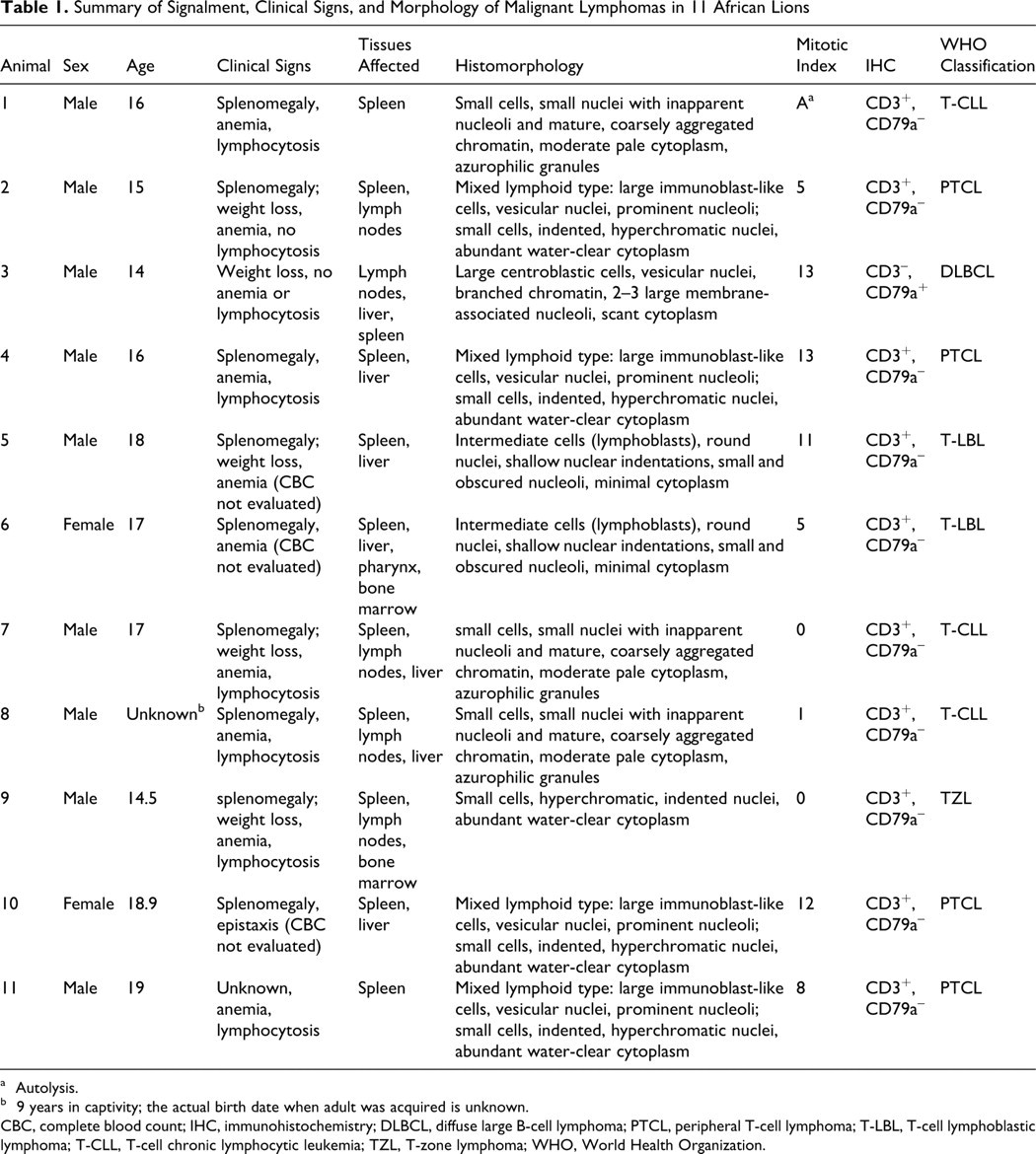
a Autolysis.
b 9 years in captivity; the actual birth date when adult was acquired is unknown.
CBC, complete blood count; IHC, immunohistochemistry; DLBCL, diffuse large B-cell lymphoma; PTCL, peripheral T-cell lymphoma; T-LBL, T-cell lymphoblastic lymphoma; T-CLL, T-cell chronic lymphocytic leukemia; TZL, T-zone lymphoma; WHO, World Health Organization.
Three animals were diagnosed with lymphoma after being found dead. The remaining animals were anesthetized for a physical examination that was either a routine examination or was performed to diagnose abnormalities. Lymphoma was diagnosed using various methods, including a complete physical examination, complete blood count, blood chemistry, ultrasound, fine needle aspiration, biopsies, and radiographs. Nine of the 11 animals had evidence of mild to severe anemia, and 6 of the 11 animals had lymphocytosis suggesting possible leukemia. Two of the 11 animals did not show lymphocytosis, and the remaining 3 animals were not evaluated. Bone marrow was microscopically examined in 6 of the 11 animals, with only 2 animals, lions (No. 6 and 9) having histological evidence of neoplastic lymphoid cells in the bone marrow.
Three animals died the day after a physical examination was performed due to complications from splenectomy (n = 2) or complications relating to ovariohysterectomy and enterotomy (n = 1). Five animals diagnosed with lymphoma were treated with various steroids including prednisone, dexamethasone, stanozolol, and Solu-Delta-Cortef and survived 1–504 days, with the longest survival associated with inclusion of a chemotherapeutic protocol. 7 Three lions were treated with chemotherapy protocols, of which 2 animals were treated with chlorambucil and 1 animal was treated with lomustine and adriamycin. One animal treated with chlorambucil survived 1 day, and the other animal survived 210 days. The animal treated with lomustine and adriamycin survived the longest at 504 days. 7 Tissues for microscopic examination were collected during surgery or 1–24 hours after death.
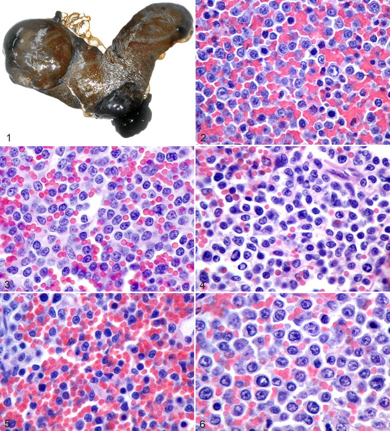
Malignant lymphoma was most commonly observed in the spleen (Fig. 1) in 10 lions (91%). Other affected tissues included the liver (7 lions, 64%), lymph nodes (5 lions, 45%), pharynx (1 lion, 9%), and bone marrow (2 lions, 18%). The tumor topography and the histomorphologic and immunohistochemical findings are summarized in Table 1.
All malignant lymphomas were classified using the current veterinary and human WHO classification of hematopoietic neoplasms. 6,17 Ten lions had T-cell lymphomas (CD3+, CD79a–) and 1 lion was diagnosed with a B-cell lymphoma (CD3–, CD79a+).
Four malignant lymphomas were classified as peripheral T-cell lymphomas (PTCL) (lion Nos. 2,4,10, and 11). These neoplasms were markedly heterogeneous with small foci of necrosis surrounded by mixed inflammation. Neoplastic cells varied from large immunoblast-like cells with vesicular nuclei and prominent nucleoli to fewer small cells with hyperchromatic, indented nuclei, and abundant water-clear cytoplasm (Fig. 2). Anisokaryosis and poikilocytosis were common. A fine, cohesive fibrovascular network was observed in all 4 lions, and the mitotic index was moderate to high (5–13/hpf).
Two malignant lymphomas were identified as T-cell lymphoblastic lymphomas (T-LBL) and were composed of diffuse sheets of lymphoblasts of intermediate nuclear size (lion Nos. 5 and 6). These cells had minimal cytoplasm and round nuclei with less distinctly stained chromosomes and shallow nuclear indentations and small and obscured nucleoli (Fig. 3 ). Both T-LBL had a high mitotic index (5 and 11/hpf), and an occasional “starry-sky” appearance was observed. Bone marrow was observed to be affected in lion No. 6, strongly suggesting the presence of a leukemic state.
Three malignant lymphomas were classified as chronic T-cell lymphocytic leukemia (T-CLL), a slowly progressive T-cell disease (lion Nos. 1, 7 and 8). These cases were characterized by small cells with a moderate volume of pale cytoplasm with variable numbers of azurophilic granules and small-sized nuclei with inapparent nucleoli and mature, coarsely aggregated chromatin (Fig. 4 ). Two of these 3 neoplasms had a low mitotic index (0 and 1/hpf), whereas the third could not be evaluated because of autolysis. Under the Revised European-American Lymphoma (REAL) classification these cases would have been characterized as T cell chronic lymphocytic leukemia/prolymphocytic leukemia (T-CLL/PLL). However, the WHO classification eliminated the T-CLL component, leaving only the entity of T cell prolymphocytic leukemia (T-PLL). 4 Based on the clinical presentation, hematology and microscopic examination of peripheral blood and affected tissues of the these 3 cases, a diagnosis of T-CLL appeared most appropriate. 4 However, the lack of bone marrow examination considerably weakens such a diagnosis.
One malignant lymphoma was classified as a T-zone lymphoma (lion No. 9). This is a specific entity within the category of peripheral T-cell lymphomas that according to the Kiel classification commonly exhibits an indolent behavior as has been previously reported in an African lion. 7 The neoplasm was composed mainly of cells with small nuclei and abundant water-clear cytoplasm (Fig. 5 ). Nuclei commonly had shallow membrane indentations with dense chromatin, obscure nucleoli, and rare mitotic figures (<1/hpf). Neoplastic cells expanded the paracortex without effacing the medullary architecture or subcapsular sinuses and resulted in peripheralization of B follicles toward stromal trabeculae. Postcapillary venules were highly prominent.
The single B-cell lymphoma was consistent with a diffuse large B-cell lymphoma (DLBCL) (lion No. 3). The neoplasm was composed of a relatively uniform population of large centroblastic cells with vesicular, uniformly shaped nuclei and scant cytoplasm (Fig. 6). Nuclei contained branched chromatin and had 2–3 large nucleoli that were closely associated with the nucleolemma. Mitoses were frequent at 13/hpf, and tingible-body macrophages were randomly distributed throughout the neoplasm.
FeLV and FIV were not detected in sections of malignant lymphoma from any of the lions in this study using immunohistochemistry. Latent infection or infections with FeLV or FIV strains that may not be detectible by the antibodies used in this study cannot be excluded. According to the provided zoo records, 1 female lion (lion No. 6) had antibodies against FeLV by serology. All lions were negative for antibodies against FIV; however, no information regarding the test format was provided.
Discussion
Malignant lymphoma is the most common malignant neoplasm diagnosed in domestic cats. 3 –9 Malignant lymphomas have also been diagnosed with some frequency in captive exotic felids, especially lions. 2,7,12 The majority of the lions in this study were adult, male lions that were older than 14 years. All lions in this study were captive born and not thought to be related, although exact genetics could not be determined. Most lions were diagnosed at an advanced stage of disease precluding successful treatment, so the survival time of lions with malignant lymphoma was usually short. Weight loss and splenomegaly were the most commonly observed clinical signs, and malignant lymphoma should be considered as a differential diagnosis in lions with similar clinical presentation.
The cause of malignant lymphomas in many species is unknown. An association between FeLV or FIV and malignant lymphoma has been established in domestic cats. 1 In previous years, approximately 70% of domestic cats with malignant lymphomas have shown FeLV antigenemia, but this rate varies with the anatomic form and subtype of lymphoma as well as the strain of virus. In contrast, FeLV has not been detected in free-ranging African lions. 8,16
There is also evidence that FIV plays a role in the development of malignant lymphomas in domestic cats. 1 A species-specific strain of FIV, FIV-Ple, has been found in captive and free-ranging lions at a incidence as high as 90% in some lion populations. 13,16 However, FIV-Ple has not been confirmed as a cause of malignant lymphoma in lions, and only a single case of small cell lymphoma in a lion with concurrent lentivirus infection has been reported. 12 In the lions reported here, all were negative for FIV by IHC. One animal was serologically positive for FeLV, but all lions were negative for FeLV by IHC. Although FeLV and FIV appear to be inconsequential in the development of malignant lymphoma in African lions, latent infection or other FeLV or FIV strains that may not be detectible through immunohistochemistry could not be ruled out.
DLBCLs are the most common type of lymphoma identified in dogs and cats. In contrast, only 1 case of DLBCL was observed in lions in this study. Interestingly, 10 of the 11 lions in this study had T-cell lymphomas. Furthermore, the spleen appeared to be the primary site of neoplastic proliferation in all T-cell lymphomas. This study indicates that male African lions appear more likely to develop T-cell lymphoma than other felid species, both exotic and domestic. The cause for such an apparently high incidence of T-cell lymphoma in lions compared with other mammalian species is unknown. Although T-cell lymphomas are anecdotally reported to carry a worse treatment prognosis than B-cell lymphoma, it is likely that delayed diagnosis is the primary reason for the poor survival in the animals included in this study. 14
In conclusion, T-cell lymphomas appear to be the most common form of lymphoma in African lions, and possible contributing causes are unknown. Affected animals tend to be older adult males and have clinical signs of splenomegaly and weight loss. There is no apparent viral association related to lymphoma in lions in contrast to reports in the domestic feline population.
Footnotes
Acknowledgements
The authors thank the following zoos for their participation in these feline lymphoma studies: Audubon Zoo, Brookfield Zoo, Buffalo Zoo, Cincinnati Zoo and Botanical Gardens, Detroit Zoo, Ellen Trout Zoo, Granby Zoo, Indianapolis Zoo, Knoxville Zoo, Los Angeles Zoo, Minnesota Zoo, National Zoo, Philadelphia Zoo, Potter Park Zoo, San Antonio Zoo, San Francisco Zoo, Toledo Zoo, Toronto Zoo, Tulsa Zoo, Wildlife Way Station, and the Woodland Park Zoo. The authors also thank Erin N. Wendt for assistance with this project.
The authors declared that they had no conflicts of interest with respect to their authorship or the publication of this article.
The authors declared that they received no financial support for their research and/or authorship of this article.