
Abstract
Objective
To evaluate the effect of a new negative-pressure drainage system in thoracoscopic lung cancer surgery; thereby, providing a new option for postoperative drainage.
Methods
We retrospectively analyzed data for 200 patients who underwent thoracoscopic surgery between May 2018 and October 2019. According to the thoracic drainage method, the patients were divided into the thoracic tube group and the new system group. The epidemiological and clinicopathological data were compared before operation, and the clinical effect of thoracic drainage was compared after operation.
Results
There was no significant difference in epidemiological and clinicopathological data between the two groups. There was also no significant difference in drain removal time, hospital stay, and complication rates between the two groups. However, the incidences of pleural effusion and poor incision healing in the new system group were lower than in the thoracic tube group. Visual analog scale (VAS) scores in the new system group were lower than those in the thoracic tube group at each postoperative interval; therefore, the new system group required less analgesia.
Conclusion
The new system was not inferior to thoracic tubes regarding the drainage effect after thoracoscopic lung cancer surgery. Hence, the system is an alternative to traditional thoracic tubes.
Keywords
Introduction
Lung cancer is the leading cause of cancer-related morbidity and mortality worldwide. The overall incidence of lung cancer has recently increased, and surgery is the main treatment for this disease. 1 With developments in minimally invasive surgery, thoracoscopic surgery has become a standard surgical treatment for lung cancer. Thoracoscopic surgery avoids the trauma caused by traditional open-chest surgery, but achieves comparable treatment efficacy. 2 Placing an indwelling rigid thoracic tube after thoracoscopy remains the standard operating procedure. However, postoperative pain, poor healing of the tube exit wound, effects on pulmonary retention, delayed early ambulation, and other factors are common clinical complications of thoracic tube placement, which is contrary to the principles of rapid rehabilitation surgery that have received widespread attention. 3 With the rise of rapid rehabilitation surgery, the influence of drainage mode selection on postoperative recovery and patients’ quality of life has gained attention. Maintaining effective drainage and reducing patient injury is a popular research area. Traditional silicone tubes with side holes or grooves can drain pleural effusion well through a low-pressure system, but their role in draining chest pneumatosis is controversial, especially with continuous air leakage after lung surgery, which should be treated with chest tubes. Hence, we evaluated the use of a multi-groove silicone tube combined with a unidirectional negative-pressure drainage system in single-operating-port thoracoscopic lung cancer surgery.
Materials and Methods
The clinical data of 200 patients who underwent thoracoscopic lung cancer surgery at our hospital between May 2018 and October 2019 were retrospectively analyzed. One hundred consecutive patients were admitted from May 2018 to February 2019, and traditional chest tubes were placed after surgery. Another 100 consecutive patients were admitted from March 2019 to October 2019, and a multi-groove silicone tube combined with a unidirectional negative-pressure drainage system was used for postoperative drainage. The patients were identified as having malignant pulmonary lesions before surgery according to the eighth edition of the National Comprehensive Cancer Network (NCCN) lung cancer diagnosis and treatment specifications. Inclusion criteria: 1) preoperative chest enhanced computed tomography (CT) showing malignant intrapulmonary lesions, or bronchoscopy biopsy-confirmed malignancy; 2) preoperative whole-body examinations ruled out signs of systemic metastasis, such as with abdominal ultrasonography, bone emission CT scan, whole-body positron emission tomography-CT; 3) patients were able to tolerate surgery according to the results of lung function, blood gas analysis, and clinical lung function evaluation; and 4) frozen intraoperative pathological examination confirmed the diagnosis of lung cancer. Patients were excluded from the study if they were converted to thoracotomy or could not tolerate general anesthesia.
All patients accepted thoracoscopic single-port surgery for the treatment of lung cancer. An approximately 3-cm incision was made in the fourth intercostal space between the axillary midline and anterior axillary line for the operating port for upper lobectomy, and an incision in the fifth intercostal space between the axillary midline and anterior axillary line was made for middle and lower lobectomy. An approximately 1-cm incision was made in the seventh intercostal space on the axillary midline for the observation port. According to the patient’s tumor characteristics, various resection methods were used, such as wedge resection, segmental resection, and lobectomy, with or without systemic lymph node dissection or sampling
After the operation was completed in the new system group, several intermittent grooves approximately 4 mm long and 2 mm wide were made in two soft silicone tubes parallel to the tube body. These tubes were then inserted through the observation port. One tube was located in the posterior mediastinum from the upper diaphragm to the apical chest cavity, and the other was extended through the anterior mediastinum to overlap the previous tube. The overlap area was at the top of the chest cavity, and the tubes formed a closed loop for drainage. If necessary, the overlap area could be adjusted to the surgical area or the main postoperative observation and drainage area, such as the bronchial stump, area of lymph node dissection, or area of postoperative suspected leakage or major drainage. The anesthetist was asked to inflate the remaining lungs for re-expansion. The drainage tubes were fixed in a suitable position, and two monomial negative-pressure bulbs were connected at the ends of the silicone tubes to induce low negative pressure. Meanwhile, in the chest tube group, a traditional 28-F rigid chest tube was inserted through the observation port and advanced to the top of the chest through the posterior mediastinum. The composition and morphology of the drainage tube, intraoperative placement, and postoperative status are shown in Figures 1 to 4.
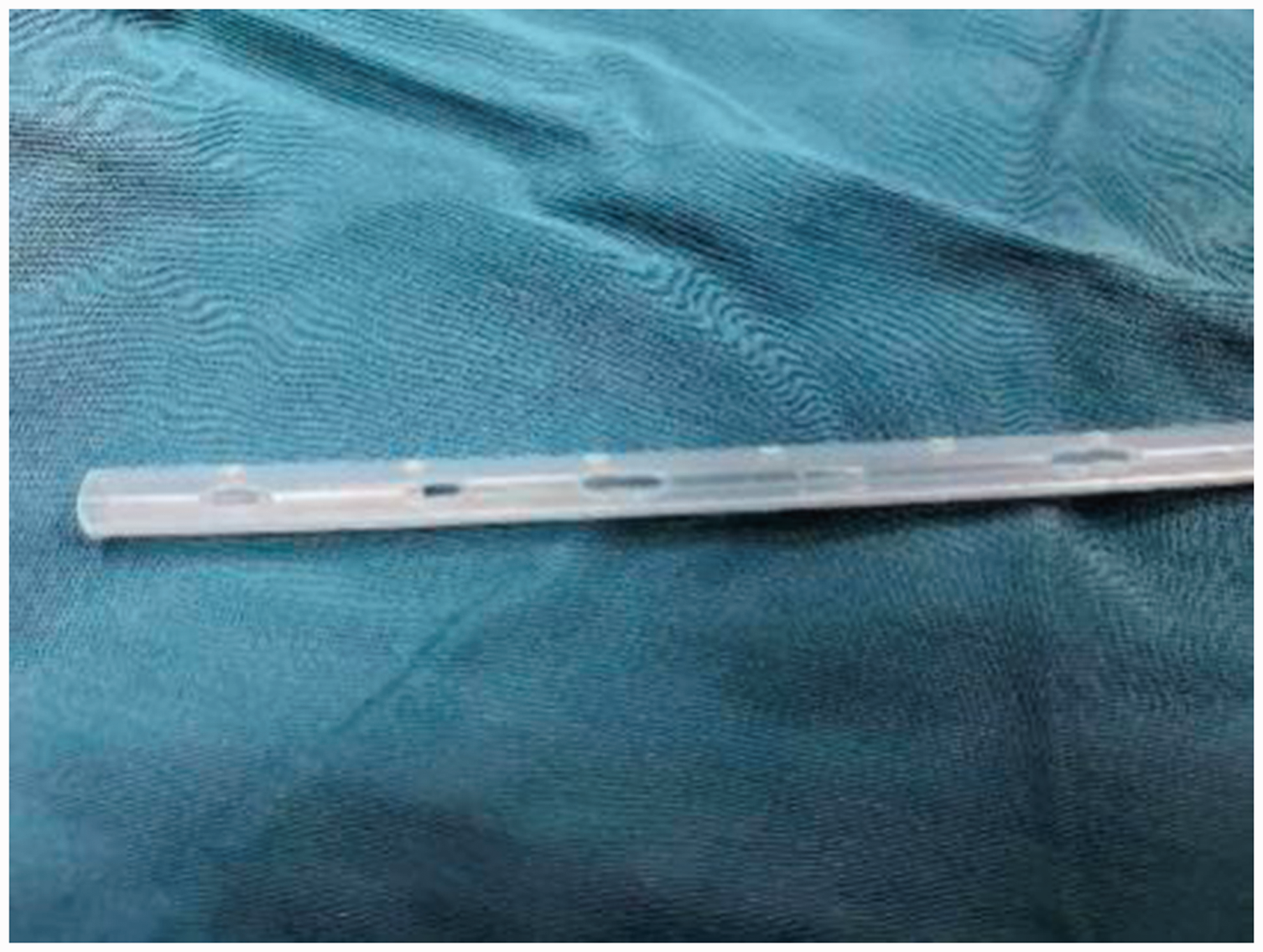
Silicone drain with fine grooves.

Silicone drain and unidirectional negative-pressure bulb.

Placing the silicone drains in the thorax.

The unidirectional negative-pressure bulb drainage system.
Postoperatively, the patients were treated as per routine thoracic surgery protocols. Within 24 hours after the surgery, nurses checked the chest drainage every 2 hours, recorded the drainage volume, and maintained the negative-pressure drainage state in the new system group, and recorded the drainage volume of pleural effusion 24 hours after the surgery. According to the patient’s visual analog scale (VAS) score,4,5 the degree of pain was scored from 1 to 10. The VAS score 24 hours after surgery was recorded by one doctor and one nurse, and the values were averaged for the statistical analysis. Lung re-expansion and the presence of pleural effusion and subcutaneous emphysema were identified by bedside chest radiographs or chest CT examination. VAS scores were recorded 24 hours after surgery, after tube removal, and at discharge, by the doctor and nurse. The total hospitalization duration was also recorded.
The incidence of complications, such as pneumonia, pleural effusion, and poor wound healing were recorded, with poor wound healing defined as incision rupture or not healed within 2 weeks after tube removal. Each administration of 40 mg parecoxib sodium injection for temporary analgesia was recorded.
Discharge indications: imaging examination of the patient indicated that the lungs had re-expanded, blood laboratory test results and body temperature were normal, and no discomfort was reported 24 hours after tube removal. Indications for tube removal: total drainage volume in 24 hours <200 mL, with hemorrhagic or yellowish effusion, no air leakage, and bedside chest radiographs suggested lung re-expansion. Indications for secondary treatment (thoracocentesis (chest tap)): the patient had a chief complaint of discomfort, and imaging indicated atelectasis, pneumothorax, or pleural effusion. All clinical data collection procedures were approved by the Ethics Committee of Changzheng Hospital, and patients gave written informed consent.
All data were analyzed using the statistical analysis software SPSS, version 19.0 (IBM Corp., Armonk, NY, USA). If both groups of data were normally distributed and passed the homogeneity test of variance between groups, then the data between groups could be compared. The measurement data were expressed as mean ± standard deviation (X ± s). Two-sample t-tests were used for comparisons between the two groups. Numerical data were expressed as number (n) and compared by the chi-square test between groups. Fisher’s exact test was used to calculate the p-value if the chi-square test was unsuitable. Two-tailed t-tests were used for all intergroup comparisons, and the significance level was set at p < 0.05.
Results
All data between the two groups were normally distributed. There were no significant differences for age, gender, pathological type, and histological features between the two groups (Table 1). No serious complications, such as pleural hemorrhage and bronchopleural fistula, occurred in either group, perioperatively. The total drainage volume in the chest tube group ranged from 815 to 1117 mL, with a median of 927 mL; the drainage volume in the new system group ranged from 908 to 1235 mL, with a median of 935 mL. The comparison of the clinical data between the two groups showed that the mean postoperative VAS score in the new system group was significantly lower than that of the conventional group at each stage (p < 0.05), and the frequency of analgesic drug use was correspondingly lower, indicating that patients were more tolerant of the new drainage method. No statistical difference was found for drain removal time and hospitalization duration between the two groups (Table 2). There was also no significant difference in the incidence of various complications (i.e., atelectasis, subcutaneous emphysema, and other lung air performance indicators) between the two groups. The new system group had a higher rate of improvement than the thoracic tube group, although the difference was not significantly different.
The patients’ characteristics and operative factors.

*: Fisher’s exact test. Values are presented as mean ± standard deviation or number (%).
ns, not significant.
Postoperative data.

*: Fisher’s exact test. Values are presented as mean ± standard deviation or number (%).
VAS, visual analog scale.
Discussion
Although minimally invasive thoracoscopic surgery has revolutionized thoracic surgery, postoperative indwelling closed thoracic drainage is a “routine procedure”. Hence, postoperative pain, poor wound healing, and infection, which hinder early postoperative recovery, must be resolved. 6 With the recent development of rapid rehabilitation in thoracic surgery, the postoperative management of thoracic drainage tubes and appropriate thoracic drainage have become important issues. 7
Traditional chest tube placement addresses postoperative drainage of chest effusion and lung air leakage, promotes pulmonary re-expansion, and thus, rebuilds the negative pressure in the chest cavity. 8 Previously, chest surgeons routinely placed two chest tubes after surgery, one to drain air toward the top of the chest and the other to drain fluid collecting under the diaphragm. The traditional hard, rigid silicone thoracic tube has the characteristics of retaining form, providing smooth drainage, and high drainage efficiency, which meets the requirements for postoperative drainage after thoracic surgery. 9 However, these characteristics also cause several problems, such as intercostal neuralgia caused by compression, poor incision healing, pulmonary atelectasis caused by continuous stimulation of lung tissue, and multiple negative pressure drainage devices, including the thoracic tubes, prevent early ambulation after surgery. 9 Recent studies have confirmed that a single thoracic tube can achieve similar drainage of effusion and air to that obtained with two thoracic tubes. Even after superior lobectomy, a single thoracic tube is sufficient for postoperative drainage; 10 however, doctors select specific drainage methods according to previous clinical experience and specific surgical conditions. Two-tube thoracic drainage, single-tube thoracic drainage, and a thoracic tube combined with negative-pressure drainage have been reported, of which two-tube thoracic drainage predominates. 11 In this study, a custom-made multi-groove silicone tube combined with a unidirectional negative-pressure bulb was used as the postoperative drainage device (two tubes and bulbs were used; both tubes exited the same port incision). According to the specific operation, patient-specific tube placement was performed in the chest cavity. Two silicone hoses were used to form a closed loop of cross-drainage in the chest cavity.
The analysis of the postoperative clinical data in this study showed that under appropriate perioperative management after thoracic surgery, patients in the new system group showed lower postoperative pain scores than those in the traditional chest tube placement group at each postoperative interval. This difference persisted even 1 month after surgery, which also reduced the frequency of clinical analgesic drugs and thus, the potential risks associated with these drugs, indicating that patients were subjectively more tolerant to the new drainage method. Chest tube-related pain occurs frequently in clinical practice and hinders early postoperative ambulation and active sputum drainage, delays the time to lung re-expansion, and indirectly increases the risk of postoperative pulmonary infection, pleural effusion, and other pulmonary complications. However, owing to the soft texture and strong plasticity, silicone tubes can avoid pain associated with intercostal drainage and indwelling drains, reduce the incidence of poor incision healing caused by soft tissue compression by the drain, and further alleviate patient’s negative psychological effects related to the operation. 12
The new system not only improved patients' drain tolerance but also performed similarly to traditional chest tubes regarding postoperative drainage. Moreover, there was no statistical difference in drain removal time, length of hospitalization, and the incidence of postoperative complications between the two groups. The new system achieved better prevention of pleural effusion, which may be related to the improved tolerance of the drain, and which allowed patients to cough and expel sputum early postoperatively, and to perform lung function exercises to promote effusion discharge. More importantly, the better effects seen with the new system may be related to the system fit and the closed loop. Under such conditions, some areas where a traditional chest tube may not be able to fully drain, such as the apical septum and interlobar fissure, can be continuously and sufficiently drained with the new system.
Another complication of thoracic surgery, namely poor air removal with continuous lung leakage, was not observed in this study. Atelectasis, subcutaneous emphysema, and other lung air performance indicators also showed no difference between the two groups. Previous studies suggested that drainage systems with low negative pressure might not be able to provide air drainage caused by continuous lung leakage; therefore, negative-pressure drainage was used, often in combination with traditional chest tubes. 13 In this study, two multi-groove silicone drainage tubes were used to improve the placement flexibility and drainage efficiency of the whole system. In addition, the connection device at the end of the negative-pressure bulb provided support for negative-pressure drainage in some extreme cases. Previous studies also suggested that continuous low negative-pressure drainage may be more conducive to reducing the incidence of pulmonary complications and accelerate patients’ recovery after lobectomy. 14
This study involved various lobectomies, including upper lobectomy, as well as pulmonary segmental resection, a possible surgical method prone to lung leakage, which matched clinical scenarios. However, larger sample sizes and prospective clinical data are needed to confirm that this new drainage system has similar effects regarding air drainage compared with traditional chest tubes.
According to our clinical experience using this new drainage system and preliminary clinical data analysis, this custom-made multi-groove silicone drain combined with a unidirectional negative-pressure bulb drainage system has the following advantages in single-operating-port lung cancer surgery: First, the patients’ postoperative pain was mild and well-tolerated. Second, this system can be placed easily by a single surgeon. Air and liquid drainage with the new system were not inferior to that with traditional chest tubes, and did not increase the incidence of complications. Third, the system management is convenient, facilitating strong interaction with patients, and the drainage system can be easily managed under guidance and supervision. Fourth, the system is simple to assemble and low in cost, which helps control medical costs. Fifth, the system can be used as a part of thoracic surgery to implement rapid rehabilitation, which has the potential to shorten the length of hospitalization, and reduce hospitalization and treatment costs.11,13 However, this was a retrospective comparative study, and our findings must be confirmed in a large-sample, prospective randomized controlled study or systematic evaluation. This study explored the application of this system only in single-port thoracoscopic lung cancer surgery. Its effect in single-operating port thoracoscopic surgery and other more complicated thoracic endoscopic surgeries, as well as whether this system can replace the traditional thoracic tube as the routine drainage system after thoracic surgery must be explored in subsequent studies.
Footnotes
Author Contributions
Ze Wang and Jian Lv were responsible for the research design and implementation. Si’ang Zhang and Wenjie Chen were responsible for data collection and the statistical analyses. Lei Xue and Bin Wu were responsible for research implementation and article review. All authors read and approved the final manuscript.
Declaration of conflicting interest
The authors declare that there is no conflict of interest.
Funding
This research received no specific grant from any funding agency in the public, commercial, or not-for-profit sectors.