
Abstract
Endophthalmitis is the most serious complication of cataract surgery. A cluster of endophthalmitis is a devastating event for surgeons. Pseudomonas aeruginosa is the main causative pathogen of Gram-negative endophthalmitis, which can be suggestive of the occurrence of an outbreak.
Ten patients diagnosed with endophthalmitis after cataract surgery performed by one surgeon were analyzed in this study. At presentation, five patients had obvious clinical findings of endophthalmitis with visual acuity of light perception, two patients had poor light perception/no light perception of vision complicated by concomitant keratitis, and three patients had earlier signs of infection (e.g., a lower degree of anterior chamber and vitreous cells, better presenting visual acuity, and greater visibility of the fundus). Investigations revealed that the source of infection was growth of P. aeruginosa on the phaco probe. All of the surgeries had been performed by the same contaminated probe without sterilization between surgeries. This finding emphasizes the importance of strict adherence to sterility protocols during high-risk surgeries such as intraocular surgeries. Additionally, this report aims to emphasize to surgeons that negligence of simple but vital steps of sterility for any reason, such as limitations in time or equipment, can lead to catastrophic events.
Introduction
Endophthalmitis may be the most severe complication of cataract surgery, occurring at an incidence rate of 0.07% to 0.12%. The most common causative agents are coagulase-negative staphylococci, which are responsible for approximately 75% of cases. 1 Although Gram-negative bacteria are found in only 6% to 29% of cases, major concerns include their more significant role in endophthalmitis outbreaks, high virulence, and undesirable visual outcomes despite immediate treatment. 2 An outbreak of this complication can be catastrophic for both patients and surgeons. Thus, recognition and correction of the cause are necessary in this situation.
Pseudomonas aeruginosa is the main causative pathogen of Gram-negative endophthalmitis, which can be suggestive of the occurrence of an outbreak. Outbreaks of postoperative endophthalmitis caused by P. aeruginosa have been reported secondary to contamination of surgical solutions, instruments, or equipment. 3 We herein report a P. aeruginosa-related endophthalmitis outbreak after phacoemulsification surgery performed by a single surgeon using one contaminated probe, leading to postsurgical infections and poor visual outcomes despite taking immediate interventions. A brief review of relevant recent reports is also presented.
Case report
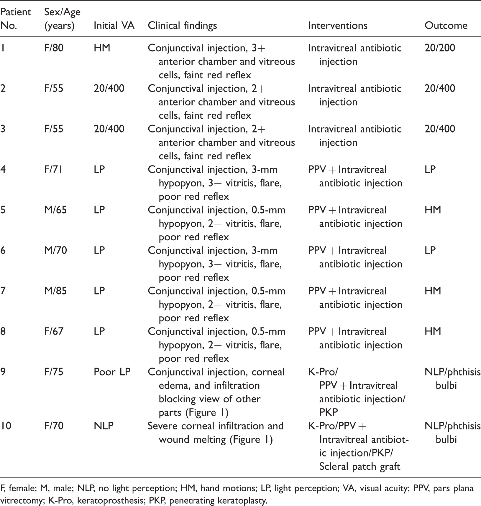
In July 2019, 10 patients diagnosed with acute endophthalmitis were referred to the emergency department of Farabi Eye Hospital. Photographs of two representative patients are shown in Figure 1. All patients had undergone uncomplicated cataract surgery with intraocular lens (IOL) implantation in a private medical center in a nearby city 2 days previously. All of the surgeries had been performed by one ophthalmologist. Three patients were male and seven were female. The patients ranged in age from 55 to 85 years, with a median of 70 years and mean of 69.3 ± 9.6 years. At presentation, five patients had visual acuity (VA) of light perception (LP) with obvious clinical signs of endophthalmitis, including corneal edema, hypopyon, and evidence of vitritis on B-scan echography. Two patients had VA of poor LP or no LP (NLP) complicated by concomitant keratitis. In these patients, severe corneal involvement and infiltration were responsible for the poor presenting VA, and keratoprosthesis (K-Pro) implantation was necessary to obtain a better view of the posterior segment during vitrectomy. The remaining three patients had earlier signs of endophthalmitis with a lower degree of vitritis. No patients had a history of intravitreal antibiotic injection.

Slit-lamp photographs of Patients 9 and 10, who presented with severe conjunctival injection and corneal edema, infiltration, and melting.
The patients were scheduled for immediate vitrectomy or intravitreal antibiotic injection within a few hours. In three patients (Patients 1–3 in Table 1) whose condition was less severe, only intravitreal vancomycin (1 mg) and ceftazidime (2.25 mg) were injected; this treatment is routinely used for patients with endophthalmitis in our department. In the other seven patients who had worse presenting VA and more severe involvement, three-port 23-gauge pars plana vitrectomy was performed by two vitreoretinal surgeons. However, in two patients with corneal involvement, vitrectomy was performed after K-Pro implantation had been completed by a cornea specialist. All of the vitrectomized eyes exhibited perivascular sheathing, hemorrhagic patches, and severe retinal necrosis, which persuaded the surgeons to use silicone oil tamponade. As noted above, none of the patients had a history of intravitreal injection of antibiotics; the perivascular sheathing had occurred secondary to endophthalmitis before the injection of vancomycin. Furthermore, intravitreal ceftazidime and vancomycin were used at the end of the vitrectomy. Vitreous sampling for microbiologic studies was performed in all 10 patients. Table 1 presents a summary of the enrolled patients’ data.
Summary of patient data.
F, female; M, male; NLP, no light perception; HM, hand motions; LP, light perception; VA, visual acuity; PPV, pars plana vitrectomy; K-Pro, keratoprosthesis; PKP, penetrating keratoplasty.
In each patient, an undiluted vitreous sample was taken before starting the vitrectomy and sent to the microbiology laboratory in a sterile container. One part of the sample was used to prepare a smear, and the rest of the sample was immediately inoculated on different culture media.
Microbiological analysis of all patients’ vitreous samples (including three non-vitrectomized patients) showed P. aeruginosa growth. Antibiotic sensitivity tests revealed susceptibility to a variety of antibiotics, including ceftazidime, gentamicin, amikacin, and ciprofloxacin.
The data used in our investigation of the source of infection were obtained via a telephone call to the referring surgeon. Microbiological evaluation was performed on samples obtained from the internal tubes of the phacoemulsification machines, povidone-iodine solution, irrigation solutions, viscoelastic devices, trypan blue, intracameral adrenaline of the same batches used for the surgery, operating microscopes, surfaces of operating tables, instrument trolleys, air-conditioning system of the operating room, and IOLs from the same lot. The evaluations revealed growth of a single organism (P. aeruginosa) on the tip of the phaco probe. The surgeon declared that he had used a single phaco probe for all patients without sterilization between operations.
Major efforts were made to collect follow-up data for as long as possible; the mean follow-up time was 168 days (range, 13–223 days).The two patients who underwent K-Pro implantation plus vitrectomy had VA of NLP at their 6-month follow-up visit, two patients had LP vision, and three patients had a final VA of hand motions. Of the three patients who received intravitreal antibiotics alone, one patient gained best-corrected VA of 20/200, whereas the final VA of the other two patients was 20/400. The disease course did not lead to evisceration or enucleation in any of the patients.
The reporting of this study conforms to the CARE guidelines. 4
Discussion
Endophthalmitis may be the most serious complication of intraocular surgeries such as cataract extraction. A cluster of endophthalmitis is a devastating complication for patients, surgeons, and clinics. Apart from presenting a negative image for surgeons and clinics, it causes many legal troubles, including license revocation for the operating surgeon and the clinic. Interestingly, several reports have described endophthalmitis outbreaks after other ocular procedures such as intravitreal injection of triamcinolone or bevacizumab.5,6 Previous outbreaks of postoperative endophthalmitis have had numerous causes, including defective perioperative eye preparation 7 ; contaminated surgical instruments, ophthalmic solutions, and IOLs; and contamination of the atmosphere of the operation room due to an inappropriate ventilation system. 8 The medicolegal aspects of practice have recently become more considerable. These aspects can involve the patients, physicians, hospitals, and other healthcare workers. Surgeons should be aware of these medicolegal issues and the consequences of incompetence and negligence.
Unusual conditions should raise suspicion about an outbreak, including detection of the same organism (especially an unusual organism) as the causative pathogen, clearly demonstrating the relationship between the cases of endophthalmitis and one member or factor of the surgery. These unusual conditions include an unexpectedly high rate of postoperative endophthalmitis, especially the cases occurred over a very short timeframe.
If an outbreak occurs, the origin of contamination should be sought using microbiological evaluation of samples from suspected sources such as the operating room, members of the surgical team, lenses, solutions, and equipment. Occasionally, however, no obvious or identifiable source is found. In some cases, the cause is an inadequate ventilation system that provides a poor air change rate per hour. In the present study, the phaco probe was found to be the source of infection. Unfortunately, the surgeon had used the same phaco probe without sterilization between surgeries.
Pseudomonas species are not part of the normal flora of the conjunctiva or periocular skin. Thus, most outbreaks may implicate an exogenous source. 1 There are currently 139 articles on postoperative Pseudomonas endophthalmitis. A few reports of endophthalmitis outbreaks caused by P. aeruginosa similar to the outbreak described in the present report are available in the literature. Outbreaks of P. aeruginosa are mostly attributed to contaminated ophthalmic solutions and devices. 2 Because of the high virulence of this bacterium, the visual outcome is usually disappointingly poor despite prompt therapeutic interventions. A summary of recent relevant studies on post-cataract surgery endophthalmitis outbreaks caused by P. aeruginosa is provided in Table 2.
Short review of recent related studies on post-cataract surgery endophthalmitis outbreaks caused by Pseudomonas aeruginosa.

F, female; M, male; VA, visual acuity; HM, hand motions; LP, light perception; CF, counting fingers; NLP, no light perception; PPV, pars plana vitrectomy; IOL, intraocular lens; PCR, polymerase chain reaction.
The visual outcome of our patients was not good. None of the vitrectomized eyes had a final VA better than hand motions. In two patients who underwent K-Pro implantation plus vitrectomy, graft failure occurred and the VA of NLP did not allow for more interventions. Severe chorioretinal atrophy in the necrotic areas and optic nerve atrophy contributed to the poor vision in the other patients. Despite these disappointing visual outcomes, neither evisceration nor enucleation was required in our patients. This is most probably because of the very prompt surgical intervention and vitrectomy within a few hours after admission to our hospital. In the non-vitrectomized patients, the endophthalmitis did not progress, and the intravitreal injection of antibiotics decreased the severity of the endophthalmitis and resulted in fairly good visual improvement. In 2003, Eifrig et al. 13 investigated the treatment outcomes of endophthalmitis caused by P. aeruginosa and found that the initial rate of enucleation or evisceration was 25% (7 of 28 eyes). This finding was not compatible with our study, which can be attributed to the different sample sizes and inclusion criteria. In their study, only 9 of 28 eyes developed endophthalmitis following cataract surgery; the rest of the patients had a history of corneal ulceration (n = 7), penetrating keratoplasty (n = 5), bleb association (n = 2), placement of a glaucoma drainage implant (n = 2), pars plana vitrectomy (n = 1), iris cyst removal (n = 1), and trauma (n = 1). Moreover, the above-mentioned study is relatively old, 13 and great technical and technological improvements in recent years might have led to the better outcomes in the present study.
In conclusion, despite all preventive policies in cataract surgery in the modern era, endophthalmitis is still a potentially catastrophic event. The importance of appropriate sterilization of instruments and devices is highlighted in this report. Other notable points of this study are the role of timely intravitreal antibiotic injection and vitrectomy in the management of postoperative endophthalmitis. Investigation of clusters of endophthalmitis is critical for preventive policy-making.
Footnotes
Availability of data and materials
The data in the study are available from the Farabi Eye Hospital medical records. The data are also available from the corresponding author on reasonable request.
Declaration of conflicting interest
The authors declare that there is no conflict of interest.
Ethics approval and consent
This study was conducted in accordance with the Declaration of Helsinki. All patients provided written informed consent for participation and publication of their personal data. Ethics approval was not required because of the nature of this study (case report).
Funding
This research received no specific grant from any funding agency in the public, commercial, or not-for-profit sectors.