
Abstract
Objective
Pediatric lumbar disc herniation (LDH), although uncommon, causes significant pain, discomfort, and sometimes disability. We examined the efficacy of percutaneous endoscopic lumbar discectomy (PELD) for pediatric LDH and the degree of lumbar disc degeneration at 1 year after PELD.
Methods
We retrospectively reviewed the data of pediatric patients with LDH who underwent PELD from December 2007 to July 2018. The patients’ symptoms, physical examination findings, clinical images, visual analog scale (VAS) scores, Oswestry Disability Index (ODI), and perioperative results (blood loss, length of hospital stay, and complications) were obtained from the medical records. Lumbar disc degeneration was graded using the modified Pfirrmann grading system at the 1-year postoperative magnetic resonance imaging (MRI) examination.
Results
Six boys and four girls who underwent PELD were evaluated. The patients’ mean age was 15.6 years (range, 13–17 years). The mean VAS score for low back pain, mean VAS score for lower limb pain, and mean ODI preoperatively and 1 year postoperatively were 6.2 and 0.3, 6.9 and 0.5, and 20 and 0.1, respectively. MRI showed significant disc degeneration after PELD.
Conclusions
Treating pediatric LDH with PELD is safe and effective. It relieves pain and reduces disability. However, lumbar disc degeneration still occurs.
Keywords
Introduction
Lumbar disc herniation (LDH) is the most common vertebral column disease in advanced-age adults. It leads to back pain, radicular pain, and eventual neurological deficits due to nerve root compression. LDH is rare in children. In previous reports from Spain and Italy, the incidence of pediatric LDH ranged from 0.4% to 3.8% in the pediatric group1,2; however, it was higher (8%–22%) in the Japanese population. 3 Disc degeneration is considered the main factor underlying the development of LDH in adults, while trauma is considered the main etiology of pediatric LDH. Other risk factors for LDH include a family history, high lumbar load, and strenuous physical exertion.4–7
Because of the low incidence of LDH in children, few reports have discussed management of pediatric LDH with minimally invasive surgery. The purpose of this article is to review the literature and discuss the efficacy of percutaneous endoscopic lumbar discectomy (PELD) for pediatric LDH and disc degeneration on magnetic resonance imaging (MRI) after PELD.
Patients and methods
Patients
The study followed the principles outlined in the Declaration of Helsinki and was approved by the hospital’s institutional review board (CE19367A#1), which waived the requirement for written consent from the participants. We retrospectively reviewed the data for patients aged <18 years who underwent PELD performed by a single neurosurgeon from December 2007 to July 2018. The inclusion criteria were dominant single-level LDH verified by MRI, low back and lower limb pain with muscle weakness, and poor response to conservative treatment for at least 3 months.
Surgical technique
All patients were given a prophylactic antibiotic (1000 mg cefazolin sodium; if the patient’s body weight was >80 kilograms, 2000 mg cefazolin sodium was used) within 30 minutes preoperatively.
The interlaminar approach was applied in patients with L5–S1 disc herniation. Surgery was performed with the patient prone on a radiolucent table under intravenous anesthesia with clear consciousness. 8 The skin incision was made in the craniocaudal aspect of the middle of the interlaminar window, as close to the median as possible. A two-channel dilator was bluntly inserted into the lateral edge of the interlaminar window. A working sleeve with an 8.0-mm outer diameter and beveled opening was then directed toward the ligamentum flavum. The rest of the procedure was performed under direct visual control and constant irrigation. A lateral incision window of approximately 6 to 8 mm was made in the ligamentum flavum to expose the neural structures and epidural fat tissue. The working sleeve with beveled opening could be turned and used as a nerve hook. Using the joystick principle, the medial, lateral, cranial, and caudal mobility within the spinal canal could be manipulated to search for and remove the protruding disc using the controlling optics and bipolar system. All operating instruments and the endoscopic system were supplied by Richard Wolf GmbH (Knittlingen, Germany). The high-resolution endoscope had a 4.1-mm intra-endoscopic working channel. The direction of view was 25°. The 8.0-mm outer diameter and beveled opening of the working sleeve together enabled the creation of visual and working fields in an area without a clear, anatomically preformed cavity. In addition, a high-radiofrequency and low-temperature bipolar probe (Elliquence, LLC, Baldwin, NY, USA) was used.
The transforaminal approach was applied in patients with L4–5 disc herniation. Surgery was performed with the patient prone on a radiolucent table under intravenous anesthesia with clear consciousness. The insertion site was localized at the highest lamina level of the symptomatic side using the fluoroscopic lateral view. A spinal cannula was gently inserted into the dorsal aspect of the L4–5 disc. A guiding wire was placed along the spinal cannula, and the spinal cannula was removed. A skin incision of about 8 mm in length was made, and the two-channel dilator was inserted into the disc space along the guiding wire. The working sleeve and endoscope were then inserted as described above. Under direct visual control, the bulging disc was removed and the posterior annulus fibrosus achieved adequate decompression.
Outcome evaluation
A visual analog scale (VAS) and the Oswestry Disability Index (ODI) (Chinese Version 2.1) were used to evaluate the patients’ low back and lower limb pain. The parameters used to grade lumbar disc degeneration on MRI were the disc height index (DHI) 9 and the Modified Pfirrmann grading system. 10 The cross-sectional area (CSA) of the multifidus muscle was obtained to evaluate muscle mass. 11
Statistical analyses
All variables are presented as mean ± standard deviation. Student’s t test was used to analyze the variables, and a p value of <0.05 was considered statistically significant. All analyses were conducted using Microsoft Excel 2010 software (Microsoft, Inc., Redmond, WA, USA).
Results
Patient demographics
A total of 741 patients underwent PELD from December 2007 to July 2018. Among these, 11 pediatric patients were identified (7 boys and 4 girls). One boy was excluded from the study because he had previously undergone microdiscectomy by another surgeon. The remaining 10 patients comprised 6 boys and 4 girls ranging in age from 13 to 17 years (mean, 15.6 years). The patients’ characteristics are shown in Table 1.
Patients’ characteristics.

M, male; F, female; Rt, right; Lt, left.
Symptoms and clinical outcomes
The preoperative symptoms were low back and lower limb pain with muscle weakness and poor response to conservative treatment for at least 3 months. All patients exhibited weakening in their legs, although they had no leg muscle wasting and their muscle tone was normal. The intraoperative blood loss was minimal. The mean postoperative length of stay was 1 day. No complications occurred in this small series. The mean VAS score for low back pain was 6.2 preoperatively and 0.3 at 1 year postoperatively. The mean VAS score for lower limb pain was 6.9 preoperatively and 0.5 at 1 year postoperatively (Figure 1). The mean ODI was 20 preoperatively and 0.1 at 1 year postoperatively (Figure 2). The pain greatly improved after the operation, both in the low back and in the lower limb.

Mean visual analog scale scores for low back pain and lower limb pain.
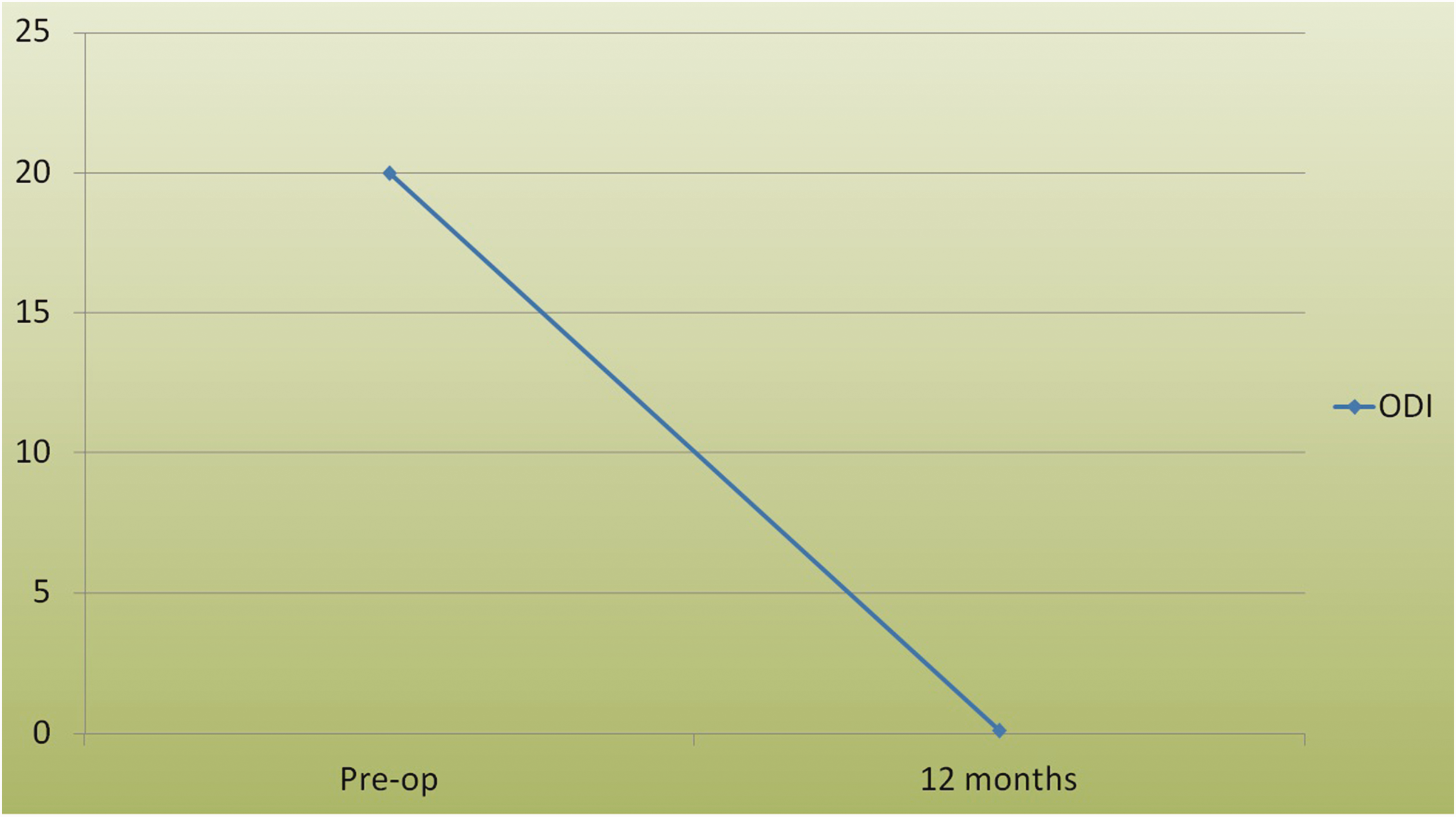
Mean ODI.
Radiological findings of disc degeneration and muscle mass
The DHI was used to evaluate disc degeneration. The DHI was obtained from MRI in our study (Figure 3(a) and (b)). The mean preoperative DHI was 0.2416 ± 0.0265, and the mean 1-year postoperative DHI was 0.2372 ± 0.0328; the decrease in the DHI was not statistically significant.

Details of sagittal measurements. (a) The sagittal view was obtained from the closest section of all sagittal views to the midline of the vertebral body. (b) Measurements used to determine the disc height index (DHI). a: anterior disc height, b: middle disc height, c: posterior disc height, d: sagittal diameter of the overlying vertebral body. DHI = [(a + b + c) / 3]/d.
The modified Pfirrmann grading system was used to evaluate disc degeneration. The mean preoperative and 1-year postoperative modified Pfirrmann scores were 3.0 ± 1.0541 and 4.8 ± 1.3984, respectively (p = 0.005), indicating significant disc degeneration on MRI after PELD.
We also measured the CSA of the multifidus muscle in patients who underwent the interlaminar approach PELD for L5–S1 LDH. The CSA of the multifidus muscle was measured at the supradiscal and infradiscal levels (Figure 4). The preoperative and 1-year postoperative CSA of the multifidus muscle at the supradiscal level on the operative side was 6.735 ± 1.8908 cm2 and 6.7067 ± 1.6549 cm2, respectively, with no significant difference. The average rate of decrease was 1.74%. The preoperative and 1-year postoperative CSA of the multifidus muscle at the infradiscal level on the operative side was 6.65 ± 2.1109 cm2 and 6.74 ± 2.2205 cm2, respectively, also with no significant difference. The average rate of increase was 3.49%. The preoperative and 1-year postoperative CSA of the multifidus muscle at the supradiscal level on the nonoperative side was 7.0967 ± 1.9424 cm2 and 7.46 ± 2.0194 cm2, respectively, with no significant difference. The average rate of increase was 6.12%. Finally, the preoperative and 1-year postoperative CSA of the multifidus muscle at the infradiscal level on the nonoperative side was 6.9583 ± 2.0293 cm2 and 7.1533 ± 2.0930 cm2, respectively, also with no significant difference. The average rate of increase was 4.49%.

Details of cross-sectional area (CSA) measurements. (a) The supradiscal level CSA was obtained from the closest section of all axial views parallel to the lower endplate of the upper level vertebral body. (b) The infradiscal level CSA was obtained from the closest section of all axial views parallel to the upper endplate of the lower level. (c) The CSA of the multifidus muscle was measured according to the area enclosed by the green line.
Discussion
Because of its rarity and unique clinical manifestations, pediatric LDH is usually misdiagnosed or the diagnosis is delayed. LDH leading to pain in the low back and lower limbs is also rare in children. The characteristics of LDH in adolescents are a soft protruded disc, no severe spinal degeneration, typical discogenic pain, a relatively short symptom duration, frequent association with back trauma, and, in some cases, a concomitant degenerative process and bony spur formation.4,12,13 Trauma is considered the main etiology of pediatric LDH. Other risk factors for LDH include a family history, high lumbar load, and strenuous physical exertion.4–7 One study showed that a high number of hours spent sitting significantly increased the prevalence of disc herniation, but episodes of low back pain, smoking status, obesity, and hours spent standing were not significant risk factors. 5
Although trauma is considered the main etiology, many patients do not have a history of trauma. In the present case series, three patients had a history of trauma related to physical exercise, but seven patients denied a history of trauma. In addition, newly developed LDH at the proximal two levels was noted on the 1-year postoperative follow-up MRI examination in one 16-year-old girl (Figure 5). The patient denied trauma or heavy lumbar loading and was very careful during daily activity. According to previous studies, the incidence of LDH differs between groups, and the risk factors include a family history. Patel et al. 14 reported that the first-degree and third-degree relatives of patients with LDH had a relative risk of 4.15 and 1.46, respectively. In another study by Kurihara and Kataoka, 3 the incidence rate reported in Japan was significantly higher than that reported in Caucasian populations. The authors hypothesized that the smaller Japanese skeleton may have a correspondingly smaller spinal canal than in Caucasians, leading to a greater tendency to develop sciatica among Japanese patients. This hypothesis could not explain the development of newly diagnosed LDH in our patient. However, others have shown an association of genetic factors with lumbar disc degeneration.15–17 Multiple genes have been found to be associated with disc degeneration with a moderate level of evidence, including asporin, collagen XI alpha 1, growth differentiation factor 5, sickle tail, thrombospondin 2, and matrix metalloproteinase 9 genes. Another report also mentioned this genetic association. 18 A myriad of genes are involved in separate processes that predispose to LDH. It is estimated that the condition has an approximately 75% heredity origin. Genes found to significantly increase the risk of LDH include those encoding structural proteins, matrix metalloproteinases, apoptosis factors, growth factors, and single-nucleotide polymorphisms in the vitamin D receptor gene resulting in inflammatory cytokine imbalance.

Magnetic resonance imaging (MRI) series in a 16-year-old girl. (a) Sagittal view on preoperative MRI. (b) Axial view of L5–S1 on preoperative MRI. (c) Sagittal view on 1-year postoperative MRI. (d) Axial view of L5–S1 on 1-year postoperative MRI.
The goals of pediatric LDH treatment are to relieve symptoms, allow early return to routine life, and prevent further lumbar disc degeneration. Conservative treatment is the first-line treatment, but the literature shows that it tends to be less effective in the pediatric population than in adults. 19 The efficacy of PELD for pediatric LDH is well established.20–22 However, to the best of our knowledge, no studies have focused on pediatric disc degeneration after PELD. Limited parameters have been used to evaluate disc degeneration, including the DHI and modified Pfirrmann grading system have been used to evaluate disc degeneration.9,10 One study showed a significant decrease in the DHI after conventional microdiscectomy. 9 In the present study, the DHI showed no obvious decrease in pediatric patients who underwent PELD, but significant disc degeneration was still noted according to the modified Pfirrmann grading system. Muscle atrophy after microdiscectomy was also noted in a previous study. 11 Kim et al. 23 reported that less damage to the paraspinal muscle was positively associated with postoperative trunk muscle performance. In the present study, we noted a tendency toward increased muscle mass at the infradiscal level on the operative side after PELD, although no statistical significance was found. A possible reason for this increase is that our study included pediatric patients. Normal growth related to puberty in these patients would cause their muscle mass to increase over time. There was no significant difference in the change in muscle mass between the operative and nonoperative side. Therefore, PELD did not interrupt the normal growth of the multifidus muscle.
This study had two main limitations. First, it was a retrospective study. Second, it included few patients because of the rarity of pediatric LDH. A larger prospective study is needed to establish the efficacy of PELD in pediatric LDH.
Conclusion
The treatment goals for pediatric LDH are to relieve symptoms and prevent further disc degeneration. Conservative treatment is the first-line treatment, but the literature shows that this treatment is less effective in the pediatric population. The role for surgical intervention in pediatric LDH is well-established. Minimally invasive surgery is the preference, and PELD was found to be a safe and efficacious choice in this small series. Further study is needed to establish the genetic association.
Footnotes
Authors’ contributions
R.H.L., H.T.C., and H.K.T. conceived and designed the study; R.H.L., H.C.C., H.C.P., and H.K.T. acquired the data; R.H.L., H.C.C., C.C.C., T.Y.C., and H.K.T. analyzed and interpreted the data; R.H.L. drafted the manuscript; H.C.C., H.C.P., H.T.C., C.C.C., C.Y.T., T.Y.C., and H.K.T. critically revised the manuscript for important intellectual content; R.H.L. and H.K. T. performed the statistical analysis; H.C.P. provided administrative, technical, or material support; and H.C.C. and H.K.T. supervised the whole process. All authors approved the final manuscript.
Availability of data and material
The data used to support the findings of this study are included within the article.
Declaration of conflicting interest
The authors declare that there is no conflict of interest.
Funding
This research received no specific grant from any funding agency in the public, commercial, or not-for-profit sectors.