
Abstract
Objective
This study aimed to investigate the protective effects of naringin on myocardial deformation and oxidative responses in rats with sepsis-induced myocardial dysfunction (SIMD).
Methods
Global and segmental layer-specific longitudinal strain (LS) was assessed by speckle tracking echocardiography. Serum levels of creatine kinase, lactate dehydrogenase, superoxide dismutase, and malondialdehyde were measured. The activity of cleaved caspase-3 was determined by immunohistochemistry. Protein expression levels of Kelch-like ECH-related protein 1 (Keap1), nuclear erythroid factor 2-related factor 2 (Nrf2), and heme oxygenase-1 (HO-1) were measured by western blotting.
Results
Naringin inhibited the lipopolysaccharide-induced decrease in global and layer-specific LS of the left ventricle. Naringin also increased superoxide dismutase expression and decreased malondialdehyde, creatine kinase, lactate dehydrogenase, and cleaved caspase-3 expression in rats with SIMD. Furthermore, naringin increased Nrf2 and HO-1 protein expression levels, and decreased Keap1 protein expression levels in rats with SIMD.
Conclusion
Layer-specific LS analysis of myocardial function by speckle tracking echocardiography can reflect early changes in myocardial systolic function. Naringin may possess a protective effect through moderating lipopolysaccharide-induced myocardial oxidative stress via the Keap1/Nrf2/HO-1 pathway in rats with SIMD.
Keywords
Introduction
Sepsis is a life-threatening organ dysfunction syndrome caused by susceptibility of the host to severe infections. 1 Septic shock and multiorgan failure caused by sepsis are the leading cause of death and critical illness worldwide in the adult intensive care unit. 2 Sepsis-induced myocardial dysfunction (SIMD), caused by bacterial endotoxin lipopolysaccharide (LPS), directly increases mortality of patients with sepsis. 3 Therefore, reducing myocardial dysfunction of sepsis in patients is critical for prolonging life and preventing morbidity.
Several mechanisms of SIMD have been hypothesized, but excessive production of proinflammatory factors and cytotoxic free radicals are the most widely accepted mechanisms. After LPS stimulation, myocardial cells can produce reactive oxygen species, which elicit the oxidative stress response, impairing mitochondrial function. 4 Additionally, LPS can stimulate immune cells and this produces a variety of pro-inflammatory factors. 5 Moreover, overexpression of nitric oxide can affect cardiac L-type calcium channels, inhibit mitochondrial function, and induce apoptosis of cardiac myocytes. 6 Therefore, inhibition of oxidative stress may be an effective treatment for LPS-induced cardiac injury. Heme oxygenase-1 (HO-1) is a heat shock protein that can be induced by a variety of substances or stimulants. HO-1 also has a protective effect on the cardiovascular system. 7 Induction of HO-1 is related to activation of the transcription factor nuclear erythroid factor 2 (NE-F2)-related factor 2 (Nrf2). 8 Under normal circumstances, Nrf2 binds to Kelch-like ECH-related protein 1 (Keap1) in the cytoplasm. When intracellular redox imbalance occurs, free Nrf2 binds to antioxidant response elements in the nucleus. Activation of Nrf2 regulates the biological activity of many phase II detoxification enzymes, such as HO-1. Previous studies have also suggested that activation of the Nrf2/HO-1 pathway attenuates hypoxia-stimulated H9c2 cell apoptosis. 9
Naringin is a natural flavonoid compound found in grapefruit and related citrus plants. Naringin possesses natural antioxidant, anti-inflammatory, anti-apoptotic, and cardiovascular protective effects.10–12 Naringin regulates oxidative stress-induced apoptosis and the inflammatory response through intrinsic and extrinsic pathways, thereby effectively protecting pancreatic β-cells in insulin-deficient diabetic mice. 13 Naringin also ameliorates arsenic-induced cardiotoxicity, which is related to the Nrf2/HO-1 pathway. 14 Previously, we reported the effect of naringin on the inflammatory response of SIMD and its molecular mechanism. 15
Speckle-tracking echocardiography (STE) is an angle-independent ultrasound technique for evaluating global and segmental deformation of the myocardium. A recent development in layer-specific STE analysis allows quantitative analysis of deformation of the myocardium’s three layers, including the endocardium, myocardium, and epicardium. This new sensitive indicator is effective for detecting cardiac dysfunction in various heart diseases, and it may be sensitive enough to detect subtle abnormalities in cardiac function over and above global strain.16–18 We previously reported changes in global layer-specific strain of the left ventricle (LV) in rats with SIMD, followed by the therapeutic effect of naringin. 15 In this study, we aimed to investigate the protective effects of naringin on myocardial deformation and oxidative responses in rats with SIMD.
Methods
Experimental animals
The experimental protocol was approved by the Ethics Committee of Shengjing Hospital of China Medical University (No: 2018PS05K). Adult male Sprague–Dawley rats (HFK Bioscience Co., Beijing, China) aged 8 to 9 weeks and weighing 295 to 305 g were bred in a pathogen-free laboratory (Certificate No: SYXK [Liao] 2017-0004). Rats were housed at 22°C to 24°C with 70% humidity. Rats were kept in a 12-hour/12-hour light/dark cycle for at least 7 days.
Rat models and treatment protocols
Rats were randomly divided into four groups as follows (n = 15 in each group). In the control group, rats were administered physiological saline (same volume as naringin) by oral gavage for 7 days. Approximately 1 hour after the last administration on day 7, rats were intraperitoneally injected with physiological saline (same volume as LPS). In the LPS group, rats were administered with physiological saline (same volume as naringin) by oral gavage for 7 days. Approximately 1 hour after the last administration on day 7, rats were intraperitoneally injected with LPS (Escherichia coli O111: B4; Sigma-Aldrich, St. Louis, MO, USA) at a dose of 10 mg/kg. 19 Naringin treatment groups included the LPS + naringin 50 mg/kg/day (Nar50) group and the LPS + naringin 100 mg/kg/day (Nar100) group. Rats were administered with naringin (Sigma-Aldrich) by oral gavage at a dose of 50 or 100 mg/kg/day for 7 days.20,21 Approximately 1 hour after the last administration on day 7, rats were intraperitoneally injected with LPS (10 mg/kg). Six hours after administration of LPS, 22 the rats were anesthetized, and then the following operations were performed (Figure 1).

Schematic diagram of the experimental procedures.
Conventional echocardiography
For evaluation of echocardiography, we used a GE Vivid E9 ultrasound machine (GE Healthcare, Horten, Norway), which was equipped with a 12S transducer with a frequency of 9 to 12 MHz. The following echocardiographic parameters were measured in each group: left ventricular end-diastolic diameter (LVEDd), left ventricular end-systolic diameter (LVEDs), left ventricular ejection fraction (LVEF), peak early diastolic velocity (E) of mitral inflow, early diastolic velocity at the septal side of the mitral annulus (E′), and the E/Eʹ ratio. These parameters were measured three times by conventional ultrasound and tissue Doppler imaging, and then an average value was taken.
Global and segmental layer-specific longitudinal strain analysis
We acquired five consecutive cardiac cycles of the LV long axis three-chamber view, the apical four-chamber view, and the apical two-chamber view. STE quantitative analysis was performed on an offline software station (EchoPAC V201; GE Healthcare, Horten, Norway). The clearest image of the endocardium was selected and the endocardial contour was manually outlined. The region of interest was adjusted to match the outline of the endocardium. The LV wall was automatically divided into three myocardial layers by the analysis software, which was used to assess the longitudinal strain (LS) of the endocardial layer, the mid-myocardial layer, and the epicardial layer. The LV was divided into 18 myocardial segments, which included six segments (anteroseptal, anterior, lateral, inferior, posterior, and septal) at the basal, mid-ventricular, and apical levels. The peak systolic LS of the three layers of the myocardium were obtained from the three apical views. The global longitudinal strain (GLS) was obtained from the mean values of myocardial deformation at three levels involving the endocardial layer (GLS-endo), mid-myocardial layer (GLS-mid), and epicardial layer (GLS-epi). The mean value of all myocardial segments was represented as ventricular GLS. The transmural gradient was the absolute value of the difference between GLS-endo and GLS-epi.
Intra-observer and inter-observer agreement
Twenty rats were randomly selected and re-measured by two experienced ultrasound physicians who were blinded to the grouping of the rats. Intra-observer reproducibility of GLS-endo, GLS-mid, and GLS-epi was performed by the same ultrasound physician using off-line data at different time points. Inter-observer reproducibility of these parameters was performed by two ultrasound physicians who repeated measurements from the same images.
Estimation of myocardial injury markers
After ultrasonic examination under anesthesia, the chest of the rats was opened to expose the heart. Blood was drawn from the right ventricle into a heparin-coated tube, and plasma was obtained by centrifuging the blood at 1370 × g for 10 minutes at room temperature. Serum levels of creatine kinase (CK) and lactate dehydrogenase (LDH) were determined using an automatic biochemical analyzer (IDEXX Laboratories Inc., Westbrook, ME, USA).
Immunohistochemistry
Myocardial tissues were prepared into 4-µm sections, then deparaffinized and boiled in citric acid buffer (pH 6.0) for antigen retrieval. Primary antibody against cleaved caspase-3 (1:200 dilution; Wanleibio, Shenyang, China) was applied at 4°C overnight. Secondary antibody was applied at room temperature for 25 minutes. After fully dipping sections in phosphate-buffered saline, horseradish enzyme-labeled streptavidin was added for 30 minutes. Finally, 3,3ʹ-diaminobenzidine was used as a chromogen, and hematoxylin was used for counterstaining. Caspase-3 expression was scored by the staining intensity (negative, mild, moderate, or high) and the positive percentage in heart tissue sections as previously described. 15
Analysis of antioxidation
The activity of superoxide dismutase (SOD) in rat serum was determined by the xanthine oxidase method according to the manufacturer’s instructions (Jiancheng, Nanjing, China). Serum samples were taken from a −20°C freezer and thawed on ice. According to the instructions, the reagents were prepared and added to a 96-well plate, mixed, and incubated at 37°C for 20 minutes. After the reaction, the optical density value was measured at 450 nm. An activity unit of SOD was the corresponding enzyme amount when the SOD inhibition rate reached 50%.
Malondialdehyde (MDA) content in rat serum was determined by the thiobarbituric acid method. The reagents were added to centrifuge tubes according to the manufacturer’s instructions (Jiancheng), mixed, and bathed in 95°C water for 40 minutes. The tubes were taken out and cooled with flowing water, and then centrifuged at 1370 × g for 10 minutes. The supernatant of each tube was collected and the absorbance value was measured at 532 nm.
Western blot analysis
Myocardial tissue stored at −80°C was lysed and homogenized in ice-cold radioimmune precipitation lysis buffer, which was supplemented with 1% v/v phenylmethyl sulfonyl fluoride (Beyotime, Shanghai, China). Nuclear protein component was extracted according to the manufacturer’s instructions (Invents, Biotechnologies, Beijing, China). The tissue homogenate was centrifuged at 14,000 × g for 20 minutes at 4°C. Equal amounts of denatured proteins were electrophoresed on sodium dodecyl sulfate-polyacrylamide gel electrophoresis and then transferred to polyvinylidenefluoride membranes (Millipore, Bedford, MA, USA). After blocking with 5% skimmed milk for 2 hours at room temperature, the membranes were then incubated with primary antibodies against Keap1 (1:1000 dilution; Abcam, San Diego, CA, USA), Nrf2 (1:500 dilution; Abcam), HO-1 (1:500 dilution; Abcam), Lamin B (1:1000 dilution; Wanleibio, Shenyang, China), and β-actin (1:10,000 dilution; Proteintech, Chicago, IL, USA) at 4°C overnight and incubated with a secondary antibody (1:20,000 dilution; Proteintech) coupled with horseradish peroxidase. Membranes were finally visualized using an enhanced chemiluminescence kit (GE Healthcare Life Sciences, Pittsburgh, PA, USA).
Statistical analysis
Values are presented as mean ± standard deviation. For all analyses, IBM SPSS 19.0 software (IBM Corp., Armonk, NY, USA) was used. Comparison of data among the four groups was performed by one-way ANOVA. Reproducibility was assessed by Bland–Altman analysis. The level of significance was set at P < 0.05.
Results
Detection of LV deformation by conventional echocardiography in rats with SIMD
Four (6.67%) rats were excluded owing to a poor echo window, including one in the control group, one in the LPS group, and two in the LPS + Nar100 group. Finally, 56 rats underwent echocardiographic analysis. Table 1 shows comparison of conventional echocardiographic parameters among the four groups of rats. There were no significant differences in body weight, heart rate, and E of mitral inflow among the four groups. LVEF and LVEDs were not significantly different in the LPS + Nar50 group compared with the LPS group. LVEF and Eʹ were significantly lower in the LPS group compared with the control group (both P < 0.01). LVEDd, LVEDs, and the E/Eʹ ratio were significantly higher in the LPS group compared with the control group (all P < 0.05), and these were significantly attenuated by naringin administration, especially in the LPS + Nar100 group (all P < 0.05).
Comparison of echocardiographic parameters in rats with sepsis-induced myocardial dysfunction.
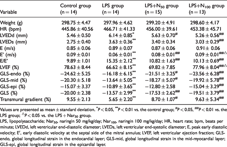
Values are presented as mean ± standard deviation. *P < 0.05, **P < 0.01 vs. the control group; #P < 0.05, ##P < 0.01 vs. the LPS group; △P < 0.05 vs. the LPS + Nar50 group.
LPS, lipopolysaccharide; Nar50, naringin 50 mg/kg/day; Nar100, naringin 100 mg/kg/day; HR, heart rate; bpm, beats per minute; LVEDd, left ventricular end-diastolic diameter; LVEDs, left ventricular end-systolic diameter; E, peak early diastolic velocity; E′, early diastolic velocity at the septal side of the mitral annulus; LVEF, left ventricular ejection fraction; GLS-endo, global longitudinal strain in the endocardial layer; GLS-mid, global longitudinal strain in the mid-myocardial layer; GLS-epi, global longitudinal strain in the epicardial layer.
Changes in layer-specific LS in rats with SIMD
GLS in all three layers and the transmural LS gradient were significantly lower in the LPS group compared with the control group (all P < 0.01). GLS in all three layers and the transmural LS gradient were significantly attenuated by naringin administration, except for GLS-epi in the LPS + Nar50 group (Table 1, Figure 2).
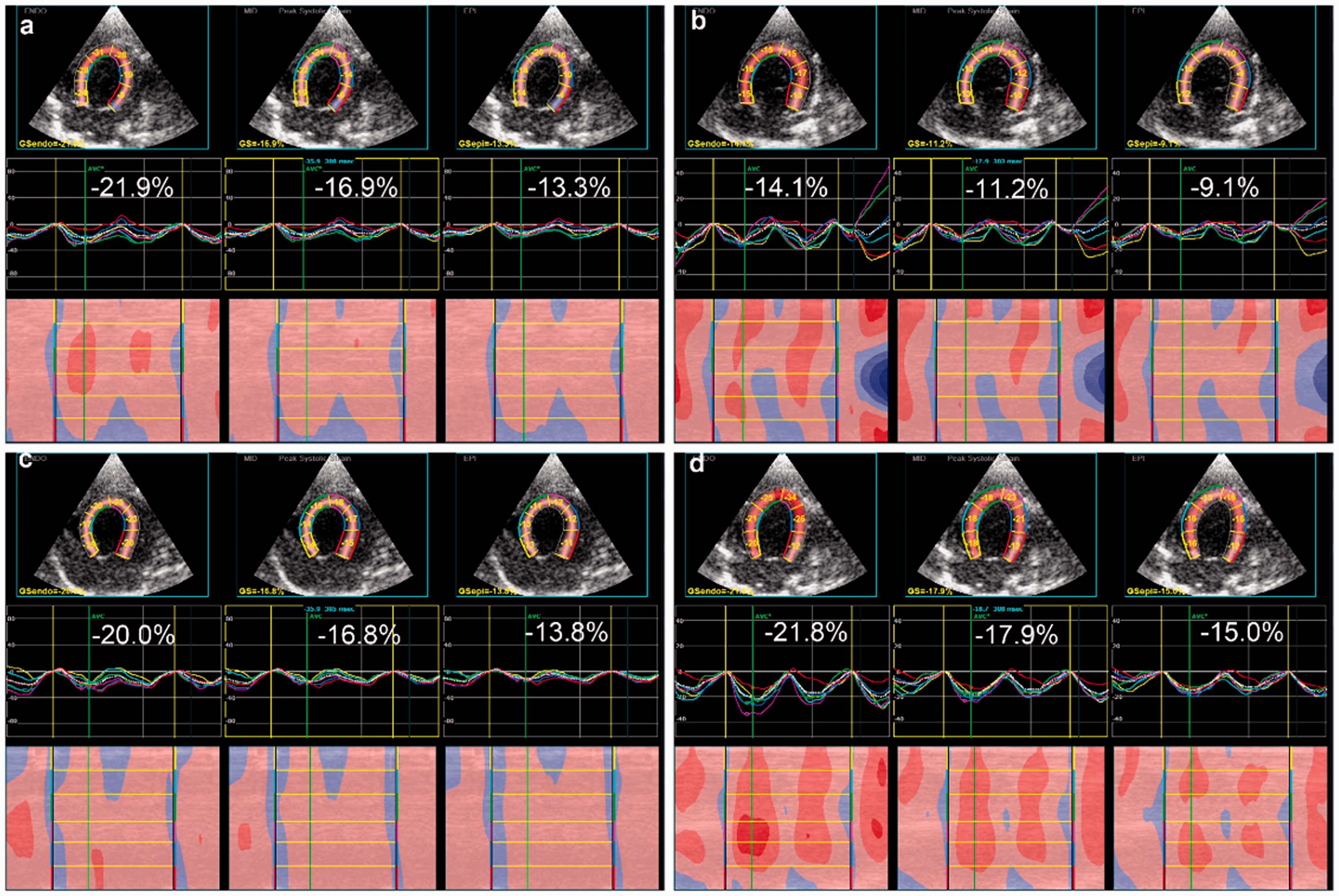
Longitudinal layered strain in the control (a), LPS (b), LPS + Nar50 (c), and LPS + Nar100 (d) groups. Endocardial, mid-myocardial, and epicardial strain is arranged from left to right in each graph. The related strain curves for the six segments within the longitudinal-axis view are shown. Color lines indicate regional strain and white dotted lines indicate the global (average) strain.
Layer-specific LS myocardial deformation parameters at three ventricular levels were evaluated (Table 2). Peak systolic LS-endo, LS-mid, and LS-epi at the LV basal, mid-ventricular, and apical levels were significantly lower in the LPS group compared with the control group (all P < 0.05). These values recovered after naringin administration, especially in the LPS + Nar100 group (all P < 0.05 in the LPS + Nar100 group vs the LPS group). Myocardial strain was highest in the endocardial layer, and gradually decreased from the endocardial layer to the epicardial layer. As shown in Figure 3, peak systolic LS-endo, LS-mid, and LS-epi in the four groups gradually increased from the basal to the apical levels (all P < 0.05), with the highest values in the apical level and the lowest in the basal level.
Effects of naringin on left ventricular three-layer longitudinal strain at the basal, mid-ventricular, and apical levels in rats with sepsis-induced myocardial dysfunction.

Values are presented as mean ± standard deviation. *P < 0.05, **P < 0.01 vs. the control group; #P < 0.05, ##P < 0.01 vs. the LPS group; △P < 0.05 vs. the LPS + Nar50 group.
LPS, lipopolysaccharide; Nar50, naringin 50 mg/kg/day; Nar100, naringin 100 mg/kg/day; LS-endo, longitudinal strain in the endocardial layer; LS-mid, longitudinal strain in the mid-myocardial layer; LS-epi, longitudinal strain in the epicardial layer.

Longitudinal layered strain of the left ventricle at the basal, mid-ventricular, and apical levels in the control group (a), LPS group (b), LPS + Nar50 group (c), and LPS + Nar100 group (d). Values are presented as mean ± standard deviation. ⋆P < 0.05, ⋆⋆P < 0.01 vs. the basal level; ★P < 0.05, ★★P < 0.01 vs. the mid-ventricular level.
The consistency limits of GLS-endo, GLS-mid, and GLS-epi for the intra-observer measurements were −3.92 to 4.50, −2.83 to 3.37, and −1.82 to 2.29, respectively, and those for the inter-observer measurements were −5.04 to 5.64, −5.49 to 5.16, and −3.69 to 4.29, respectively (Figure 4).
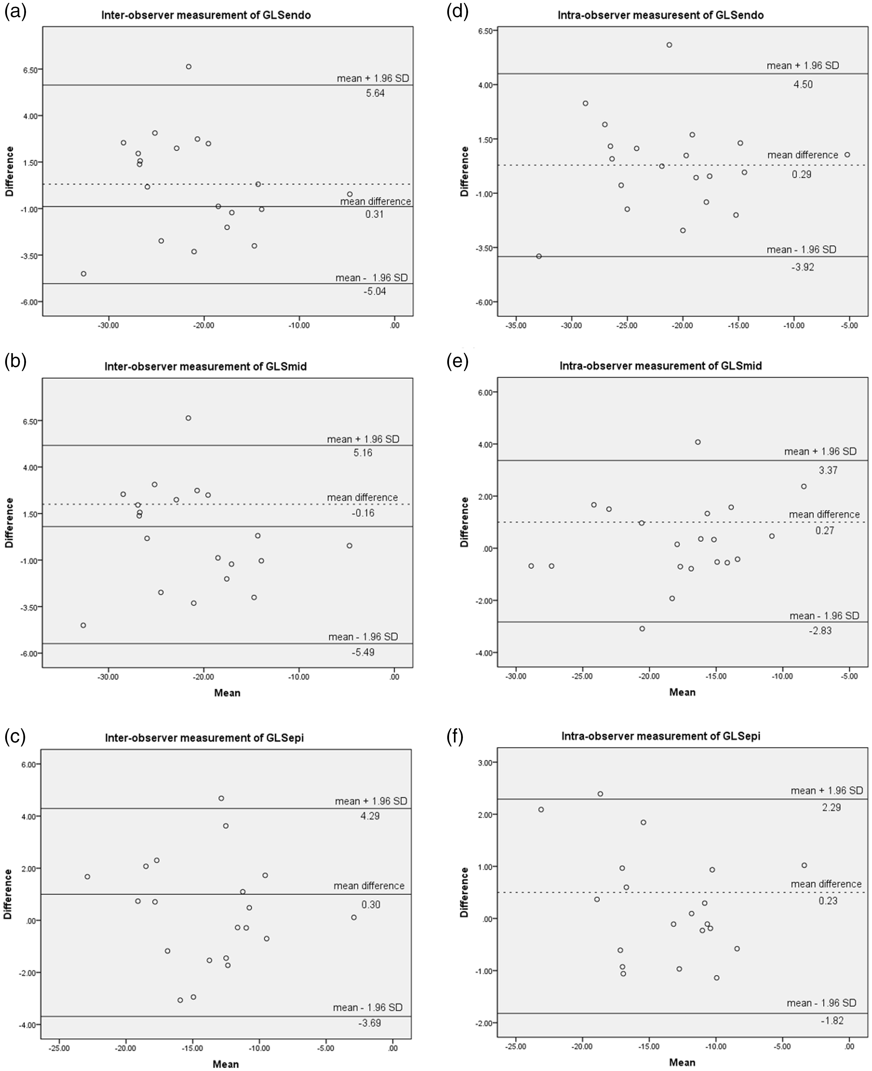
Bland–Altman analyses of GLS-endo, GLS-mid, and GLS-epi for inter-observer (a), (b), and (c) and intra-observer (d), (e), and (f) measurements, respectively.
Naringin alleviates myocardial apoptosis in rats with SIMD
Expression of cleaved caspase-3 activity was significantly higher in the LPS group compared with the control group (P < 0.01, Figure 5). As expected, pretreatment with naringin inhibited the activity of cleaved caspase-3 in a dose-dependent manner (both P < 0.01). These results suggest a role of naringin in attenuating myocardial apoptosis in rats with SIMD.

Immunohistochemistry shows that cleaved caspase-3 levels are decreased in rats with sepsis-induced myocardial dysfunction treated with naringin (light microscopy, hematoxylin and eosin stain, × 400). Control group (a), LPS group (b), LPS + Nar50 group (c), LPS + Nar100 group (d), and immunohistochemical scores of cleaved caspase-3 (e). Values are presented as mean ± standard deviation. **P < 0.01 vs. the control group; ##P < 0.01 vs. the LPS group; △△P < 0.01 vs. the LPS + Nar50 group.
Naringin increases SOD activity and decreases MDA, and CK and LDH levels in rats with SIMD
As myocardial marker enzymes, the myocardial injury parameters CK and LDH are usually used to measure the severity of ischemic heart disease. Expression of CK (Figure 6a) and LDH (Figure 6b) was significantly higher in the LPS group compared with the control group (both P < 0.01). However, naringin pretreatment dramatically reduced CK and LDH levels compared with LPD treatment (both P < 0.01). The activity of SOD (Figure 6c) and MDA (Figure 6d) in serum was examined to verify the antioxidant activity of naringin. SOD activity was significantly lower and the content of MDA in serum was lower in the LPS group compared with the control group (both P < 0.01). Pretreatment with naringin reversed the changes in MDA content and SOD activity by LPS in a dose-dependent manner (all P < 0.01).

Effects of naringin on CK (a), LDH (b), SOD (c), and MDA (d) in rats with sepsis-induced myocardial dysfunction. Values are presented as mean ± standard deviation. **P < 0.01 vs. the control group; ##P < 0.01 vs. the LPS group; △△P < 0.01 vs. the LPS + Nar50 group.
Effects of naringin on Keap1/Nrf2/HO-1 expression in rats with SIMD
To estimate naringin’s capability to activate Nrf2 signaling in myocardial tissues, Keap1, Nrf2, and HO-1 protein levels were assessed (Figure 7). Challenge with LPS increased Keap1, Nrf2, and HO-1 protein levels (all P <0.01 vs controls). Nrf2 and HO-1 protein levels were significantly higher, and Keap1 protein levels were significantly lower in the naringin + LPS groups compared with the LPS group (all P < 0.05).

Effects of naringin on Nrf2 (a, b), Keap1 (a, c), and HO-1 (a, d) expression in rats with sepsis-induced myocardial dysfunction. Values are presented as mean ± standard deviation. **P < 0.01 vs. the control group; ##P < 0.01 vs. the LPS group.
Discussion
Oxidative stress is a risk factor for myocardial dysfunction caused by sepsis. 19 Naringin is a flavonoid glycoside that has many pharmacological and therapeutic effects. 23 However, there have been no reports on the molecular mechanism of naringin’s protective effect on oxidative stress damage and LV segmental layer-specific LS in rats with SIMD. To the best of our knowledge, this study is the first to show the mechanism of naringin’s effect on oxidative stress damage in rats with SIMD.
Real-time tracking of the location of different pixels within myocardial tissues in two-dimensional images was reviewed by STE analysis in our study. STE quantitatively analyzes speckle echoes to provide information about the global and segmental function of the LV. A previous study showed that STE parameters were more sensitive than traditional methods when evaluating LPS-induced myocardial dysfunction in mice. 24 We found that the three layers of peak systolic GLS and conventional LV functional characteristics in the LPS group were lower than those in the control group. In fact, the greatest decrease in GLS occurred in the endocardial layer. GLS in the naringin treatment groups was higher than that in the LPS group, but improvement of conventional LV functional characteristics was not obvious in the low dose naringin group. This finding indicated that longitudinal movement injury of the myocardium in rats treated with LPS was alleviated by naringin. Therefore, naringin improved LV systolic dysfunction in rats with SIMD. Conventional echocardiography showed decreased diastolic function in the LPS group (Eʹ was decreased and E/Eʹ was increased), which was significantly attenuated by naringin administration. However, LVEF and LVEDs were not significantly different in the LPS + Nar50 group compared with the LPS group. STE is more sensitive than conventional echocardiography in evaluating the efficacy of naringin.
Recently, STE has become able to automatically grade each segment according to the speckle tracking quality. This is achieved by dividing the LV wall thickness into the endocardial layer, mid-myocardial layer, and epicardial layer, so that the three layers can be quantified separately. 25 Layer-specific quantification of myocardial function has been investigated in many cardiovascular diseases.25,26 However, previous studies on LV strain of SIMD mainly focused on global strain, without further analysis of myocardial damage in different layers of the LV. Layer-specific STE might have higher sensitivity than global strain for detecting subtle myocardial functional abnormalities.17,18 In the current study, multilayer strain in all three layers and a transmural LS gradient were significantly decreased after LPS administration. Moreover, myocardial strain was highest in the endocardial layer, and gradually decreased from the endocardial layer to the epicardial layer. Potential causes for these findings might be as follows: 1) increased stress of the end-diastolic wall on the subendocardial myocardium, 2) differences in coronary artery perfusion and metabolism between the subendocardial and subepicardial myocardium, and 3) the subendocardial myocardium has a large motion range and consumes a lot energy.27,28 Myocardial strain at the three levels of the LV was lowest at the base, which increased gradually from the base to the apex. The LV myocardium moves toward the bottom of the heart during systole. Therefore, the role of ventricular systolic activity increases gradually from the base to the apex of the myocardium. We found that treatment with naringin resulted in a significant increase in LV strain parameters. Careful assessment of the endocardial layer might improve the accuracy of diagnosing SIMD and the efficacy of naringin.
Oxidative stress plays a crucial role in cardiovascular disease and cardiac dysfunction. 29 The imbalance between oxidation and antioxidant factors in the heart’s defense system is caused by excessive production of free radicals and consumption of antioxidants. 30 SOD is an important myocardial antioxidant enzyme that scavenges oxygen free radicals. 31 MDA is a common biomarker of lipid peroxidation and an indirect marker of cellular oxidative damage. 32 An abnormal increase in MDA and reduction in SOD may be an important factor leading to myocardial injury caused by superoxide anion accumulation. 33 In this study, we found that naringin reduced MDA levels and increased SOD levels in rats with SIMD. This observation suggested that naringin played a certain protective role in LPS-induced cardiac injury by decreasing the release of oxidative mediators. After naringin pretreatment, the effect of LPS on serum CK and LDH levels in rats was also significantly reduced. Apoptosis is a programmed cell death that is critical for tissue development and homeostasis. Caspase-3 is the main mediator of apoptosis and necrosis. In our study, pretreatment with naringin inhibited the activity of cleaved caspase-3 in a dose-dependent manner. These results indicate that naringin has a certain protective effect on LPS-induced myocardial injury.
Nrf2, as a redox-sensitive transcription factor, plays a major role in redox homeostasis and interacts with many protective factors. 34 Nrf2 is activated after release from Keap1 in the presence of oxidative stress. Activation of Nrf2 can regulate the expression of the downstream molecule HO-1, which has detoxification and antioxidant properties. As an endogenous antioxidant, HO-1 reduces oxidative stress and degrades heme into carbon monoxide, biliverdin, and ferrous iron, thereby protecting cells and tissues from oxidative stress. 35 Previous studies have shown that naringin inhibits arsenite-induced cardiotoxicity by regulating the transforming growth factor-β/Smad-3 and Nrf2/HO-1 pathways. 14 In LPS-induced acute lung injury in mice, inhibition of oxidative stress was largely dependent on the Keap1/Nrf2 signaling pathway. 36 In the present study, protein expression levels of Keap1, Nrf2, and HO-1 were significantly higher in the LPS group compared with the control group. However, pretreatment with naringin significantly increased the expression of Nrf2 and HO-1, and decreased that of Keap1 in rats with SIMD. These results suggest that naringin is related to the Keap1/Nrf2/HO-1 signaling pathway in reducing oxidative stress in rats with SIMD.
This study has the following limitations. 1) In this study, an M12S transducer was used for STE, not a special small animal probe, but the chest wall of rats was thin with good resolution. The heart is smaller, heart rate is faster, and the boundaries of each section of the LV short axis are not obvious in rats compared with infants. Therefore, only LS was selected for segmental analysis. 2) For each rat, the images were obtained by a single sonographer using a single acquisition echocardiography machine. In the future, variability in rats from different acquisition devices, different cardiac cycles, or multiple sonographers should be evaluated. 3) In this study, we used two-dimensional and not three-dimensional echocardiography. Therefore, out-of-plane motion may have occurred, which may have affected measurements. However, we only selected images that were suitable for tracking.
In conclusion, this is the first study to show that naringin preserves heart function in rats with SIMD by increasing cardiac antioxidant expression associated with the Keap1/Nrf2/HO-1 signaling pathway. Layer-specific strain analysis of myocardial function could reflect changes in myocardial systolic function at an early stage, providing an experimental basis for treating myocardial injury with naringin.