
Abstract
Objective
To determine the ability of shear wave elastography to measure the stiffness of the knee menisci in healthy adults.
Methods
This observational cross-sectional study evaluated knee joints in healthy adults. Shear wave elastography was used to evaluate the anterior horn of the medial menisci bilaterally. The correlations between the mean elasticity bilaterally and age, weight, height and body mass index (BMI) were calculated using Pearson’s correlation coefficient test.
Results
A total of 34 knee joints in 17 healthy subjects were evaluated. The mean ± SD shear elastic modulus of the anterior horn of the right medial meniscus was 24.86 ± 6.35 kPa and of the anterior horn of the left medial meniscus was 23.86 ± 4.49 kPa. A significant inverse correlation was observed between the right medial meniscus elasticity and height. Other demographic factors showed no significant relationship to the anterior horn of the right medial meniscus elasticity. A significant inverse correlation was observed between the anterior horn of the left medial meniscus elasticity and age, while a significant positive correlation was observed between left medial meniscus elasticity and BMI.
Conclusion
These preliminary results suggest that shear wave elastography could be a potential tool to aid in studying the stiffness of the knee menisci.
Introduction
The menisci are semi-circular fibro-cartilaginous structures that function within the knee joint to protect the underlying articular cartilage. They also act as weight absorbers and provide stability.1,2 Tension/compression nonlinearities are among the complex biomechanical properties of the menisci. Considering the fact that the menisci have a limited ability to heal, studying the changes of the biomechanical meniscal properties occurring with degenerative osteoarthritis can help in the prevention and treatment of degeneration. 1 Some studies have been performed on the normal human meniscus and demonstrated a decrease in the compressive modulus with the increase in meniscal degeneration.1–5 Arthroscopy is considered the gold standard for the diagnosis of meniscal pathologies, but the cost, invasive nature and the need for a high skill level are among its drawbacks. Magnetic resonance imaging (MRI) is a non-invasive imaging technique, with more than 90% accuracy in the diagnosis of meniscal injuries, but it is an expensive modality, with a long acquisition time and some patients experience claustrophobia. 6 High resolution ultrasound is a non-invasive, cheap and fast technique, but its diagnostic accuracy is clearly less than arthroscopy and MRI. 6 It has been used for more than 20 years in the assessment of soft tissues and the musculoskeletal system. 6 Shear wave elastography was introduced as a new era of ultrasound where the transducer induces an acoustic pulse that stimulates the target tissue and propagates transverse shear waves with particle displacement and consequent attenuation by different tissues. 7 This displacement is measured as a numerical value and is related to the Young modulus formula. 7 It provides a semiquantitative and qualitative evaluation of tissue elasticity without manual compression artefacts. Another type of elastography exists known as strain elastography, which depends on mild manual pressure on the probe that gives an impulse and measures tissue stiffness.7–12 The objective of this current study was to determine the ability of shear wave elastography to measure the stiffness of the knee menisci in healthy adult subjects.
Subjects and methods
Study design
This observational cross-sectional study evaluated knee joints in healthy adult subjects in the Department of Internal Medicine, College of Medicine, Prince Sattam Bin Abdulaziz University, Al-Kharj, Kingdom of Saudi Arabia between September 2019 and October 2019. The inclusion criteria were as follows: (i) male or female with regular exercise habits; (ii) age range 24–30 years. The exclusion criteria were as follows: (i) history of knee trauma; (ii) previous lower limb surgery; (iii) osteoarthritis; (iv) inflammatory arthritis; (v) rheumatic diseases. For each participant, data including sex, age, weight, body mass index (BMI) and height were recorded.
This study was approved the Institutional Review Board of the College of Medicine, Prince Sattam Bin Abdulaziz University, Al-Kharj, Kingdom of Saudi Arabia (no. PSAU/COM/RC/IRB/A/27). Written informed consent was obtained from all study participants.
Study procedure
The anterior horn of the medial menisci of both knees was scanned in the coronal view using an L18-4, M HZ linear-array transducer (EPIQ Elite SW 5.0.1 ultrasound system; Philips Healthcare, Best, the Netherlands). All examinations were performed by two experienced radiologists (M.B. with 19 years of experience; and A.E. with 15 years of experience). All subjects were scanned in the supine position with the knee joint in a 30° flexion. Each subject was scanned three consecutive times with the probe removed from the skin between measurements to assess intra-observer reliability. To increase the reliability of the reported stiffness values, a confidence map was used to mask areas below a specific confidence level. After identifying the anterior horn of the medial meniscus, the probe was held stationary for 5s and a 2-mm diameter region of interest (ROI) circle was placed over the centre of the examined meniscus and away from its surface to avoid artefactual elasticity measurements from surrounding tissues. After viewing the colour map, real-time shear wave images were recorded with colour-coding. The readings consisted of median elasticity, maximum elasticity and mean elasticity with standard deviation were reported in kPa. The colour scale was mapped to a 0 kPa to 200 kPa range. The spectrum of scale colours ranged from blue for softer tissues through red for stiffer tissues (Figure 1).

Representative elastographic measurements taken of the coronal view of the anterior horn medial meniscus shear wave elastography showing the confidence map on the left and a colour map with the measurement of stiffness on the right in kPa. The colour scale was mapped to a 0 kPa to 200 kPa range. The spectrum of scale colours ranged from blue for softer tissues through red for stiffer tissues. The colour version of this figure is available at: http://imr.sagepub.com.
Statistical analyses
All statistical analyses were performed using IBM SPSS Statistics for Windows, Version 21.0 (IBM Corp., Armonk, NY, USA). All data were presented as mean ± SD and range. Intra-observer variability was measured using Kohen's Kappa test. Independent sample t-test was used to assess the differences between mean elasticity of the right and left medial menisci. The correlations between the mean elasticity bilaterally and age, weight, height and BMI were calculated using Pearson’s correlation coefficient test. The sample size was estimated to be 25 prior to the start of the study, but fewer subjects fulfilled the optimal measurement criteria and signed informed consent. A P-value ≤0.05 was considered statistically significant.
Results
This observational cross-sectional study evaluated 34 knee joints in 17 healthy adults (1 male, 16 females). The study subjects had a mean ± SD age of 27.35 ± 1.77 years (range, 24–30 years), a mean ± SD height of 155.29 ± 5.78 cm (range, 144–165 cm), a mean ± SD weight of 54.89 ± 10.03 kg (range, 43–84 kg) and a mean ± SD BMI of 22.60 ± 3.11 kg/m2 (range, 18.7–30.9 kg/m2) (Table 1). The intra-observer reliability calculations resulted in an overall intra-class correlation coefficient of 0.87. The inter-rater reliability between the two observers was 0.78.
Demographic characteristics of the study population of healthy adult subjects (n = 17).

The mean ± SD shear elastic modulus of the anterior horn of the right medial meniscus was 24.86 ± 6.35 kPa (range, 15.3–36.1 kPa) and of the anterior horn of the left medial meniscus was 23.86 ± 4.49 kPa (range, 14.6–37.6 kPa). The values of the mean shear wave elastic modulus at the right and left sides were compared in each subject and no significant statistical differences were observed. A significant inverse correlation was found between the elasticity of the right medial meniscus and height (P < 0.05) (Table 2). No significant correlation was found between the elasticity of the right medial meniscus and weight, BMI or age. A significant inverse correlation was found between the elasticity of the left medial meniscus and age (P > 0.001). A significant positive correlation was found between the elasticity of the left medial meniscus and BMI (P < 0.05). No significant correlation wad found between the elasticity of the left medial meniscus and weight or height.
Correlation between mean elasticity of the bilateral medial menisci and the demographic characteristics of the study population of healthy adult subjects (n = 17).
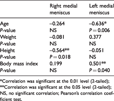
*Correlation was significant at the 0.01 level (2-tailed); **Correlation was significant at the 0.05 level (2-tailed); NS, no significant correlation; Pearson’s correlation coefficient test.
Discussion
This current study used quantitative shear wave elastography to evaluate the anterior horn of the medial menisci in healthy adult subjects. The relationship between elasticity and height, weight, BMI and sex was also studied. A significant inverse correlation was observed between the right medial meniscus elasticity and height. Other demographic factors showed no significant relationship to the right medial meniscus elasticity. A significant inverse correlation was observed between the left medial meniscus elasticity and age, while a significant positive correlation was observed between left medial meniscus elasticity and BMI. Previous studies evaluated the mean stiffness of some components of the musculoskeletal system using shear wave elastography. For example, the elasticity of the Achilles tendon in the resting position was 51.5 kPa, while the elasticity of the supraspinatus tendon was 29.1 kPa.13–17 The elasticity of the gastrocnemius was 16.5 kPa in the resting position and 225.4 kPa during contraction. 16 A few studies have evaluated ligaments such as the transverse carpal ligaments (55.2 kPa).13–17 Important peripheral nerves have also been evaluated by elastography such as the median nerve, which had a mean elasticity at the carpal tunnel reaching 32 kPa. 18 The values of mean elasticity of the menisci obtained in this current study would be expected to increase or decrease in different types of pathologies based on the degree of stiffness. The anatomical position of the medial meniscus was a considerable challenge confronting this current study. In order to optimize the images, a minimum amount of transducer pressure was used, together with a thick layer of gel for each scan, since precompression of the tissue can impact their stiffness. Two previous studies evaluated the knee joint using elastography, but both dealt with the femoral condyle cartilage and both were undertaken using strain elastography.19,20 Only one previous study investigated the knee menisci using shear wave elastography in five healthy volunteers. 21 This previous study demonstrated comparable results to the current study, with a mean ± SD stiffness of the medial meniscus of 30.6 ± 3.5 kPa and a mean ± SD lateral meniscus stiffness of 23.3 ± 3.1 kPa compared with 24.86 ± 6.35 kPa at the right anterior horn medial meniscus and 23.86 ± 4.49 kPa at the left anterior horn medial meniscus in the current study. This previous study also demonstrated a correlation between stiffness measurements in the menisci and the degree of meniscal degeneration. 21
This study had several limitations. First, the sample size was small, which decreases the reliability and validity of the data together with the accuracy of the elasticity measurements. Secondly, all of the participants were females except one, which limits the generalization of these results across the two sexes. Thirdly, the mean ± SD age was 27.35 ± 1.77 years, so the study subjects were not representative of the general population because they were all under 30 years of age. Fourthly, the study only involved the anterior horn of the medial meniscus. Fifthly, the reliability of the correlation analysis with the demographic characteristics needs to be increased by analysing a larger sample size. Future studies using a larger sample size, with more evenly distributed sexes and a wider age range, would increase the accuracy, reliability and reproducibility of these measurements.
In conclusion, these preliminary results suggest that shear wave elastography could be a potential tool to aid in studying the stiffness of the knee menisci. Future studies should consider including patients with different common knee pathologies in addition to normal menisci.
Footnotes
Acknowledgements
The authors are grateful to the Deanship of Scientific Research at Prince Sattam bin Abdulaziz University.
Author contribution
MA Bedewi codesigned the study, conducted the research and was the major contributor in drafting, writing and editing the manuscript. AA Elsifey and AK Saleh assisted in the interpretation of the data. T Alfaifi codesigned the study. All authors read and approved the final manuscript.
Declaration of conflicting interest
The authors declare that there are no conflicts of interest.
Funding
This research received no specific grant from any funding agency in the public, commercial, or not-for-profit sectors.