
Abstract
Objective
To explore the roles of human mesenchymal stem cell (hMSC) death-associated protein kinase 1 (DAPK1) in modulating CD4+ T lymphocyte proliferation.
Methods
Human MSCs and peripheral blood mononuclear cells were isolated and cocultured in vitro for 3 days. Lentiviral-mediated RNA interference (LV-sh-DAPK1) was used to silence DAPK1 expression in hMSCs. Expression of DAPK1 was assessed by western blotting. Transcriptional levels of DAPK1, transforming growth factor-β1, indoleamine 2,3-dioxygenase, inducible nitric oxide synthase, interleukin (IL)-6, suppressor of cytokine signaling 1, IL-10 and cyclooxygenase-2 were investigated by quantitative PCR. Levels of IL-10 were assessed by ELISA. Proliferation of CD4+ T cells was assessed by flow cytometry.
Results
DAPK1 was abundantly expressed in ex vivo-expanded hMSCs and expression was positively correlated with hMSC suppression of CD4+ T cell proliferation. Silencing of DAPK1 in hMSCs reduced the ability of these cells to inhibit CD4+ T cell proliferation and resulted in decreased IL-10 levels compared with untreated controls. Exogenous supplementation with recombinant human IL-10 in DAPK1-silenced hMSCs restored immunosuppression of CD4+ T cells.
Conclusions
The DAPK1-IL-10 axis mediates a novel immunoregulatory function of hMSCs toward CD4+ T cells.
Keywords
Introduction
Mesenchymal stem cells (MSCs), originally isolated from the bone marrow stroma by Friedenstein et al., 1 are multipotent progenitor cells that can differentiate into adipocytes, chondrocytes and osteocytes as well as myocytes, astrocytes, neurons, endothelial cells and lung epithelial cells.2–4 MSCs can also be isolated from other tissues such as cord blood, placenta, amnion, and adipose tissue. 5 No single antigen is exclusively expressed by MSCs. Generally, MSCs have been identified in vitro based on adherence to plastic, multipotent differentiation, expression of cell surface markers such as CD90, CD29, CD73, and CD105, and absence of markers such CD45 and CD34.6,7
In addition to their the stem/progenitor properties, MSCs have been shown to mediate broad immunoregulatory functions including inhibition of T lymphocyte proliferation. 8 The immunomodulatory roles of MSCs depend on transforming growth factor-β1 (TGF-β1), indoleamine 2,3-dioxygenase (IDO), interleukin (IL)-10, cyclooxygenase-2 (COX-2), inducible nitric oxide synthase (iNOS), IL-6, and suppressor of cytokine signaling 1 (SOCS1).9–12 Although these factors contribute to the immunomodulatory properties of MSCs, the precise mechanisms underlying the action of MSCs on immune cells remain to be elucidated.
Death-associated protein kinase 1 (DAPK1), originally identified in an unbiased genetic screen for positive regulators of interferon (IFN)-γ-induced cell death, 13 is a member of the Ca2+-calmodulin-regulated kinases that includes two other closely related homologues: Zip kinase [ZIPK, also known as Dlk (DAP-like kinase) or DAPK3] and DAPk-related protein 1 (DRP-1, also known as DAPK2). 14 Subsequent reports demonstrated that all three members of this kinase family possessed tumor and metastasis suppressor properties, 15 generating significant interest in the structures, functions, and physiological roles of the DAPKs as well as their relationships to human disease. The multidomain structure of DAPK1 includes a Ca2+/calmodulin domain proximal to the N-terminal catalytic domain, a stretch of ankyrin repeats, a cytoskeleton-binding domain and a C-terminal death domain. 14 Various modes of programmed cell death have been linked to DAPK1. In addition to the IFN-γ pathway, DAPK1 is also involved in cell death mechanisms associated with Fas, tumor necrosis factor (TNF)-α, TGF-β, ceramide, caspase and p53.15–17 Furthermore, DAPK1 promotes cell death resulting from ischemia-reperfusion events in both kidney and brain tissue in mouse models.18,19
DAPK1 has also been implicated as a modulator of inflammation in multiple cell types. Interestingly, DAPK1 can regulate inflammation either positively, through nucleotide-binding domain and leucine-rich repeat pyrin domain containing 3 (NLRP3) inflammasome formation and production of the proinflammatory cytokine IL-1β in macrophages, 20 or negatively, through the inhibition of nuclear factor kappa-light-chain-enhancer of activated B cells (NF-κB) 21 and expression of a subset of late-onset inflammatory proteins in monocyte lines treated with IFN-γ. 22 DAPK1 can attenuate inflammatory responses in the lung induced by lipopolysaccharide (LPS) 23 and in intestinal epithelial cells by controlling STAT3 activity. 24 Likewise, transgenic murine T cells expressing a dominant negative form of DAPK1 and murine T cell hybridomas in which endogenous DAPK1 expression was silenced exhibited hyperactivation of NF-κB and enhanced activation in response to T cell receptor signals. 21 However, to the best of our knowledge, the role of DAPK1 in the inflammatory responses of MSCs has not been studied either in vitro or in vivo.
In the present study, we examined the role of DAPK1 in human MSC (hMSC) inhibition of CD4+ T cell activation. We found a positive association between hMSC immunosuppression of CD3/28-stimulated CD4+ T cells and the expression level of DAPK1 in hMSCs. Silencing of DAPK1 in hMSCs greatly decreased the immunosuppression of CD4+ T cells and decreased production of soluble IL-10 compared with untreated hMSCs. Exogenous supplementation with recombinant human IL-10 in DAPK1-silenced hMSCs restored their immunosuppressive effects towards T cells. Our results reveal a novel role of DAPK1 in hMSC inhibition of CD4+ T cell activation.
Materials and methods
Isolation and preparation of hMSCs
All procedures were carried out with approval of the Ethics Committee of Sun Yat-sen University (Guangzhou, China). All healthy donors signed informed consent forms. Bone marrow was aspirated and MSCs were isolated strictly according to an internationally standardized procedure. 25 The fourth passage of hMSCs was used in all experiments. After immunophenotyping of a marker panel [CD29-phycoerythrin (PE), clone: MAR4; CD90-fluorescein isothiocyanate (FITC); clone: 5E10, CD105-FITC, clone: 266; CD45-FITC, clone: 30-F11; CD34-allophycocyanin, clone: 563; and human leukocyte antigen - antigen D related (HLA-DR)-PE, clone: G46-6] by flow cytometry and confirmation of differentiation potential (osteogenesis, chondrogenesis, or adipogenesis), MSCs were used for subsequent experiments. All antibodies were purchased from BD Biosciences (San Jose, CA, USA).
Trilineage differentiation potential assays of hMSCs
Osteogenic differentiation
HMSCs were cultured in six-well plates (1 × 10⁵ cells/well) in 3 mL/well of Dulbecco’s modified Eagle medium (DMEM, Invitrogen, Carlsbad, CA, USA) containing 10% fetal bovine serum (FBS, Gibco, Carlsbad, CA, USA), 50 mg/L ascorbic acid (Sigma, St. Louis, MO, USA), 10 mM-glycerophosphate (Sigma), and 100 nM dexamethasone (Sigma). Half of the medium volume was replaced every 3 days until 21 days of induction. Alizarin red staining was used to confirm the osteogenic ability of hMSCs.
Chondrogenic differentiation
HMSCs (2.5 × 10⁵ cells) were centrifuged at 600 ×g for 5 minutes in a 15-mL conical tube. The cells were cultured in high-glucose DMEM containing 1% ITS-Premix (Corning, Corning, NY, USA), 1 mM sodium pyruvate (Sigma), 50 mg/L ascorbic acid (Sigma), 10 ng/mL TGF-β3 (R&D), and 100 nM dexamethasone (Sigma). The medium was replaced every 3 days. The total culture duration was 21 days and was followed by toluidine blue staining.
Adipogenic differentiation
HMSCs were cultivated in six-well plates (1 × 10⁵ cells/well) in DMEM supplemented with 10% FBS, 10 g/mL insulin (Sigma), 0.2 mM indomethacin (Sigma), 0.5 mM 3-isobutyl-1-methylxanthine (Sigma), and 1 M dexamethasone (Sigma). After 3 days of induction, the media was replaced with DMEM containing 10 g/mL insulin for 1 day, then the medium was replaced with the induction medium described above. After three cycles of media changes, hMSCs were cultured in DMEM containing 10 g/mL insulin until day 21. Oil red O staining was performed following fixation with 4% paraformaldehyde.
CD4+ T cell proliferation assay
Detailed steps for this assay can be found in previous reports.11,26 Peripheral blood mononuclear cells (PBMCs) were isolated and incubated with 5 µM carboxyfluorescein diacetate succinimidyl ester (CFSE, Life Technologies, Carlsbad, CA, USA) for 30 minutes and washed three times with phosphate-buffered saline (PBS) containing 10% FBS. HMSCs were cocultured with PBMCs pretreated with purified anti-CD3 (0.2 g/mL, BD Biosciences) and anti-CD28 (1 g/mL, BD Biosciences) antibodies at a 1:20 ratio of MSCs:PBMCs. Cells were cocultured in RPMI-1640 medium in a final volume of 3 mL for 5 days. PBMCs were harvested by centrifugation at the end of the coculture. PE-CD4 (clone: RPA-T4, BD Biosciences) was incubated with PBMCs and the proliferation of CD4+ T cells was measured by flow cytometry (FACSVerse, BD Biosciences).
Immunofluorescent staining of DAPK1 in hMSCs
Fourth-passage hMSCs were fixed in 4% paraformaldehyde at room temperature for 15 minutes, then incubated with 0.1% Triton X-100 for 10 minutes. The cells were blocked for 1 hour with goat serum, then incubated at 4°C overnight with a rabbit antibody against DAPK1 (#3008s, Cell Signaling Technology, Danvers, MA, USA; 1:500 dilution). Tetramethylrhodamine-conjugated goat anti-rabbit secondary antibody (A-21428, Thermo Fisher Scientific, Rockford, IL, USA; 1:1000 dilution) was then added and incubated for 1 hour at room temperature in the dark. Nuclei were stained with 4′,6-diamidino-2-phenylindole (DAPI) for 5 minutes (D9542, Sigma, 1:1000 dilution). The MSCs were examined using a laser-scanning confocal microscope (LSM510, Carl Zeiss, Jena, Germany).
Western blotting
Cytosolic lysates were prepared from fourth-generation hMSCs and from hMSCs infected for 3 days with lentiviruses expressing sh-DAPK1 or a control lentivirus. Protein concentrations were measured in duplicate using a bicinchoninic acid assay. Equal amounts of protein were separated by 10% SDS-PAGE (Bio-Rad, Hercules, CA, USA). Proteins were transferred electrophoretically to nitrocellulose membranes (Millipore, Billerica, MA, USA) and detected using the following antibodies: rabbit anti-DAPK1 (Cell Signaling Technology, #3008s), mouse anti-β-actin (Sigma-Aldrich, A1978), and mouse anti-glyceraldehyde 3-phosphate dehydrogenase (GAPDH) (Millipore, MAB374). The membranes were then incubated with alkaline phosphatase-conjugated secondary antibodies targeting either mouse IgG (BA1050, Boster, Pleasanton, CA, USA) or rabbit IgG (BA1054, Boster). Blots were developed using an enhanced chemifluorescence substrate (GE Healthcare, Piscataway, NJ, USA). Densitometry was performed using ImageJ software (NIH, Bethesda, MD, USA).
Transfection of hMSCs with short hairpin (sh)RNAs
Lentiviral vectors (LV3-H1/GFP-Puro) encoding shRNAs targeting DAPK1 were packaged by GenePharma (Shanghai, China). The shRNA sequences were as follows: sh-DAPK1#1, 5′-GCGAGCTGTTTGACTTCTTAG-3′ and sh-NC#2, 5′-TTCTCCGAACGTGTCACGTTTC-3′. sh-NC was a negative control. hMSCs were plated in a six-well plate at a density of 2 × 10⁵ cells per well 24 hours before transfection, then incubated with 5 µL (1 × 10⁹ transforming units/mL) of lentiviral vectors encoding sh-DAPK1 or sh-NC along with Polybrene (final concentration: 5 µg/mL, GenePharma) in DMEM containing 10% FBS at 37°C for 24 hours. Subsequently, 3 mL of fresh medium containing 10% FBS was added and the culture was incubated for an additional 72 hours. The cells were collected for DAPK1 detection by quantitative real-time PCR (qPCR) and western blotting to confirm the depletion of DAPK1.
qPCR
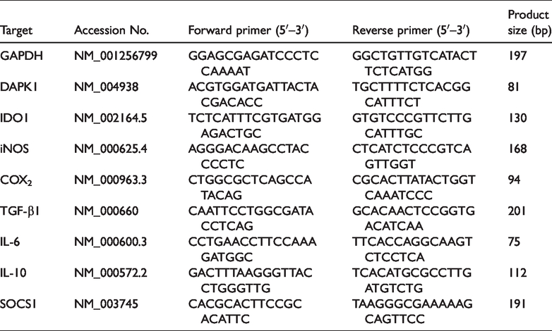
Trizol (Invitrogen) was used to extract total RNA from collected cells. cDNA synthesis was performed using a Prime Script RT reagent kit (Takara Bio Inc., Shiga, Japan) according to the manufacturer’s instructions. qPCR was performed on a Light Cycler 480 Real-Time PCR System (Roche, Basel, Switzerland) using SYBR Premix Ex Taq (Takara). The thermal cycling parameters were: 30 s at 95°C followed by 40 cycles of 5 s at 95°C and 20 s at 60°C. Relative changes in mRNA levels of target genes were assessed using the 2(−ΔΔct) method and normalized to levels of the housekeeping gene GAPDH. The sequences of primers used in the qPCR assay are listed in Table 1.
Primers used for qPCR in this study.
CCK-8 assay
hMSCs were seeded in 96-well plates at a concentration of 1 × 10⁴ cells/mL in 100 μL fresh medium (low-glucose DMEM containing 10% FBS). Each sample was seeded in three wells in parallel. Cell viability curves for hMSCs were acquired using a Cell Counting Kit-8 (Dojindo Molecular Technologies, Rockland, MD, USA) according to the manufacturer’s protocol to determine absorbance at 490 nm on days 1 to 5. Fresh medium was used as a negative control.
Cell death assay
MSCs were harvested using digestion with 0.125% trypsin. After centrifuging at 300 ×g for 5 minutes, the MSCs were resuspended in 100 µL of PBS containing 5 µL anti-Annexin-V and 5 µL 7-aminoactinomycin D (Cat. 556570 and 559763, BD Biosciences) and incubated for 20 minutes. Cell death of hMSCs was measured by flow cytometry (FACSVerse).
ELISA
IL-10 concentrations in hMSC culture supernatants 3 days following lentiviral infection (sh-DAPK1, sh-NC and untreated control cells) were measured using a human IL-10 Quantikine ELISA Kit (R&D Systems, Minneapolis, MN, USA) according to the manufacturer’s protocol.
Statistical analysis
The experiments and analysis were performed by different authors. All values in the text and figure legends represent means ± standard errors of the means (SEMs). Differences between groups were assessed using two-tailed Student’s t tests or analysis of variance in SPSS version 13.0 (SPSS Inc., Chicago, IL, USA). Values of p < 0.05 were considered statistically significant.
Results
Characterization and differentiation potential of hMSCs
Freshly isolated hMSCs at the fourth passage displayed adherence and expansion in culture and showed a fibroblast-like morphology under a light microscope (Figure 1b). Flow cytometry showed that the hMSCs expressed high levels of the typical lineage markers CD29, CD44, CD90 and CD105, but not of CD34, CD45 and HLA-DR (Figure 1a). The osteogenic, adipogenic and chondrogenic differentiation capacities of the hMSCs were assessed by the presence of bone nodules, lipid vesicles, and polysaccharide amines stained with Alizarin Red, Oil Red O, and toluidine blue, respectively, after 21 days of induction (Figure 1b).

Characterization and differentiation potential of hMSCs.
Expression of DAPK1 in hMSCs correlates with inhibition of CD4+ T cell proliferation
To better understand the contribution of DAPK1 in hMSCs to immunoregulation, we first evaluated DAPK1 expression using immunofluorescence and western blotting. hMSCs expressed DAPK1 abundantly in the cytoplasm under ex vivo-expanded conditions (Figure 2a), and showed the same expression pattern as in other cell types.14,16 Intriguingly, the expression of DAPK1 was clearly heterogeneous in the fourth passage of hMSCs from six healthy donors when cultured under the same conditions (Figure 2b). However, cell viability as shown by CCK-8 assay revealed no differences among these hMSCs (Figure 2c). hMSCs can suppress in vitro T-lymphocyte proliferation induced by cellular or nonspecific mitogenic stimuli. Determining whether DAPK1 plays a role in this process was the primary goal of our study. We found a negative correlation between the expression of DAPK1 and proliferation of CD4+ T cell after co-culturing hMSCs and PBMCs pretreated with anti-CD3/CD28 antibodies (Figure 2d and 2e). Thus, DAPK1 underlies the immunosuppressive effects of MSCs on T cell activation.

DAPK1 expression and its association with inhibition of CD4+ T cell proliferation by hMSCs.
Disruption of DAPK1 in hMSCs leads to reduced immunosuppression of CD4+ T cells
To further confirm that DAPK1 expressed by hMSCs suppressed the activation of T cells, T cell proliferation following CD3/28 stimulation was assessed in the presence of hMSCs infected with lentiviruses expressing sh-DAPK1-GFP (sh-DAPK1) or sh-NC-GFP (sh-NC) (Figure 3a). After 3 days of infection, expression of sh-DAPK1 in hMSCs reduced DAPK1 mRNA by ∼90% (Figure 3c) and DAPK1 protein by more than 60% compared with cells infected with lentiviruses expressing sh-NC and control cells (Figure 3b and 3c). hMSCs transduced with sh-DAPK1 showed similar viability in the CCK-8 assay compared with hMSC/sh-NC cells and untreated hMSCs (Figure 3d). Moreover, flow cytometry confirmed that DAPK1 silencing did not significantly alter cell death (Figure 3e). The hMSCs were co-cultured with PBMCs pretreated with CFSE and CD3/CD28. Five days later, the PBMCs were observed under a light microscope and collected. Flow cytometry was used to detect CD4+ T cell proliferation. Silencing of DAPK1 in hMSCs resulted in reduced suppression of CD4+ T cell proliferation compared with control and sh-NC-treated hMSCs (Figure 3f and 3g).

Silencing of DAPK1 expression in hMSCs reduces their inhibition of CD4+ T-cell proliferation.
Silencing of DAPK1 resulted in downregulated IL-10 production
We next examined the contribution of DAPK1 to the expression of molecules involved in MSC-mediated CD4+ T cell suppression. Previous studies demonstrated that MSCs secrete soluble factors that play important roles in MSC immunosuppressive properties, such as IDO, TGF-β, IL-10 and nitric oxide.9–12 We performed gene expression analyses on hMSCs transduced with sh-DAPK1 and sh-NC as well as on control hMSCs. After 3 days of infection, levels of IL-10 mRNA were significantly decreased and IL-10 secretion was diminished in hMSCs transduced with sh-DAPK1 compared with hMSCs transduced with sh-NC or control hMSCs (Figure 4a and 4h). By contrast, the transcriptional levels of TGF-β1, IDO, iNOS, IL-6, SOCS1, and COX2 showed no significant differences when DAPK1 was silenced (Figure 4b, 4c, 4d, 4e, 4f and 4g).

Downregulation of IL-10 production in hMSCs following DAPK1 silencing.
IL-10 mediated DAPK1-related hMSC immunosuppression of CD4+ T cells
We further explored whether decreased IL-10 levels mediated the DAPK1-related immunosuppressive effects of hMSCs on CD4+ T cells. We supplemented transduced hMSC-PBMC co-cultures with exogenous recombinant human IL-10 (1 ng/mL) and assessed the proliferation of CD4+ T cells after 5 days by flow cytometry. In the presence of exogenous IL-10, the proliferation of CD4+ T cells in co-cultures with hMSCs transduced with sh-DAPK1 were restored almost to the same levels as those of T cells co-cultured with hMSCs transduced with sh-NC (Figure 5a and 5b). Taken together, these data suggest that IL-10 mediates hMSC immunosuppression of CD4+ T cells in a DAPK1-dependent manner.

IL-10 mediated DAPK1-related hMSCs immunosuppression of CD4+ T cells.
Discussion
Our results demonstrate that DAPK1 plays an important role in hMSC suppression of CD4+ T-lymphocyte proliferation. First, we investigated the expression of DAPK1 in MSCs, and found that DAPK1 was strongly expressed in the cytoplasm. Furthermore, there was a negative correlation between DAPK1 expression in MSCs and proliferation of CD4+ T cells in co-cultures of hMSCs and PBMCs. Silencing of DAPK1 in hMSCs resulted in decreased suppression of CD4+ T cell proliferation. Decreased secretion of the soluble factor IL-10 by hMSCs may play an important role in loss of immunosuppression following DAPK1 silencing.
To the best of our knowledge, the contribution of DAPK1 to immunoregulation by MSCs has not previously been demonstrated. A previous study used RT-PCR to demonstrate that MSCs from the peripheral blood of acute lymphoblastic leukemia patients prominently expressed the DAPK1 gene. 27 Our results demonstrated high DAPK1 expression at the protein level in ex vivo-expanded hMSCs isolated from bone marrow (Figure 2a and 2b) and documented a correlation between DAPK1 expression level and MSC immunosuppression of CD4+ T cells (Figure 2d and 2e). These results prompted us to focus on the contribution of DAPK1 to the immunoregulatory functions of hMSCs.
Initially, DAPK1 was identified as a mediator of several cell death programs, including apoptosis, autophagy-associated cell death, and programmed necrosis. 14 Overexpression of DAPK1 led to cell death, while silencing it protected cells from death during oxidative stress. 28 In our experiments, silencing DAPK1 expression in hMSCs had no effect on cell proliferation or cell death (Figure 3d and 3e). We were unable to successfully infect hMSCs with LV-DAPK1, a DAPK1-overexpressing lentivirus, because the infection efficiency of this lentiviral vector was too low; this may have been related to the large molecular weight of DAPK1 (160 kDa) and consequently the large vector size of LV-DAPK1, which may have reduced transduction efficiency. In addition to the role of DAPK1 in cell death, several studies have demonstrated that DAPK1 also participates in both acute and chronic inflammatory processes in multiple cell types including neutrophils, monocytes, macrophages, dendritic cells, and epithelial cells.23,24 Previous studies reported that DAPK1 negatively regulated human T cell activation by suppressing the translocation of key NF-κB-activating molecules into membrane rafts. 21 Our results suggest that the suppressive functions of hMSCs toward PBMC are linked to DAPK1 because disruption of DAPK1 expression in hMSCs weakens immunosuppression of PBMCs (Figure 3f and 3g). Previous studies have reported that DAPK1 was involved in inflammatory processes triggered by LPS, TNF-α, IFN-γ, and Toll-like receptor 4 through the NF-κB, mammalian target of rapamycin complex 1 (mTORC1), IFN-γ and activated inhibitor of translation (GAIT) complex pathways.22,23,29,30 It remains to be seen whether any of these pathways, or perhaps a novel mechanism, mediate DAPK1-induced suppression of PBMC activation in hMSCs.
The broad immunoregulatory properties of hMSCs depend on the release of soluble factors such as IL-10 and TGF-β. The release of these soluble factors is enhanced following stimulation of hMSCs with TNF-α and IFN-γ, but unstimulated hMSCs also produce these mediators. 31 In previous studies of human MSCs, it was reported that IDO and IL-10 may serve as pivotal molecules suppressing T cell proliferation. 26 In our experiments, knockdown of DAPK1 in hMSCs deceased the secretion of the soluble factor IL-10 into culture supernatants, whereas the expression of IDO and TGF-β did not change markedly compared with controls (Figure 4h, 4b and 4c). IL-10, an anti-inflammatory cytokine, can contribute to down-regulation of T cell proliferation and other effector responses. 26 Previous studies have reported that silencing of DAPK1 expression and lack of IL-10 secretion occurred concurrently in resected stage I non-small-cell lung cancer patients, 32 suggesting that disruption of DAPK1 may reduce the production of IL-10 in tumors. However, this relationship was not explored at a mechanistic level. In our experiments, levels of IL-10 secreted by hMSCs were distinctly decreased following shRNA silencing of DAPK1, further validating the relationship between DAPK1 and IL-10 in hMSCs. However, the detailed regulatory mechanisms linking DAPK1 and IL-10 in MSCs as well as in tumors require more exploration.
Taken together, our results indicate that a DAPK1–IL-10 pathway in hMSCs mediates the inhibition of T cell activation. Furthermore, differences in immunosuppression by hMSCs are consistent with the heterogeneity of DAPK1 expression observed in hMSCs, suggesting that DAPK1 expression level in hMSCs may be a marker for the immunosuppression potential of hMSCs. MSCs have been used therapeutically in clinical trials of tissue injury, transplantation, and autoimmunity. Our study reveals a novel role of DAPK1 in regulating the immunomodulatory activities of hMSCs, providing additional insights into the potential clinical applications of MSCs. In addition to the immunoregulatory role of DAPK1 in MSCs, we are also probing its function in osteogenesis. In preliminary experiments, a significant enhancement in osteogenesis was confirmed (unpublished data) following silencing of DAPK1 in hMSCs. It remains to be further explored whether DAPK1 is involved in diseases such as ankylosing spondylitis and osteoporosis.
Footnotes
Declaration of conflicting interest
The authors declare that there is no conflict of interest.
Funding
This work was supported by the National Natural Science Foundation of China (grant number 31700884), the Guangdong Province Science and Technology Plan (grant number 2017A020215070), and the Sun Yat-sen University Young Teachers Cultivation Project (grant number 17ykpy42).