
Abstract
Objectives
Gastric cancer (GC) is the leading cause of cancer-related deaths worldwide; however, the underlying molecular mechanisms of GC remain unclear. This study investigated the role of the miR-877–AQP3 axis in GC tumorigenesis.
Methods
The levels of miR-877 expression were measured in GC tissues and cell lines by qRT-PCR. Functional assays were performed to elucidate the role of miR-877 in GC development.
Results
Our results showed that miR-877 levels were lower in GC tissues and cell lines compared with the corresponding controls. Additionally, reduced miR-877 levels were associated with unfavorable prognoses. Increased miR-877 expression suppressed proliferation, invasion, and epithelial-mesenchymal transition, while promoting apoptosis in GC cells. Luciferase reporter assays showed that aquaporin 3 (AQP3) was a direct downstream target of miR-877. Overexpression of AQP3 partially rescued the tumor suppressive effects of miR-877 in GC cells. Moreover, miR-877 was negatively correlated with AQP3 mRNA expression in GC tissues.
Conclusions
This study demonstrated that miR-877 plays a suppressive role in GC tumorigenesis by regulating AQP3.
Introduction
Gastric cancer (GC) is one of the most widespread gastrointestinal malignant tumors and remains an important public health problem in China.1,2 Treatments for GC include surgery, radiotherapy, and chemotherapy. Nevertheless, the prognosis of GC remains poor even with the best treatment approaches. 3 GC patients usually do not experience obvious signs or symptoms at early stages; thus, most cases are diagnosed at advanced stages, leading to unfavorable clinical outcomes. Currently, the molecular events that drive GC carcinogenesis are unclear. Therefore, it is important to elucidate the molecular mechanisms of the initiation and progression of GC.
MicroRNAs (miRNAs) are a group of endogenous small non-coding RNAs of 18 to 25 nucleotides in length. 4 MiRNAs post-transcriptionally silence their target genes to regulate protein expression, thereby influencing a majority of physiological processes such as cell proliferation, survival, differentiation, and apoptosis. 5 Dysregulation of miRNAs has been comprehensively described in various types of cancer, including GC.6,7 MiRNAs can either act as an oncomiR or tumor suppressor gene in GC tumorigenesis, depending on the specific downstream targets and pathways. For instance, miR-1254 is significantly downregulated in GC tissues and cell lines, and overexpression of miR-1254 suppresses the proliferation, migration, and invasion capacity of GC cells in vitro and inhibits tumorigenesis in vivo, indicating that miR-1254 plays a tumor suppressive role in GC. 8 Conversely, miR-589 is overexpressed in GC tissues and cell lines, and upregulation of miR-589 promotes malignant behaviors in GC cells, suggesting miR-589 plays an oncogenic role in GC. 9
MiR-877 is located on 6p21.33 and plays important regulatory roles in many biological processes. Previous studies have demonstrated that miR-877 is actively involved in the development of multiple tumor types.10–12 However, its role in GC progression has been poorly studied. In this study, we show that miR-877 is downregulated in GC tissues and cell lines. Additionally, functional experiments showed that ectopic miR-877 expression inhibited the malignant phenotypes of GC cells by downregulating AQP3.
Materials and methods
Tissue samples
This study was approved by the Institutional Review Board of the Second Affiliated Hospital, Zhejiang University. In total, 120 GC tissues and matched adjacent normal tissues (ANTs) were collected from the Department of General Surgery, the Second Affiliated Hospital of Zhejiang University. All specimens were snap frozen in liquid nitrogen and stored at −80°C until RNA extraction. Written informed consent was obtained from each participant for using their samples. Clinicopathological information of the included GC patients are summarized in Table 1. Overall survival (OS) was defined as the time from randomization to death from any cause.
Correlations between miR-877 expression and the clinicopathological parameters of GC patients.

GC, gastric cancer.
Cell culture
The human gastric epithelial cell line, GES-1, and five human GC cell lines, GC9811, BGC823, SGC-7901, MKN-45 and HGC-27, were obtained from American Type Culture Collection (Manassas, VA, USA). All cell lines were cultured in Dulbecco’s Modified Eagle’s Medium (Invitrogen, Carlsbad, CA, USA) supplemented with 10% fetal bovine serum,100 U/mL penicillin, and 100 μg/mL streptomycin. Cells were incubated in 5% CO2 at 37°C.
Cell transfection
The miR-877 mimics and negative control (NC) mimics were purchased from Genepharma (Sigma Aldrich, St. Louis, MO, USA). Transfections were performed using Lipofectamine RNAiMAX according to the manufacturer's instructions (Invitrogen, Carlsbad, CA, USA). Transfected cells were cultured for 48 hours before functional assays.
Lentivirus construction and infection
The full-length AQP3 cDNA was cloned into the pGCL-GFP vector, and the constructs were validated by sequencing. The recombinant lentiviral plasmid and packaging plasmid were co-transfected into 293T cells to generate lentiviral particles. Media containing the lentiviral vector particles were harvested 72-hours after transfection. The scramble lentiviral vector LV-CTRL was used as a control. GC cells were stably infected with lentivirus at a multiplicity of infection of 30.
Apoptosis assay
Apoptosis assays were performed according to the manufacturer’s instructions. Briefly, cells were seeded into a white opaque 96-well plate, and then equal amount (100 μL) of Caspase-Glo® 3/7 reagent (Promega, Madison, WI, USA) was added to each well and incubated for 2 hours at room temperature protected from light. The luminescence intensity was measured by a microplate reader.
Cell counting
Cells were plated at a density of 1 × 10⁵ cells/well in 12-well plates. After incubation for 0, 24, 48, 72, and 96 hours, the number of viable cells was counted using a hemocytometer by the trypan blue exclusion assay.
Cell Counting Kit-8 assay
Cells were seeded into 96-well plates at an initial density of 3 × 10⁴ cells/mL with 100 μL of culture medium per well. After incubation for 0, 24, 48, 72, and 96 hours, 10 µL of Cell Counting Kit-8 reagent (Dojindo Molecular Technologies, Inc., Kumamoto, Japan) was added to each well. Absorbance was measured at 450 nm using a microplate reader.
Invasion assay
The invasion capacity of cancer cells was evaluated by the Matrigel invasion assay using Matrigel-precoated Transwell plates (BD Biosciences, Franklin Lakes, NJ, USA). Transfected cells were seeded in the upper chambers at a density of 2 × 10⁵ cells/per well, while the lower chambers were filled with 0.8 mL of complete culture media. After a 48-hour incubation, the non-invasive cells that remained in the upper chambers were removed with a cotton swab, while the invading cells were treated with 4% paraformaldehyde for 20 minutes, followed by staining with 1% crystal violet for 30 minutes at room temperature. The number of invading cells was counted in at least in five random fields using a light microscope.
RNA extraction and qRT-PCR
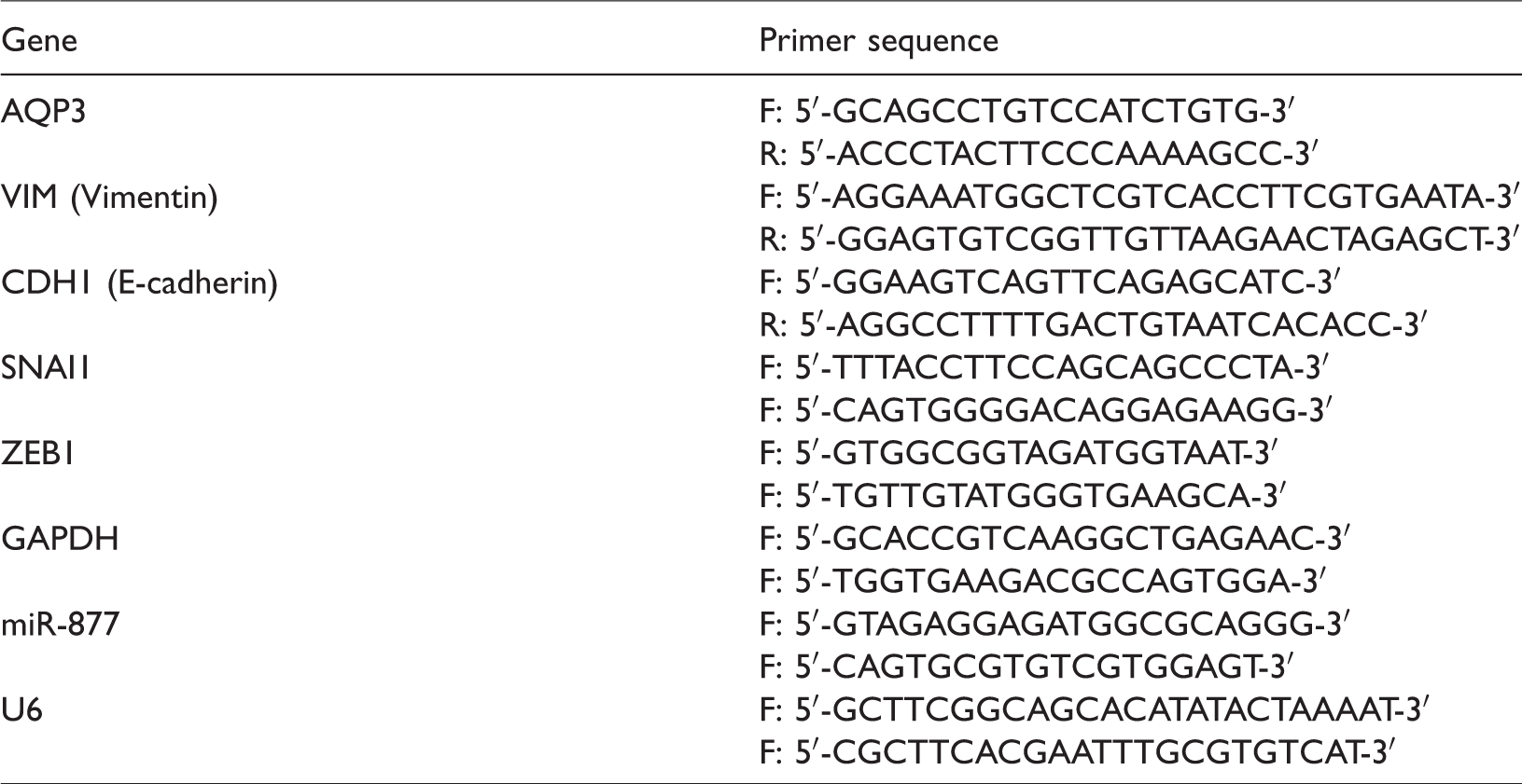
Total RNA from tissue samples and cultured cells was extracted using the RNAiso Plus reagent (TaKaRa, Dalian, China) according to manufacturer’s protocol. First strand complementary DNAs were synthesized with PrimeScript™ RT Master Mix (TaKaRa). Real time PCR was performed with SYBR Premix ExTaq II (Takara) in an ABI Prism 7500 Sequence Detection System (Applied Biosystems, Foster City, CA, USA). Endogenous U6 snRNA and GAPDH were used as the internal controls for miRNAs and mRNAs, respectively. The relative expression of miRNAs and mRNAs were calculated by the 2−ΔΔCt method. The primers used in this study are listed in Table 2.
Primer sequences for qRT-PCR.
Western blotting
RIPA lysis buffer was used to extract total protein contents from samples. Total protein was separated by 10% SDS-PAGE and transferred to a PVDF membrane. After blocking with 5% non-fat milk, the membranes were probed with the AQP3 primary antibody (Santa Cruz Biotechnology, Santa Cruz, CA, USA) at 4°C overnight. Following washing with TBST buffer, horseradish peroxidase (HRP)-conjugated secondary antibody was incubated with the membrane for 1 hour at room temperature. The enhanced chemiluminescent detection reagent (Pierce, Rockford, IL, USA) was applied to detect the signals.
Luciferase reporter assay
Regions of the AQP3 sequence that encompassed either the wild-type (AQP3-WT) or corresponding mutant miR-877 target site (AQP3-MUT) were respectively amplified by PCR and cloned into the pmirGLO plasmid (Promega). For luciferase assays, miR-877 mimics, NC mimic, and pmirGLO-AQP3-WT or pmirGLO-AQP3-MUT were transfect into 293T cells using Lipofectamine 2000 (Invitrogen). After a 48-hour incubation, the Dual-Luciferase Reporter Assay System (Promega) was used to evaluate the relative luciferase activity.
Statistical analysis
All statistical analyses were performed with GraphPad 7.0 (GraphPad Software Inc., La Jolla, CA, USA). For data with a distribution, statistical analyses were conducted using the Student’s t-test or one-way ANOVA. The chi-square test was performed to evaluate the association between miR-877 expression and clinicopathological parameters. The potential targets of miR-877 were predicted using TargetScan Human 7.1 (http: //www.targetscan.org/vert_71/). Pearson’s correlation coefficient was used to calculate the correlations between miR-877 and AQP3 in the tissue samples. Kaplan–Meier plots and the log-rank test were used to assess differences in OS. P-values of <0.05 represented statistically significant differences.
Results
MiR-877 levels were reduced in GC tissues and cell lines
Our data showed that miR-877 expression was downregulated in GC tissues compared with paired non-tumor tissues (***P < 0.001) (Figure 1a). Additionally, miR-877 showed significantly lower expression in GC cell lines compared with the normal gastric epithelium cell line GES-1 (***P < 0.001) (Figure 1b). MiR-877 was most downregulated in SGC-7901 and MKN-45 cells, so these two cancer cell lines were chosen for subsequent in vitro studies.

MiR-877 expression was reduced in GC tissues and cell lines. (a) MiR-877 expression was significantly downregulated in GC tissues compared with adjacent normal tissues. (b) MiR-877 was decreased in GC cell lines compared with the gastric epithelial cell line. (c) Survival analysis showed that patients in the low miR-877 group had a significantly shorter overall survival than those in the high miR-877 group. GC, gastric cancer.
Low miR-877 expression was associated with unfavorable prognosis
The GC patients were divided into high (n = 56) and low (n = 64) miR-877 expression groups. The chi-square analysis showed that low miR-877 expression was strongly correlated with lymph node metastasis (P = 0.0353) and advanced tumor stage (P = 0.0025). However, it was not associated with sex (P = 0.3197), age (P = 0.2174), tumor size (P = 0.5170), distant metastasis (P = 0.3063), or tumor grade (P = 0.2679) (Table 1). Survival analysis showed that patients in the low miR-877 group had a significantly shorter OS than those in the high miR-877 group (P = 0.0074) (Figure 1c).
MiR-877 suppressed proliferation and invasion but promoted apoptosis in GC cells
MiR-877 expression was increased in GC cell lines transfected with miR-877 mimic compared with those transfected with NC mimic (***P < 0.001) (Figure 2a). Apoptosis assays showed that relative caspase-3 and -7 activities were significantly higher in the miR-877 mimic group than in the NC mimic group (**P < 0.01, ***P < 0.001) (Figure 2b). CCK-8 assays revealed that optical density values were lower in cancer cells transfected with miR-877 mimic compared with those transfected with NC mimic (*P < 0.05, **P < 0.01, ***P < 0.001) (Figure 2c). Similarly, cell counting assays showed that the number of viable cells was significantly lower in the miR-877 mimic group compared with the NC mimic group (*P < 0.05, **P < 0.01, ***P < 0.001) (Figure 2d). Matrigel invasion assays showed that miR-877 overexpression significantly attenuated the invasive capabilities of GC cell lines (***P < 0.001) (Figure 2e).

MiR-877 overexpression suppressed the proliferation and invasion of GC cells. (a) MiR-877 expression was significantly higher in miR-877 mimic-transfected cells than in NC mimic-transfected cells. (b) Increased luminescence was found when GC cells were treated with miR-877 mimic, suggesting caspase-3 and -7 activities were upregulated. (c) The optical density values were lower in the miR-877 overexpression group. (d) Cell counts were significantly reduced in miR-877 mimic-transfected cells. (e) The number of invading cells was lower in the miR-877 overexpressing group. GC, gastric cancer; NC, negative control.
AQP3 is a direct downstream target of miR-877
Bioinformatics analysis showed that AQP3 was a potential downstream target of miR-877 (Figure 3a). For wide type AQP3, the reporter assays demonstrated that the relative luciferase activity was significantly lower in miR-877 mimic-transfected 293T cells compared with NC mimic-transfected cells. However, no significant difference was found for mutated AQP3 (***P < 0.001) (Figure 3b). Additionally, overexpression of miR-877 significantly reduced AQP3 expression in GC cells at both the mRNA and protein levels (**P < 0.01, ***P < 0.001) (Figure 3c–3d). GC patients with higher AQP3 mRNA expression had a worse OS rate than those with lower AQP3 mRNA expression (P = 0.0013) (Figure 3e). Moreover, an inverse correlation was found between miR-877 and AQP3 expression in GC tissues (r=−0.6195, P < 0.001) (Figure 3f).

AQP3 is a direct downstream target of miR-877. (a) The 3′-UTR region of AQP3 was highly complementary to the seed sequence of miR-877. (b) For wide type AQP3, the relative luciferase activity was significantly lower in miR-877 mimic-transfected cells than in NC mimic-transfected cells. No significant difference in luciferase activity was found for the mutated AQP3 between the miR-877 overexpression group and the control group. (c) AQP3 mRNA expression was significantly reduced in GC cells following miR-877 overexpression. (d) AQP3 protein expression was downregulated in GC cells following miR-877 mimic transfection. (e) GC patients with higher AQP3 mRNA expression had a worse overall survival rate than those with lower AQP3 mRNA expression. (f) A negative correlation was found between miR-877 and AQP3 in GC. NC, negative control; GC, gastric cancer.
AQP3 overexpression partially rescued the tumor suppressive effects of miR-877
We next explored whether upregulation of AQP3 could rescue the tumor suppressive effects of miR-877 in GC cells. Thus, miR-877 mimic was transfected into AQP3-overexpressing cancer cells (LV-AQP3 + miR-877 mimic group) and control cells (LV-CTRL + miR-877 mimic group). AQP3 protein expression was significantly higher in the miR-877-overexpressing cells infected with AQP3 lentivirus (LV-AQP3) compared with those infected with control lentivirus (LV-CTRL) (Figure 4a). AQP3 overexpression partially reduced the apoptosis of GC cells induced by miR-877 mimic (**P < 0.01, ***P < 0.001) (Figure 4b). Both the CCK-8 and cell counting assays showed that cells in the LV-AQP3 + miR-877 mimic group had a higher proliferation capacity than those in the LV-CTRL + miR-877 mimic group (*P < 0.05, **P < 0.01, ***P < 0.001) (Figure 4c–4d). Similarly, the number of invading cells was higher in the LV-AQP3 + miR-877 mimic group than in the LV-CTRL + miR-877 mimic group (***P < 0.001) (Figure 4e).

AQP3 overexpression partially rescued the tumor suppressive effects of miR-877. (a) AQP3 protein expression was significantly higher in the LV-AQP3 + miR-877 mimic group compared with the LV-CTRL + miR-877 mimic group. (b) Relative caspase-3 and -7 activities were significantly lower in the LV-AQP3 + miR-877 mimic group. (c) Optical density values were higher in the LV-AQP3 + miR-877 mimic group. (d) Cell counts were higher in the LV-AQP3 + miR-877 mimic group. (e) The number of invading cells was higher in the LV-AQP3 + miR-877 mimic group.
Ectopic expression of miR-877 also suppressed the expression of epithelial–mesenchymal transition (EMT) biomarkers such as vimentin, SNAI1 and ZEB1, while increasing the levels of E-cadherin (**P < 0.01, ***P < 0.001) (Figure 5a). Levels of vimentin, SNAI1, and ZEB1 were higher in the LV-AQP3 + miR-877 mimic group than in the LV-CTRL + miR-877 mimic group, while E-cadherin levels were higher in the LV-CTRL + miR-877 mimic group (***P < 0.001) (Figure 5b).

MiR-877 regulates EMT markers. (a) Ectopic expression of miR-877 affected the expression of EMT markers. (b) AQP3 overexpression partially rescued the effects of miR-877 upregulation on the expression of EMT biomarkers. EMT, epithelial–mesenchymal transition.
Discussion
These results showed that miR-877 expression was downregulated in GC tissues and cell lines. Additionally, miR-877 downregulation was associated with a worse prognosis in GC. Ectopic miR-877 expression inhibited proliferation, invasion, and EMT but promoted apoptosis in GC cell lines. Mechanistically, AQP3 was identified as a direct downstream target of miR-877. AQP3 overexpression partially rescued the tumor suppressive effects of miR-877. Moreover, a negative association was found between miR-877 expression and AQP3 mRNA expression in GC tissues. These data suggest that miR-877 plays a tumor suppressive role in GC by targeting AQP3.
EMT is closely correlated with cancer progression, tumor heterogeneity, and therapeutic resistance. Therefore, targeting EMT is a promising strategy for treating GC. A previous study showed that AQP3 promoted EMT in GC cells through the PI3K/AKT/SNAIL pathway. 13 Our results show that AQP3 is a major functional downstream target of miR-877. Additionally, miR-877 reduces the expression of EMT markers in GC cells, while AQP3 overexpression partially reversed this effect. Therefore, miR-877 might suppress GC progression by downregulating AQP3, which subsequently leads to inhibition of EMT. A single miRNA might regulate hundreds or even thousands of downstream targets. In addition to AQP3, there might be many important oncogenes or tumor suppressor genes affected by miR-877. Further studies are warranted to elucidate the downstream genes and pathways regulated by miR-877 in GC.
To the best of our knowledge, this is the first study to explore the role of miR-877 in GC. MiR-877 has been shown to play important roles in other cancer types; for instance, miR-877 expression is remarkably reduced in both blood and tissue specimens from renal cell carcinoma (RCC) patients compared with corresponding controls. Additionally, miR-877 overexpression suppresses the proliferation and migration capacity of RCC cells by downregulating eEF2K. 14 Similarly, miR-877 was downregulated in hepatocellular carcinoma (HCC) tissues and cell lines. In HCC, low miR-877 expression was correlated with a worse prognosis, and CDK14 was identified as a downstream target of miR-877. 12 Another study showed that upregulation of miR-877 could sensitize HCC cells to paclitaxel. 11 MiR-877 expression was found to be higher in BRAF-mutant colorectal cancer (CRC) tissues compared with KRAS-mutant CRC tissues, indicating that miR-877 might be involved in a specific type of CRC. 15 Currently, no study has reported that miR-877 functions as an oncogene, suggesting a conserved tumor suppressive role of miR-877 in tumorigenesis. Nevertheless, further studies are needed to explore the role of miR-877 in other types of cancer.
Aquaporins are a family of membrane proteins that control water transportation across the plasma membrane. AQP3 is widely expressed in various organs, including the gastric mucosa. 16 Abnormal AQP3 expression is closely correlated with carcinogenesis in many tumor types, including GC. 17 For instance, a recent study reported that helicobacter pylori infection upregulated AQP3 expression, promoting GC pathogenesis. 18 AQP3 promotes cisplatin chemoresistance in GC cells, suggesting that targeting AQP3 might be an effective strategy for treating GC. 19
In conclusion, we have proven that miR-877 is dysregulated in GC. Additionally, miR-877 overexpression was found to inhibit the oncogenic activities of GC cells in vitro. Moreover, AQP3 was identified as a functional downstream target of miR-877. The miR-877–AQP3 axis represents a new pathway that regulates GC progression, providing a rationale for targeting miR-877 and AQP3 for GC treatment.