
Abstract
Background
The indirect antiglobulin test (IAT) and direct antiglobulin test (DAT) have been used as common tests for transfusion. Recently, we have found that in addition to causing false increases, rheumatoid factor (RF) can also cause false decreases in immunoassays for hepatitis B surface antigen and B-type natriuretic peptide. However, it remains unclear whether RF also interferes with the IAT and DAT.
Methods
IAT models were produced by mixing IAT-positive plasma and RF-positive plasma, then one-step and two-step IATs were adopted for detection. DAT models were produced by mixing DAT-positive red blood cells (RBCs) and RF-positive plasma, followed by detection with the DAT. The DAT models were diluted using the same RF-positive plasma, and the DAT was performed again.
Results
The rate of decrease of the two-step IAT (40.63%) was significantly higher than that of the one-step IAT (31.51%). Both the rate of decrease (76.67%) and increase (16.67%) of the results of the 60 DAT models were significantly higher than those of the IAT models after two-fold dilution.
Conclusions
The RF can lead to both false decreases and false increases in IAT and DAT. And the interference effects are related to the RF content relative to the IgG-sensitized RBCs.
Keywords
Nonstandard abbreviations
IAT, indirect antiglobulin test; DAT, direct antiglobulin test; RF, rheumatoid factor; HDN, haemolytic disease of the newborn; AIHA, autoimmune haemolytic anaemia; RBC, red blood cell; Fc, fragment crystallizable; AHG, anti-human globulin; RA, rheumatoid arthritis; HCT, haematocrit; LISS, low ionic strength solution; SLE, systemic lupus erythematosus
The indirect antiglobulin test (IAT) and the direct antiglobulin test (DAT), also known as the indirect and direct Coombs tests, respectively, were first reported by Robin Coombs in 1945. These assays play important roles in the diagnosis of haemolytic disease of the newborn (HDN), autoimmune haemolytic anaemia (AIHA) and haemolytic transfusion reactions. Although the IAT and DAT have evolved from the tube method to the more sensitive gel microcolumn method, many false negatives still occur (e.g., due to the amount of IgG being inadequate to detect agglutination, low anti-human globulin [AHG] activity or poor centrifugation technique) as well as false positives (e.g., due to non-specific spontaneous agglutination of red blood cells [RBCs], over-centrifugation and sample clotting). 1 In addition, Barcellini et al. reported that false negatives in the IAT and DAT could also be caused by the presence of anti-RBC IgA and low-affinity antibody. 2 In addition to these factors, many studies have focused on endogenous interference factors, such as heterophilic antibody, human anti-animal antibody, autoimmune antibody and rheumatoid factor (RF), in under-tested samples.3–6 Similarly, for the IAT and DAT immunoassays, results should also be influenced by endogenous interference factors present in samples.
As an autoantibody against the fragment crystallizable (Fc) of human or animal degenerative IgG, RF can bind to the Fc of IgG-capture antibodies or IgG-labelled antibodies in a sandwich immunoassay, consequently resulting in a false elevation of immunoassay results.4–6 Our group has recently found that RF can also lead to both false decreases and false increases in immunoassays against hepatitis B surface antigen and B-type natriuretic peptide.4,5 However, in IATs and DATs using both the tube or the gel microcolumn methods, RBCs are agglutinated when using multi-specific AHG to detect RBC-bound IgG antibodies. Because the antigens targeted by RF are denatured IgGs, we further clarified whether RF similarly interferes with the IAT and DAT.
Materials and methods
Samples
A total of 263 samples were collected from the Union Hospital and the Wuhan Children’s Hospital, Tongji Medical College, Huazhong University of Science and Technology, from 2017 to 2018. Nineteen DAT-positive blood samples (7 male and 12 female patients; age range, 2 days–57 years; RF <20 IU/mL), 68 IAT-positive plasma samples (26 male and 42 female patients; age range, 1 day–59 years; RF <20 IU/mL), 100 negative plasma samples from healthy volunteers (45 male and 55 female patients; mean age, 48 ± 16 years; age range, 18–77 years; RF <20 IU/mL; DAT and IAT were both negative for these samples) and 76 RF-positive plasma samples from rheumatoid arthritis (RA) patients (32 male and 44 female patients; mean age, 51 ± 14 years; age range, 23–83 years; 594 IU/mL ≤ RF ≤ 12,700 IU/mL; DAT and IAT were both negative for these samples) were collected. The ethylenediaminetetraacetic acid-anticoagulated blood samples were centrifuged at 2,230 × g for 10 minutes and separated plasma was cryopreserved at −25°C for use in subsequent experiments. Isolated DAT-positive RBCs could not be cryopreserved and were immediately used for subsequent processing. The study was approved by the Ethics Committee of Wuhan Children’s Hospital, Tongji Medical College, Huazhong University of Science and Technology. All patients signed informed consent forms.
Preparation of the DAT and IAT models
In this study, 219 IAT models were produced by mixing equivalent volumes of 68 IAT-positive plasma samples with 74 of 76 RF-positive plasma samples (the ABO/Rh blood type of each IAT-positive plasma sample was consistent with that of the RF-positive plasma sample). The samples were then centrifuged at 2,230 × g for 10 minutes, and the supernatants were collected subsequent experiments. Mixed-negative plasma was produced by mixing 100 negative plasma samples from healthy volunteers. IAT control models were prepared by mixing the same IAT-positive plasma samples with mixed-negative plasma (the ABO/Rh blood type of each IAT-positive plasma sample was consistent with that of the mixed-negative plasma) at a volumetric ratio of 1:1.
In addition, 19 DAT-positive RBCs and 34 of 76 RF-positive plasma samples (the ABO/Rh blood type of each DAT-positive sample was consistent with that of the RF-positive plasma sample) were randomly mixed in amounts proportional to the DAT-positive patients’ haematocrits (HCTs) and then 60 DAT models were prepared. Control DAT models were prepared by mixing the same DAT-positive RBCs and mixed-negative plasma (the ABO/Rh blood type of the DAT-positive plasma sample was consistent with the mixed-negative plasma) in the same proportions. After the models were incubated at 37°C for 1 hour, pre-treated IgG-sensitized RBCs were separated by centrifugation at 1,200 × g for 10 minutes for subsequent experiments.
Quantitation of plasma RF and IgG concentrations
Plasma RF concentrations were quantitated using the BNII System and N Latex RF Kit (Siemens Healthcare Diagnostics Inc., Newark, USA). IgG concentrations were determined using the BNII System and N Latex IgG Kit (Siemens Healthcare Diagnostics Inc.) according to the manufacturer’s instructions.
Detection of one-step IAT
The IAT was detected using a gel card (DG gel card, Diagnostic Grifols, Barcelona, Spain) containing AHG in the gel and tested using RhD-positive “O” type 0.8% suspended RBCs (Serascan Diana3, Diagnostic Grifols) or RhD-positive “A1/B” type 0.8% suspended RBCs (Serigrup Diana A1/B, Diagnostic Grifols). First, 50 µL of reagent RBCs were dispensed into the microcolumn of the gel card. Then, 25 µL of the IAT model or IAT control model was added to the microcolumn, followed by incubation at 37°C for 15 minutes and centrifugation at 120 × g for 9 minutes (DG SPIN, Diagnostic Grifols). The gel cards were scanned using an automated analyser (WADIANA COMPACT, Diagnostic Grifols) according to the manufacturer’s operation manual. The results were reported as negative (−), weakly positive ( ± ), 1+, 2+, 3+ or 4+.
Detection of two-step IAT
First, 50 µL of IAT-positive plasma was mixed with a 0.8% suspension of reagent RBCs and incubated at 37°C for 15 minutes. Then, the treated RBCs were equilibrated in a low ionic strength solution (LISS, Diagnostic Grifols). A total of 50 µL of the suspension was added to the gel card, and then, 25 µL of RF-positive plasma or mixed-negative plasma was added and incubated at 37°C for 15 minutes before centrifuging. The methods for reading and reporting were the same as those in the one-step IAT.
DAT
The DAT was also performed using a gel card according to the manufacturer’s instructions. IgG-sensitized RBCs pre-treated in the DAT model or DAT control model equilibrates as a 1% RBC suspension in LISS. Then, 50 µL of the suspension was added to the gel card and the cards were read after centrifugation at 120 × g for 9 minutes. The methods for reading and reporting were the same as those in the one-step IAT.
Treating IAT models or DAT models with IgG-sensitized latex particles
The RF-positive plasma in the IAT or DAT models was mixed with IgG-sensitized latex particles (Jiemen Biological and Technical Co., Shanghai, China) at a ratio of 1:2, incubated at 37°C for 1 hour with gentle vibration, and centrifuged at 15,760 × g for 10 minutes. The supernatant was collected and mixed with the same IAT-positive plasma or DAT-positive RBCs. The subsequent processing method was the same as for the IAT and DAT models.
Two-fold dilution of DAT models
A subset (25 of 60) of DAT models were diluted at ratios of 1:2, 1:4, 1:8, and 1:16 using the same RF-positive plasma as in the DAT models. Control models were diluted at ratios of 1:2, 1:4, 1:8, and 1:16 using the mixed-negative plasma.
Statistical analysis
Statistical analyses was performed using SPSS software version 19.0 (IBM Corp., Armonk, NY). Non-normally distributed data were described using medians and interquartile ranges. Categorical variables were presented as numbers and percentages. Non-parametric tests were used to compare continuous data, whereas the chi-squared test was used to analyse categorical data. A two-sided p value ≤0.05 was considered statistically significant.
Results
Results of one-step IAT models
Sixty-nine IAT results (69/219, 31.5%) from 219 IAT models were lower than those of the IAT control models, with the results of 20 cases becoming negative. In addition, 16 IAT models (16/219, 7.3%) had higher results than those of the control models. A Kruskal–Wallis test showed that there were no significant differences in the ratios of RF (from RF-positive plasma) and IgG (from IAT-positive plasma) concentrations in the decrease group, increase group or consistent group of one-step IAT models (H = 3.0, p = 0.23, Table 1).
Ratios of the RF and IgG concentrations in one-step IAT models and two-step IAT models.

Data are presented as medians (interquartile ranges). #H=3.0, p=0.23; there were no significant differences in the decrease group, increase group or consistent group. §H=2.1, p=0.36; there were no significant differences in the decrease group, increase group or consistent group.
IAT, indirect antiglobulin test
Based on the differences in the magnitude of the decreased strength of the IAT results, we divided the 219 IAT models into three groups. There were no significant differences in RF concentrations among the three groups (H = 3.0, p = 0.23, Table 2).
RF concentrations of 219 IAT models.
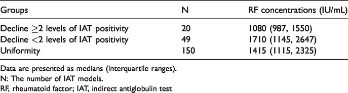
Data are presented as medians (interquartile ranges).
N: The number of IAT models.
RF, rheumatoid factor; IAT, indirect antiglobulin test
Results of the IAT models after treatment with IgG-sensitized latex particles
The median RF concentration of the 219 IAT models was 1360.0 IU/mL (interquartile range: 1030.0, 2220.0 IU/mL). The IAT models were treated with IgG-sensitized latex particles. Following treatment, the RF concentrations were 220.0 (109.0, 355.0) IU/mL. A Wilcoxon test showed that the RF concentrations of IAT models were significantly lower than those before treatment (Z = −10.591, p < 0.0005). After treatment, only five IAT models (5/219, 2.3%) had lower results than those of the control models, and three other IAT models (3/219, 1.4%) had higher results. The proportions of increasing (1.4% vs. 7.3%, x2 = 9.3, p = 0.002) and decreasing (2.3% vs. 31.5%, x2 = 66.6, p < 0.0005) results of the IAT models were significantly lower than those prior to treatment.
Results of two-step IAT models
Because of the limited sample volumes, we tested 96 of the 219 IAT models with the two-step IAT. Compared with the control models, 39 models (39/96, 40.6%) showed decreased results, with 22 samples becoming negative. In addition, the results of 15 IAT models (15/96, 15.6%) were higher than those of control models. However, 10 results (10/96, 10.4%) from 96 IAT models tested by the one-step method were lower than those of the control IAT models, and eight IAT models (8/96, 8.3%) tested by the one-step method gave higher results than the control models. The rate of decrease of the two-step IAT was significantly higher than that of the one-step IAT (40.6% vs. 10.4%, x2 = 23.0, p < 0.0005), but there was no significant difference in the increase rate (15.6% vs. 8.3%, x2 = 3.4, p = 0.065). A Kruskal–Wallis test showed that there were no significant differences in the ratios of the RF (from RF-positive plasma) and IgG (from IAT-positive plasma) concentrations among the decrease group, the increase group and the consistent groups in the two-step IAT models (H = 2.1, p = 0.36, Table 1).
Results of DAT models
Among the 60 results of DAT models, 22 (22/60, 36.7%) were lower than those of the control DAT models, while 8 (8/60, 13.3%) results were higher. Compared with the one-step IAT models, the decrease rate (36.7% vs. 31.5%, x2 = 0.6, p = 0.45) and the increase rate (13.3% vs. 7.3%, x2 = 2.2, p = 0.14) of the DAT models showed no significant differences.
The median RF concentration of the 60 DAT models was 1410.0 (1042.5, 2370.0) IU/mL. The DAT models were treated with IgG-sensitized latex particles. After treatment, the RF concentrations were 221.0 (125.0, 405.0) IU/mL. A Wilcoxon test showed that the RF concentrations of the DAT models were significantly lower than those before treatment (Z = −6.739, p < 0.0005). After treatment, there were only three DAT models (3/60, 5.0%) that had lower results than those of the control models, while one DAT model (1/60, 1.7%) had a higher result. The proportions of increasing (1.7% vs. 13.3%, x2 = 4.3, p = 0.038) and decreasing (5.0% vs. 36.7%, x2 = 18.2, p < 0.0005) results of the DAT models were significantly lower than those prior to treatment.
Results of DAT models with two-fold dilution
The results of control models did not change after two-fold dilution with the mixed-negative plasma. Following two-fold dilution in the DAT models, another 24 DAT models were found have lower results than those of the DAT control models, and two models had higher results. Both the rate of decreasing (76.7% vs. 31.5%, x2 = 39.6, p < 0.0005) and increasing (16.7% vs. 7.3%, x2 = 4.9, p = 0.027) DAT models were significantly higher than those of the IAT models (Table 3).
Rate of decrease and increase of the DAT models after dilution.

Data are presented as N (%).
#x2 = 39.6, p<0.0005; §x2 = 4.9, p = 0.027 compared with IAT models.
DAT, direct antiglobulin test; IAT, indirect antiglobulin test
Following two-fold dilution, as the relative content of RF increased, the degree of decrease of the 17 DAT models became increasingly significant compared with control models (Figure 1). The results of another eight DAT models changed in an inversely proportional trend. Of these, three DAT models that were originally higher than control models became lower than the control models, and the other five models showed a trend of initially decreasing, then increasing and finally dropping at a dilution of 1:16 (Figure 2).

After dilution, the results of control models did not change. As the relative content of RF increased, the degree of decrease in the 17 DAT models became increasingly significant compared with control models. DAT, direct antiglobulin test; RF, rheumatoid factor.

After dilution, the results of control models did not change, but the results of 8 DAT models changed with an inversely proportional trend. The results for X172+R133, X165+R133 and X133+R109 were higher in HCT samples than control models, and then became lower than the control models after dilution. The other 5 models showed a trend of initially decreasing, then increasing and finally dropping at dilutions of 1:16. DAT, direct antiglobulin test; HCT, haematocrit.
Discussion
In this study, the results of 69 IAT models were lower than those of the IAT control models, and 16 IAT models had higher results. We mixed all 219 IAT models with IgG-sensitized latex particles to remove RF. After RF removal, the results of 69 IAT models (the results of which had been lower than those of the control models) were higher than prior to removal of RF, and 64 results were equivalent to those of the control models. The results of 13 IAT models (the results of which had been higher than those of the control models) were equivalent to those of the control models. The results of the remaining models were unchanged. Thus, changes in the results of the 85 IAT models were caused by RF, which not only resulted in a false elevation of IAT results but also caused false decreases and even false negatives.
However, most of the 219 IAT models showed no interference of RF, even in some plasma models with very high RF concentrations. We therefore analysed the correlation between the RF concentration and interference in these 219 models, which were divided into three groups based on the different results of negative interference. There were no significant differences in RF concentrations among the three groups. In addition, we also analysed the ratios of the RF and IgG concentrations in the one-step IAT models and two-step IAT models. There were no significant differences in these ratios among the decrease group, increase group and consistent group. Therefore, it can be concluded that interference was not significantly related to the RF concentration or RF/IgG ratio in the IAT models. However, among the different IAT models containing the same RF-positive plasma, some showed obvious interference, while some showed no interference. Interference in immunoassay results caused by RF was based on the combination of RF with IgG in the reaction system.4–6 Similarly, if RF interfered with the IAT, RF had to first bind to RBC-bound IgG antibodies. In the reaction process of the IAT, two steps are involved in RF binding to the RBC membrane. First, anti-RBC IgG antibodies in plasma bind to the RBC membrane, and then, RF is able to bind to RBC-bound IgG antibodies. If RF fails to bind to RBC-bound IgG antibodies within a limited incubation time, no corresponding interference will occur. To test this hypothesis, we designed two-step IAT to ensure that IgG anti-RBC antibodies in plasma were bound to RBCs before RF reacted with the IgG antibodies coating them, to increase the opportunities for RF to bind to RBCs. The results demonstrated that in 96 IAT models, the incidence of negative interference in the two-step IAT was significantly higher than that in the one-step IAT. Therefore, the models showing no interference in the one-step IAT occurred not because RF did not interfere with the IAT, but because RF did not bind to the RBC-bound IgG antibodies within the limited reaction time. Moreover, the RF-positive plasma added to the one-step IAT was equivalent to a two-fold dilution of IAT-positive plasma, while the RF-positive plasma added in the two-step IAT was not diluted. Therefore, one reason that the incidence of negative interference in the two-step method was higher than that in the one-step method could be because of the higher RF concentration.
Because the DAT detected RBC-bound IgG antibodies and RF-positive plasma in the DAT models was not diluted, the interference in the DAT caused by RF could be more significant according to the above conclusions. Following the detection of 60 DAT models, the incidence of negative interference in the DAT models was not significantly higher than that in the IAT plasma models. Therefore, the DAT models without interference were not because RF did not bind to RBC-bound IgG antibodies within the limited incubation time. In the DAT models, RF-positive plasma and DAT-positive RBC were mixed according to the proportion of DAT-positive patients’ HCTs. We believed that at this proportion, the content of RF in the DAT models relative to RBCs was not high. Therefore, we wondered whether the absence of interference was due to the relative insufficiency of RF in the DAT models. We then performed a dilution series on the DAT models using the corresponding RF-positive plasma to increase the RF content relative to RBCs, followed by measurement of the DAT. After dilution, the incidences of negative interference and positive interference in the DAT models were significantly higher than those in the IAT models. Therefore, we conclude that as long as RF can bind to RBC-bound IgG antibodies, there is a certain correlation between its relative content and the degree of interference. From Figure 1a, we can easily see that as the relative content of RF increases, the degree of false decreases in the DAT results is increasingly obvious, and false negatives can occur.
It is interesting that in the process of diluting the DAT models, the results of three models that were originally higher than those of the DAT control models became lower than those of the control models (Figure 2). Meanwhile, five DAT models were initially falsely decreased. However, after dilution, as the relative content of RF increased, the results did not decrease in an inversely proportional trend. The tests tended to first decrease, then increased and decrease again at high dilution factors (Figure 2). In the DAT, the AHG contained in the gel card bound to the IgG-sensitized RBCs, resulting in agglutination. However, whether agglutination occurred was not only related to the presence of IgG-sensitized RBCs but also to the ratio of AHG to IgG-sensitized RBCs, and only when the ratio was equal did agglutination occur. Like AHG, all of the RF can bind to IgG-sensitized RBCs in the reaction system, and IgM, IgG and IgA are the common subgroups of RF. 7 If the ratio of RF to IgG-sensitized RBCs is equal, RBCs can also be agglutinated. However, AHG was IgG, and its agglutination titre is lower than that of IgM. Therefore, the DAT-positive results from RF interference were more obvious than those of the control models, resulting in a false increase in the results. However, if the content of RF relative to IgG-sensitized RBCs was too high or too low, no detectable agglutination could be detected. The binding of high-content RF to IgG-sensitized RBCs hindered the binding of AHG to IgG antibodies, resulting in a false-negative DAT. An excessively low level of RF could not only bind to a portion of IgG antibodies on RBCs and prevent AHG from binding to IgG, but could also alter the original proportion of AHG and IgG-sensitized RBCs, so the expected agglutination would be weakened. Therefore, we could only observe such a phenomenon under certain specific dilution ratios of some DAT models, which showed, once again, that as long as RF could bind to IgG-sensitized RBCs, there was a correlation between relative RF content and the magnitude and type of interference. In the IAT test, both RF and AHG were used as secondary antibodies against anti-RBC antibodies. RF could change the proportion of AHG and IgG-sensitized RBCs. Therefore, the agglutination expected when the ratio of AHG and IgG-sensitized RBCs was equal would be weakened or would not occur, resulting in falsely decreased or false-negative IAT results. Similarly, if RF made the proportional relationship between AHG and IgG-sensitized RBCs more suitable for agglutination, agglutination might be enhanced, resulting in false increases or false-positive IAT results.
Even so, the interference between different DAT models had no obvious relationship with the concentration of RF, which agreed with the conclusions of our previous studies.4–5 In addition, regardless of whether the test steps were changed or the relative concentrations of RF increased, there were always some IAT models and DAT models that did not show any interference. Therefore, we returned to the question of whether RF can really bind RBC-bound IgG antibodies. According to Renaudineau et al., 8 RF in RA patients mainly reacted with Ga determinants of IgG, which are mainly present on the Fc of IgG1, IgG2 and IgG4, but not IgG3. However, in diseases such as HDN and AIHA, no matter whether IgG antibodies were bound or free, they were mainly IgG1 and IgG3.9–13 In this study, IAT-positive plasma in IAT models and DAT-positive RBCs in DAT models were derived from patients with HDN and AIHA. Therefore, the anti-RBC antibodies in models were also mainly IgG1 and IgG3. If they were IgG3, it would have been difficult for RF to bind them. Meanwhile, for different models, there was no correlation between the degree of interference and RF concentration, and the degree of interference might be related to the subtypes of the anti-RBC antibodies. If the content of IgG1 anti-RBC antibodies was high, then low-concentration RF could produce significant interference.
DAT and IAT results are important bases to support the diagnosis of AIHA. Despite the application of high-sensitivity techniques, 10% of DAT results for patients with AIHA were still negative, and only approximately 3% of DAT negative results were true negatives. 2 Most DAT-negative cases, in addition to being caused by IgA antibodies or low-affinity antibodies, potentially represented false negativity caused by RF.
One additional point needs to be highlighted—the principle of the cross-matching test is similar to that of the IAT. If IgG anti-RBC antibodies exist in the plasma of the recipient, then RBC agglutination will occur in the major cross-matching test. However, if there is also RF in the recipient’s plasma, interference would also appear in the cross-matching test. If it is a false positive, it may result in unnecessary examinations or a delay of blood dispensing; if it is a false negative, then RBCs would be wrongly transfused to the patient, resulting in a serious transfusion-related adverse event. According to our statistics, the incidence of such adverse events was 0.91% in the Wuhan Children's Hospital from January 2016 to December 2017 and 0.0104% in the Union Hospital from January to December 2017. Whether any transfusion-related adverse events were caused by incorrect transfusions due to RF interference with the cross-matching results requires further research. However, this result at least provides a warning that we should also consider whether false negatives may also occur for so-called normal clinical trial results. This is especially important when matching blood to patients with diseases such as AIHA and RA, since 57.9% of SLE patients are AIHA patients, 14 and RF is detectable in 15–35% of SLE patients . 7 In addition, RF can be detected in 70% to 90% of patients with RA. Therefore, there is a risk of RF interference in test results for this group of patients related to blood cross-matching, which requires special attention.
In summary, in the DAT model or the IAT model of our study, as long as RF in the patient's plasma was able to bind to IgG anti-RBC antibodies, interference with the IAT and DAT was related to the RF content relative to the number of anti-RBC antibody-sensitized RBCs.