
Abstract
Objectives
The objective was to investigate the effects of microRNA-421 against myocardial ischemia/reperfusion injury in C57BL/6 mice.
Methods
Male C57BL/6 mice (n = 27) were randomly divided into three groups: normal control (NC) group (sham-treated); I/R model group, which underwent the I30min/R24h model (ischemia for 30 minutes followed by reperfusion for 24 hours); and the miRNA group, which were injected with miR-421. Pathology was assessed by hematoxylin and eosin staining and myocardial infarct size was measured by triphenyltetrazolium chloride staining. The apoptosis rate was measured by TUNEL assay, and relative expression of toll-like receptor-4 (TLR4), Janus kinase 2 (JAK2), and signal transducer and activator of translation 3 (STAT3) was evaluated by immunohistochemistry. Interleukin (IL)-6, tumor necrosis factor (TNF)-α, IL-10, and high mobility group protein B1 (HMGB1) serum concentrations were measured by ELISA.
Results
Compared with the NC group, in the model group, the myocardial infarction was large; inflammatory cell infiltration was severe; apoptosis was enhanced; expression of TLR4, JAK2, and STAT3 was increased; and serum concentrations of IL-6, TNF-α, IL-10, and HMGB1 were significantly increased. In the miRNA group, the ischemia/reperfusion injury was significantly improved.
Conclusions
Overexpression of miRNA-421 could reduce ischemia/reperfusion inflammatory response, perhaps via inactivation of TLR4, JAK2, and STAT3.
Keywords
Introduction
Reperfusion therapy for myocardial infarction has advantages and disadvantages. Early and effective recanalization of coronary blood flow is the most effective method to improve the prognosis of coronary heart disease, but reperfusion itself can also cause myocardial injury. The mechanism of ischemia/reperfusion (I/R) injury is complex, and inflammatory reaction is its characteristic manifestation. 1 Toll-like receptor 4 (TLR4) is an important pathogen recognition signal molecule, and its mediated inflammatory response plays an important role in the myocardial ischemia reperfusion injury (MIRI) process. 2 MicroRNAs (miRNAs) are endogenous, regulatory, small non-coding RNAs that regulate cell differentiation, proliferation, and apoptosis by controlling the translation and expression of target genes.3–5 Based on results from previous studies, miR-421 has an important role in cell apoptosis.6–8 In those studies, different target genes of miR-421 were identified. Analysis using bioinformatics software (http://www.targetscan.org/mamm_31/) indicated that TLR4 was another target gene of miR-421. We hypothesized that overexpression of miR-421 might improve myocardial damage induced by I/R via regulation of TLR4 in an in vivo study.
Materials and methods
This animal experiment was approved by the Laboratory Animal Ethics Committee of the Affiliated Cardiovascular Hospital of Qingdao University, and it conforms to the principles of animal protection, animal welfare and ethics, and the relevant provisions of the National Laboratory Animal Welfare ethics.
Reagents
Enhanced chemiluminescence reagent (ECL) highly sensitive luminescent liquid was from Beyotime Biotech Inc. (Jiangsu, China); polyvinyl difluoride (PVDF) membrane (0.45 µm) was purchased from GenScript (Nanjing) Co. Ltd. (Nanjing, China); horseradish peroxidase (HRP)-labeled sheep anti-rabbit, as the secondary antibody, was purchased from GenScript (Nanjing) Co. Ltd.; and non-fat milk powder was from Oxoid (Basingstoke, UK). Acrylamide and N,N-methylene-bis-acrylamide, glycine, and β-mercaptoethanol were from Amresco/VWR International (Radnor, PA, USA). Alkyl sodium sulfate, persulfate amine, methyl ethylenediamine, trihydroxymethyl aminomethane, bromophenol blue, nitrocellulose membrane, and the TUNEL kit were from Sigma (St. Louis, MO, USA). The TLR4, JAK2, and STAT3 antibodies were purchased from Abcam (Cambridge, UK), and miR-421 was purchased from GenScript (Nanjing) Co. Ltd.
Animal grouping and model preparation
Twenty-seven male C57BL/6 mice (body weight: 20 to 30 g) were purchased from Qingdao University. The feeding conditions were as follows: room temperature of 21 to 25°C, 12-hour day/night cycle, with free drinking and feeding. The mice were randomly divided into three groups: mice in the NC group underwent a sham operation (n = 9); mice in the model group were treated with I30min/R24h model (ischemia for 30 minutes followed by reperfusion for 24 hours) operation (n = 9); and mice in the miRNA group were injected with 10 mL/kg miR-421 in the caudal vein and underwent the same ischemia/reperfusion protocol every day (n = 9). The I/R animal model preparation steps were as follows: 9 narcosis was induced in mice using isoflurane; then, a small incision (around 1.2 cm) was cut in the left thoracic cavity, the tissue was peeled back, and the heart gently extruded. A slipknot was made around the left anterior descending artery using 6-0 silk. The heart was then placed back in the chest using 4-0 silk and the skin sutured, leaving the 6-0 slipknot outside. After 30 minutes of ischemia, the slipknot was released to allow recovery of coronary artery perfusion. Mice in the miRNA group were injected with 0.2 mL of miR-421 in the caudal vein 7 days before model preparation. Mice in the other two groups were injected with 0.2 mL of 0.9% normal saline. All mice survived following the 24-hour reperfusion. The pathological morphology and cardiac function of mice were detected by hematoxylin and eosin (H&E) and triphenyltetrazolium chloride (TTC) staining, respectively, to verify the model.
H&E staining
After 24 hours of reperfusion, the myocardial tissue of mice was collected and paraffin-embedded sections were prepared and stained with H&E. Pathological changes were observed using an optical microscope under 200× magnification.
Evans blue and TTC staining
After reperfusion for 24 hours, mice were injected in the aorta with 1% Evans blue and then the heart was removed and cut into slices 1 mm thick. Cardiac slices were incubated in TTC solution at 37°C for 15 minutes. The infarct area (IA), area at risk (AAR), and left ventricle (LV) were measured by Photoshop CS5 software (Adobe Inc., San Jose, CA, USA).
TUNEL assay
The cardiac tissues were placed in 4% paraformaldehyde to fix, and paraffin-embedded sections were prepared according to the instructions of the terminal deoxynucleotidyl transferase mediated dUTP biotin nick-end labeling (TUNEL) kit. Cardiac tissue slices were observed under the microscope; the nucleus and cytoplasm were dense and fluorescence was positive. The cardiac tissues were stained with PBS as negative control in this experiment. At high magnification, five visual fields were randomly selected and the apoptotic cells in those fields were counted.
Immunohistochemistry (IHC) assay
Protein expression of TLR4, JAK2, and STAT3 in cardiac tissue slices (4 µm) was measured by horseradish peroxidase method as described in the literature. 10 Briefly, paraffin sections of tissue were made, followed by dewaxing, hydration, antigen repair, and incubation with 3% H2O2 at room temperature for 10 minutes to eliminate endogenous peroxidase activity. Then, sections were incubated at 37°C for 15 minutes with the addition of sealing solution, followed by discarding of the sealing solution without washing. The primary antibody (1:100) was added and incubated at 4°C overnight. The section was removed the next day, the corresponding biotin-labeled second antibody was added, and the section incubated at 37°C for 15 minutes, followed by the dripping of horseradish peroxidase-labeled working solution of Streptomyces ovalbumin and incubation at 37°C for 10 to 15 minutes. After development with DAB for 1 to 2 minutes, the reaction was stopped by rinsing with tap water, followed by washing with tap water, and restaining with hematoxylin, dehydration, and sealing with neutral gum. All pathological sections were scored by two experienced pathologists without clinical pathological data. If the results differed, they were assessed again until consensus was reached. The intensity of immunohistochemical staining was divided into five grades (from 0 to 4), and the percentage of specific pigmented cells (0% to 100%) was assessed in five fields of view: 0 (no expression, positive cell number <5%), 1 (low expression, positive cell number between 5% and 25%), 2 (middle expression, positive cell number between 25% and 50%), 3 (high expression, positive cell number between 50% and 75%), and 4 (high expression, positive cell number is ≥75%).
ELISA assay
After reperfusion for 24 hours, blood of the carotid artery was collected before cardiac tissues were harvested. Blood was left for 2 hours and then centrifuged at 2,500 × g for 10 min. The serum was used to measure concentrations of interleukin (IL)-6, tumor necrosis factor (TNF)-α, IL-10, and high mobility group protein B1 (HMGB1) by ELISA kit (Sigma, St. Louis, MO, USA).
Statistical analysis
The data of this study were analyzed using IBM SPSS Statistics for Windows, version 19.0 (IBM Corp., Armonk, NY, USA), and data are expressed as mean ± standard deviation (SD). Analysis of variance was used to analyze differences between groups. The t-test was used to analyze the variance between groups, and one-way ANOVA was used. A p-value < 0.05 indicated a significant difference.
Results
Myocardial pathology
H&E staining of myocardial tissues from mice in the NC group showed that the myocardial stripes were clear, the arrangement of myocardial fibers was regular, the nuclei were normal, and no hyperemia, hemorrhage, or inflammatory reaction was found in the interstitium. In mice of the I/R model group, the myocardial cross striation disappeared and the myocardial cells were disordered and accompanied by degeneration and necrosis. In the miRNA group, the morphological structure of myocardial tissue was intermediate between that of the I/R model group and the NC group, and the degree of degeneration and necrosis of myocardial cells were alleviated (Figure 1).

Pathology of difference groups by hematoxylin and eosin staining (200× magnification).
Myocardial infarct area
We observed no significant differences among the three groups (NC, I/R model, and miRNA) in AAR. However, the IA of mice in the I/R model group was significantly increased compared with that of the NC group (p < 0.001), and the IA of the miRNA group was significantly improved (p < 0.05). compared with that of the I/R model group. Data are shown in Figure 2.

The effect of miR-421 on myocardial infarct sizes of ischemia/reperfusion (I/R) mice. (a) NC, normal control mice (treated with sham operation). (b) Model, ischemia/reperfusion (I/R) model group mice. (c) miRNA, mice injected with miR-421 based on model; AAR, area at risk; LV, left ventricle; IA, infarct area. ***p < 0.05, compared with NC group; #p < 0.05, compared with Model group.
Cell apoptosis rate
Compared with the NC group, the number of cells positive for apoptosis in the I/R model group was significantly increased (p < 0.001); however, the apoptosis rate in the miRNA group was significantly decreased compared with that of the I/R model group (p < 0.05). The relative data are shown in Figure 3.

Apoptosis of cardiomyocytes in different groups by terminal deoxynucleotidyl transferase mediated dUTP biotin nick-end labeling (TUNEL) assay (200× magnification). NC, normal control mice (treated with sham operation); Model, ischemia/reperfusion (I/R) model group mice; miRNA, mice injected with miR-421 based on model. ***p < 0.05, compared with NC group; #p < 0.05, compared with Model group.
Relative protein expression
Compared with expression in the NC group, the expression of TLR4, JAK2, and STAT3 in the I/R model group was significantly upregulated, as shown by immunohistochemistry (p < 0.001). However, expression of these proteins in the miRNA group (with miR-421 overexpression) was significantly downregulated compared with that in the I/R model group (p < 0.05, respectively). The data are shown in Figures 4–6.
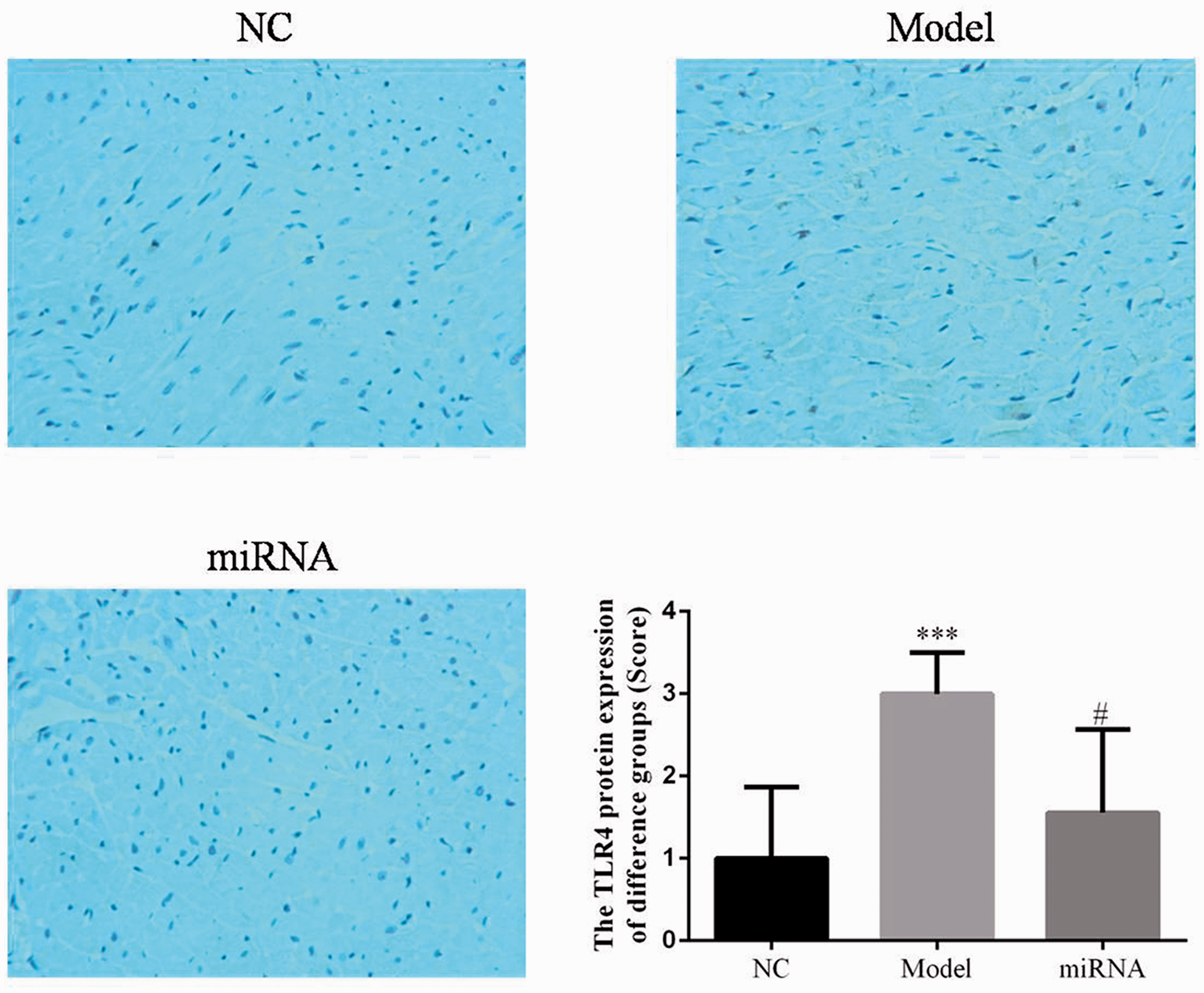
Expression of TLR4 protein in different groups by immunohistochemistry (200× magnification). NC, normal control mice (treated with sham operation); Model, ischemia/reperfusion (I/R) model group mice; miRNA, mice injected with miR-421 based on model; TLR4, toll-like receptor 4. ***p < 0.05, compared with NC group; #p < 0.05, compared with Model group.

Expression of JAK2 protein in different groups by immunohistochemistry (200× magnification). NC, normal control mice (treated with sham operation); Model, ischemia/reperfusion (I/R) model group mice; miRNA, mice injected with miR-421 based on model; JAK2, Janus kinase 2. ***p < 0.05, compared with NC group; #p < 0.05, compared with Model group.

Expression of STAT3 protein in different groups by immunohistochemistry (200× magnification). NC, normal control mice (treated with sham operation); Model, ischemia/reperfusion (I/R) model group mice; miRNA, mice injected with miR-421 based on model; STAT3, signal transducer and activator of transcription 3. ***p < 0.05, compared with NC group; #p < 0.05, compared with Model group.
Concentrations of IL-6, TNF-α, IL-10, and HMGB1
Compared with those in the NC group, concentrations of IL-6, TNF-α, IL-10, and HMGB1 were significantly increased in the I/R model group (all p < 0.05); however, in the miR-421 group, concentrations of IL-6 and TNF-α were significantly lower and those of IL-10 and HMGB1 of were significantly higher than those of the I/R model group (p < 0.05, respectively). The relative data are shown in Figure 7.

Effects of miR-421 on the serum level of inflammatory cytokines of ischemia/reperfusion (I/R) model mice. NC, normal control mice (treated with sham operation); Model, I/R model group mice; miRNA, mice injected with miR-421 based on model; IL, interleukin, TNF-α, tumor necrosis factor-α; HMGB1, high mobility group protein B1.
Discussion
TLR4 is an important pathogen pattern recognition signal molecule, and its mediated inflammatory response plays an important role in the MIRI process. 11 Defects in TLR4 signal transduction can reduce infarct size during ischemia reperfusion and reduce inflammation associated with myocardial injury, including neutrophil aggregation, oxidative stress, and activation of complement deposition. 12 In patients with coronary heart disease after reperfusion therapy, TLR4 overexpression in peripheral blood monocytes and the induced release of serum inflammatory factors aggravate myocardial injury. 13 miRNA is a highly conserved, non-coding, small single-stranded RNA in eukaryotes that can regulate gene expression and participate in cell differentiation, proliferation, and apoptosis. The expression of miRNA is specific. The results suggest that miRNA expressed in the heart is involved in cardiovascular diseases.14–16 Based on bioinformatics software, we found that miR-421 might target TLR4. In the current study, we investigated the effects and mechanisms of miR-421 in improving myocardial damage induced by I/R.
We investigated the molecular mechanism of miR-421 inhibiting and ameliorating the I/R inflammatory response and apoptosis in I/R model mice by injecting miR-421 into the tail vein. Our results showed that myocardial injury, myocarditis infiltration, and myocardial infarction were observed in I/R model mice; however, miR-421 treatment significantly decreased the infiltration of myocarditis and the degree of apoptosis of myocardial cells. Overexpression of miR-421 could work to ameliorate I/R injury by regulation of the TLR4/JAK2/STAT3 pathways.
The JAK2/STAT3 signaling pathway is involved in cell proliferation, differentiation, apoptosis, inflammation and immune regulation, and many other important biological processes. Studies have shown that inhibition of JAK2/STAT3 activation protects against fructose-induced hepatitis, and the JAK2 signaling pathway associated with TLR4 plays an important role in phagocytosis of macrophages. 17 , 18 The protective effect of berberine on myocardial I/R damage is related to JAK2/STAT3 signaling pathway and endoplasmic reticulum stress. 19 Cardioprotective preconditioning with remifentanil also activates the JAK2/STAT3 pathway. 20 In this study, by detecting the expression level of JAK2/STAT3, we found that expression of JAK2/STAT3 in the miRNA group was reduced compared with that in the I/R model group. Thus, the protective effect of miR-421 on I/R mice is related to inactivation of the JAK2/STAT3 signaling pathway.
IL-6 and TNF-α are proinflammatory factors, and IL-10 is an anti-inflammatory factor. HMGB1 is a non-histone nuclear protein widely distributed in eukaryotic cells. It maintains nucleosome structure and regulates gene transcription in cells. After cell necrosis or when immune cells are stimulated by pathogens, HMGB1 is secreted to the extracellular region and actively migrates to the target organ or tissue site. As an important inflammatory cytokine, it participates in the inflammatory response of MIRI.21–23 Serum HMGB1 concentration is closely related to the severity of myocardial ischemia and the effect of reperfusion therapy in patients with coronary heart disease. 24 Inhibition of HMGB1 activity has protective effects on animal models of infectious diseases induced by infectious factors and noninfectious injuries. 25 Our results showed that the expression of proinflammatory cytokines IL-6 and TNF-α increased in I/R model mice, while the serum anti-inflammatory factors IL-10 and HMGB1 showed a compensatory decrease. Thus, miR-421 can not only inhibit the expression of proinflammatory cytokines IL-6 and TNF-α, but also upregulate the expression of anti-inflammatory factors IL-10 and HMGB1, suggesting that miR-421 may play a cardioprotective role by inhibiting inflammation via regulation of TLR4 expression.
In conclusion, miR-421 overexpression had protective effects on myocardial ischemia/reperfusion injury in C57BL/6 mice, including reducing infarct size, improving cardiac function, inhibiting inflammatory response, and regulating the expression of inflammatory cytokines in serum. The effects of miR-421 might be related to the TLR4/JAK2/STAT3 pathway.
Footnotes
Acknowledgements
Lin-lin Guo was responsible for the study concepts and design, data analysis, manuscript preparation and editing; Ming-lei Guo is the guarantor of integrity of the entire study, definition of intellectual content, and manuscript review; Jian Yao was responsible for the literature research, data acquisition, and manuscript preparation; Yun-qi Weng was responsible for the clinical studies, experimental studies, and statistical analysis; Xue-zhi Zhang was responsible for the experimental studies, data acquisition, and data analysis. All authors approved this manuscript.
Declaration of conflicting interest
The authors declare that there is no conflict of interest.
Funding
This research received no specific grant from any funding agency in the public, commercial, or not-for-profit sectors.